

A self-assembled and H2O2-activatable hybrid nanoprodrug for lung infection and wound healing therapy
- PMID: 40365280
- PMCID: PMC12068296
- DOI: 10.7150/thno.114344
A self-assembled and H2O2-activatable hybrid nanoprodrug for lung infection and wound healing therapy
Abstract
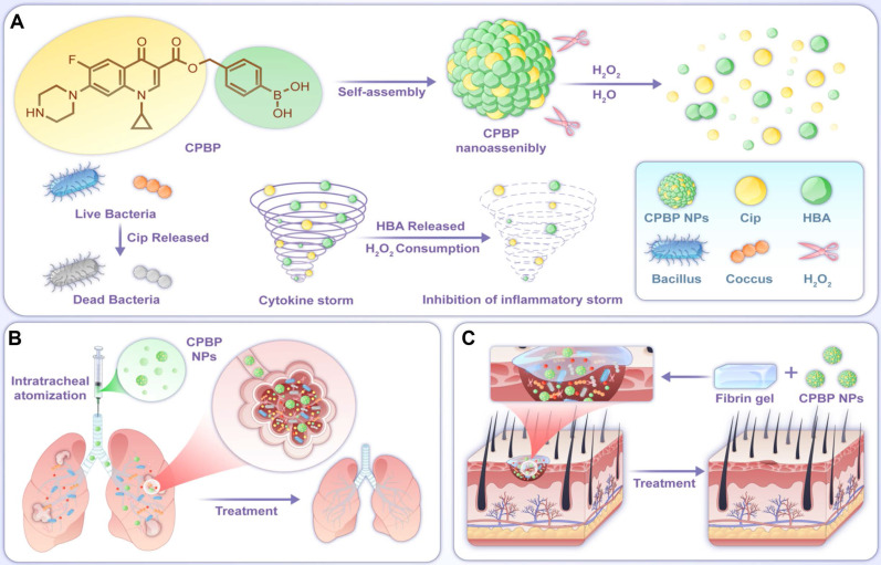
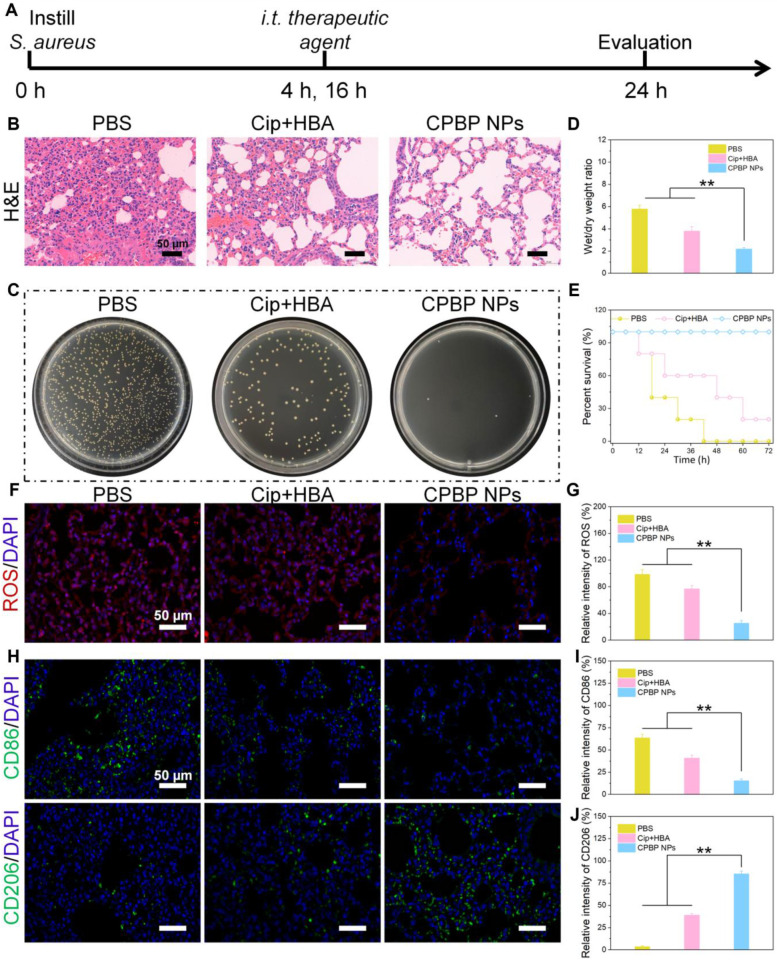
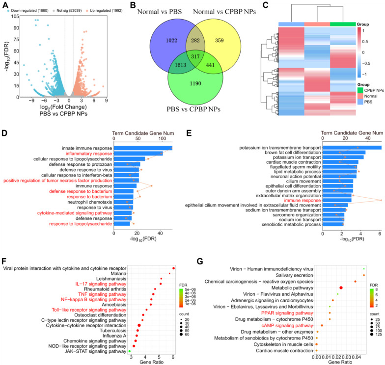
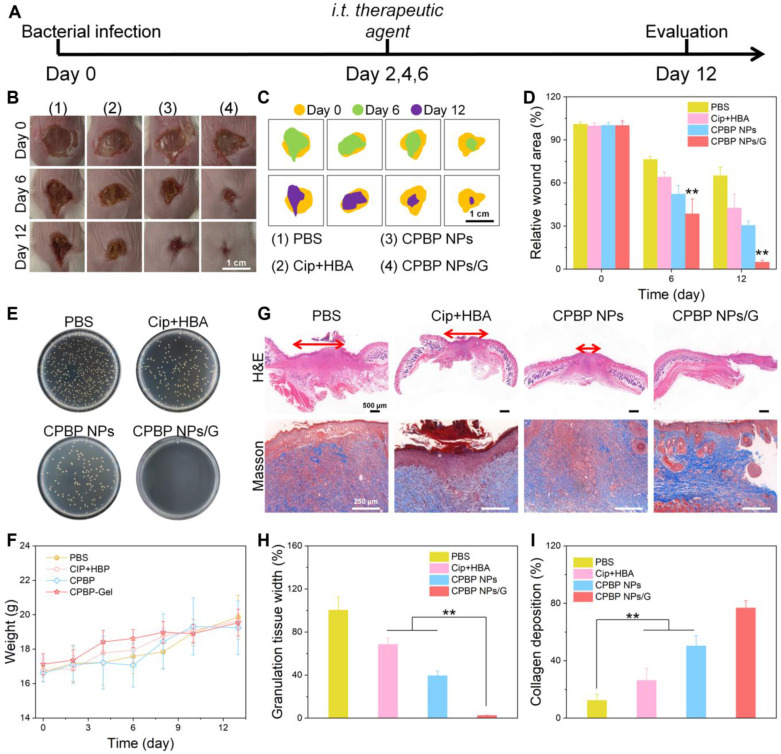
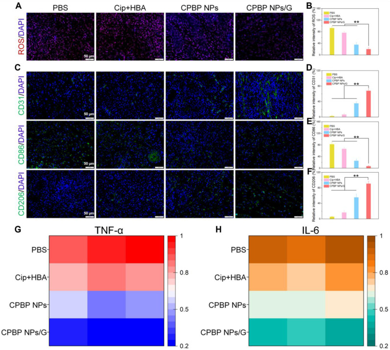
Background: The pursuit of effective antibacterial strategies aimed at mitigating pathogenic bacterial infections while minimising drug resistance remains of paramount importance. A combinational therapeutic strategy that integrates distinct treatment components can enhance overall efficacy and mitigate undesired effects, thereby exhibiting considerable promise in combating bacterial infections. Methods: In this study, a meticulously engineered self-assembling hybrid nanoprodrug (CPBP NPs) has been devised, functioning as a hybrid prodrug of Ciprofloxacin (Cip) and hydroxybenzyl alcohol (HBA). Results: CPBP molecules can generate nanoassemblies via self-assembly and subsequently undergo decomposition to synchronously release Cip and HBA upon hydrogen peroxide (H2O2) exposure. The CPBP NPs exert antibacterial and anti-inflammatory properties through the controlled release of Cip and HBA, while also facilitating the scavenging of reactive oxygen species. These CPBP NPs exhibit broad-spectrum antibacterial activity against both Gram-negative bacteria (E. coli, 98.4%) and Gram-positive bacteria (S. aureus, 98.5%). Notably, CPBP NPs not only accumulate in the lungs to facilitate organ-specific infection treatment but also expedite the healing process of infected wounds. Conclusions: Consequently, this H2O2-activatable hybrid nanoprodrug, possessing excellent biocompatibility, holds substantial promise for advancing clinical applications in managing bacterial infections.
Keywords: Lung infection; Nanoprodrug; Self-assemblies; Synergetic therapy; Wound healing.
© The author(s).
Conflict of interest statement
Competing Interests: The authors have declared that no competing interest exists.
Figures
Similar articles
-
A Zr-based metal-organic framework drug release system with long-lasting antibacterial behavior for accelerating wound healing.Dalton Trans. 2024 Dec 10;53(48):19226-19234. doi: 10.1039/d4dt02734e. Dalton Trans. 2024. PMID: 39513398
-
Coordination-Driven Self-Assembly of Metal Ion-Antisense Oligonucleotide Nanohybrids for Chronic Bacterial Infection Therapy.ACS Appl Mater Interfaces. 2024 Jun 5;16(22):28041-28055. doi: 10.1021/acsami.4c01453. Epub 2024 May 20. ACS Appl Mater Interfaces. 2024. PMID: 38767982
-
Multifunctional EGCG@ZIF-8 Nanoplatform with Photodynamic Therapy/Chemodynamic Therapy Antibacterial Properties Promotes Infected Wound Healing.ACS Appl Mater Interfaces. 2024 Sep 25;16(38):50238-50250. doi: 10.1021/acsami.4c08169. Epub 2024 Sep 16. ACS Appl Mater Interfaces. 2024. PMID: 39284745
-
Green synthesis of polyethylene glycol coated, ciprofloxacin loaded CuO nanoparticles and its antibacterial activity against Staphylococcus aureus.Sci Rep. 2024 Sep 11;14(1):21246. doi: 10.1038/s41598-024-72322-1. Sci Rep. 2024. PMID: 39261712 Free PMC article.
-
Disease-Inspired Design of Biomimetic Tannic Acid-Based Hybrid Nanocarriers for Enhancing the Treatment of Bacterial-Induced Sepsis.Mol Pharm. 2024 Oct 7;21(10):4924-4946. doi: 10.1021/acs.molpharmaceut.4c00048. Epub 2024 Aug 30. Mol Pharm. 2024. PMID: 39214595
Cited by
-
D-Tryptophan Promotes Skin Wound Healing via Extracellular Matrix Remodeling in Normal and Diabetic Models.Int J Mol Sci. 2025 Jul 24;26(15):7158. doi: 10.3390/ijms26157158. Int J Mol Sci. 2025. PMID: 40806291 Free PMC article.
References
-
- Wang W, Cui Y, Wei X, Zang Y, Chen X, Cheng L. et al. CuCo2O4 Nanoflowers with multiple enzyme activities for treating bacterium-infected wounds via cuproptosis-like death. ACS Nano. 2024;18:15845–63. - PubMed
-
- Hatlen JT, Miller GL. Staphylococcal Skin and Soft Tissue Infections. Infect Dis Clin North Am. 2021;35:81–105. - PubMed
-
- Roope SJL, Smith DR, Pouwels BK, Buchanan J, Abel L, Eibich P. et al. The challenge of antimicrobial resistance: What economics can contribute. Science. 2019;364:eaau4679. - PubMed
MeSH terms
Substances
LinkOut - more resources
Full Text Sources
Medical
Miscellaneous
