. 2025 Apr 29:16:1542296.
doi: 10.3389/fphar.2025.1542296. eCollection 2025.

Dl-3-n-butylphthalide attenuates DOX-induced cardiotoxicity in mice by inhibiting Nrf2/Keap1 complex formation

Affiliations

Dl-3-n-butylphthalide attenuates DOX-induced cardiotoxicity in mice by inhibiting Nrf2/Keap1 complex formation

Yixiao Yan et al. Front Pharmacol. .

Abstract

Introduction: Drug-induced cardiotoxicity (DICT), defined as myocardial injury caused by direct or indirect toxicity of therapeutic agents, disrupts cardiovascular homeostasis, underscoring the urgent need for preventive strategies in clinical practice. Doxorubicin (DOX), a clinically established anthracycline chemotherapeutic, induces dose-dependent cardiotoxicity driven by reactive oxygen species overproduction. Notably, Dl-3-n-butylphthalide (NBP), a bioactive phytochemical derived from celery, has shown potential in mitigating DOX-induced cardiomyopathy via its antioxidant activity. Therefore, this study aimed to investigate the protective effects of NBP on DOX-induced cardiomyopathy, with a focus on elucidating the underlying mechanisms.

Method: We developed both in vivo and in vitro models of DOX-induced cardiotoxicity. For the animal model, male C57BL/6 mice were administered with DOX (4 mg/kg, i.p.) once a week for 3 weeks. For the cell model, H9C2 myoblasts were exposed to 1 μM DOX for at least 6 h to establish acute cardiotoxicity.

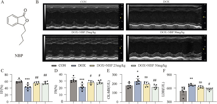
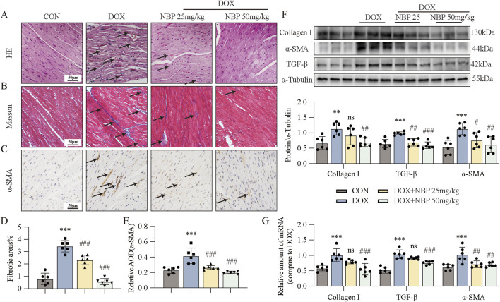
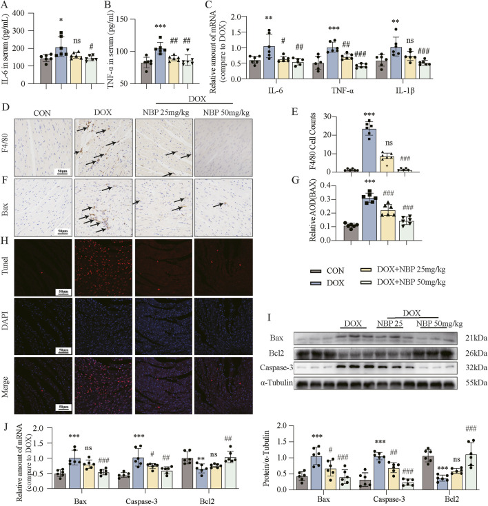
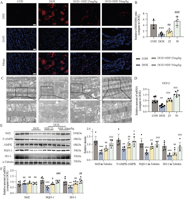
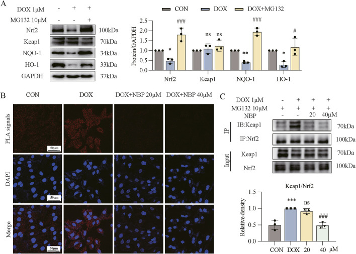
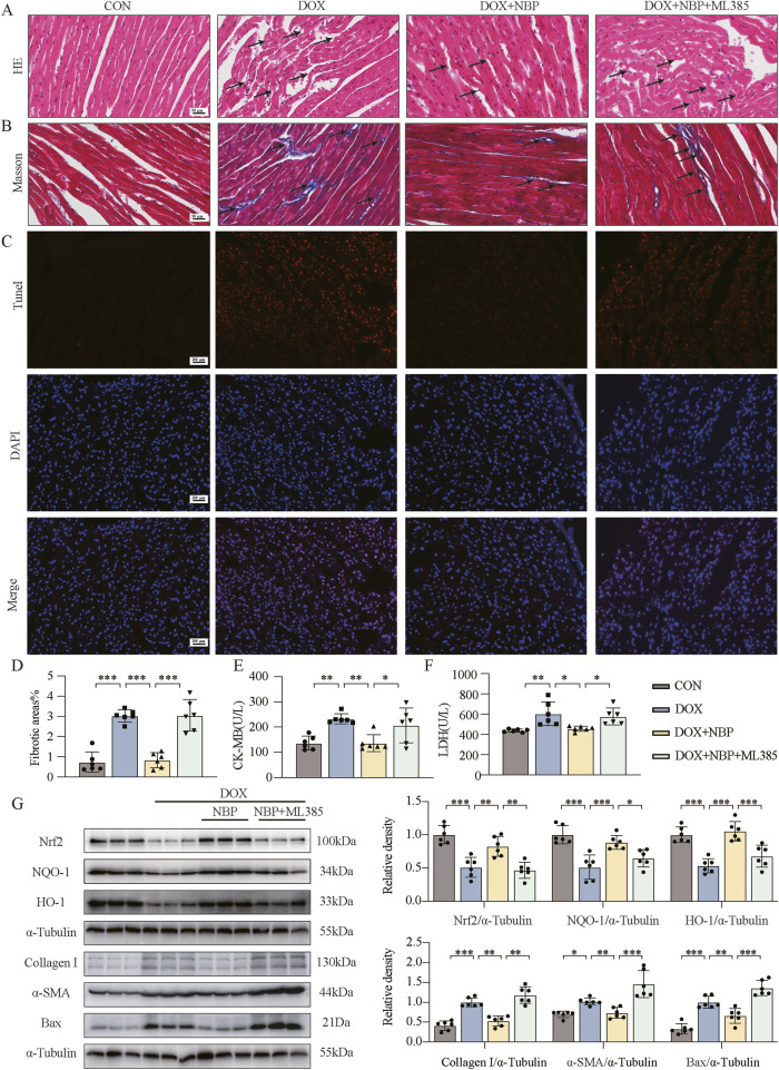
Results: Our results demonstrate that NBP significantly improves cardiac function, as evidenced by approximately 10% increase in cardiac functional parameters (ejection fraction and left ventricular shortening fraction). Besides, NBP exerts favorable effects on cardiac inflammation, apoptosis, fibrosis, and mitochondrial damage both in vivo and in vitro. Further mechanistic investigations revealed that NBP blocks the interaction between Kelch-like ECH-associated protein-1 (Keap1) and Nrf2, thereby preventing the formation of the Nrf2/Keap1 complex.

Discussion: This study indicate that NBP alleviates DOX-induced cardiotoxicity by inhibiting Nrf2/Keap1 complex formation, highlighting its potential as a therapeutic agent for DICT and suggest that Nrf2/Keap1 may be a potential therapeutic target for the management of this condition.

Keywords: C57BL/6 mice; Dl-3-n-butylphthalide (NBP); doxorubicin (DOX); drug-induced cardiotoxicity (DICT); kelch-like ECH associated protein-1 (Keap1); nuclear factor erythroid 2-related factor 2 (Nrf2).


Conflict of interest statement

The authors declare that the research was conducted in the absence of any commercial or financial relationships that could be construed as a potential conflict of interest.

Figures

FIGURE 1
FIGURE 1
Therapeutic effects of NBP on cardiac function in DOX-induced cardiomyopathy mice. (A) The molecular structure of NBP was shown. (B) Representative cardiac ultrasound images of each group were obtained. (C,D) Quantitative analysis of cardiac functional parameters, including ejection fraction (EF%) and left ventricular shortening fraction (FS%), was performed using Vevo Lab software (n = 6 per group). (E,F) Quantitative analysis of serum cardiac biomarkers was performed. The concentrations of creatine kinase-isoenzyme (CK-MB) and lactate dehydrogenase (LDH) in murine serum samples were determined using dedicated kits (n = 6 per group). The data are presented as the means±SDs. Different groups were compared with a one-way analysis of variance (ANOVA). *P < 0.05, **P < 0.01, ***P < 0.001 compared with CON. # P < 0.05, ## P < 0.01, ### P < 0.001 compared with DOX.
FIGURE 2
FIGURE 2
NBP treatment alleviated DOX-induced cardiac fibrosis. (A) Representative hematoxylin-eosin (H&E) staining myocardial tissue sections (n = 6 mice per group, 1 section per mouse) were performed to demonstrate the protective effects of NBP against DOX-induced structural alterations, with arrows indicating specific areas of myocardial fiber disorganization and structural deficits. (B) Fibrosis in the heart tissues of each group of mice was evaluated through representative micrographs obtained from Masson staining. (C) Representative micrographs of anti-α-smooth muscle actin (α-SMA) staining were obtained to visualize the accumulation of α-SMA, with arrows indicating the accumulation areas. (D,E) Quantification of fibrotic areas (%) and α-SMA abundance via Masson’s trichrome staining and anti-α-SMA staining (n = 6 mice per group, 1 section per mouse). (F) Representative Western blot analysis of collagen type I (Col-I), α-SMA, and transforming growth factor-beta (TGF-β) in heart tissues is shown, with α-Tubulin as the loading control. Bottom, densitometric quantification of the data in (F) (n = 6 per group). (G) qRT-PCR was used to quantify the mRNA levels of Col-I, α-SMA, and TGF-β1 in cardiac tissues (n = 6 per group). The data are presented as the means ± SDs. Different groups were compared with a one-way analysis of variance (ANOVA). *P < 0.05, **P < 0.01, ***P < 0.001 compared with CON. # P < 0.05, ## P < 0.01, ### P < 0.001 compared with DOX.
FIGURE 3
FIGURE 3
NBP treatment alleviated DOX-induced cardiac inflammatory and apoptotic responses. (A,B) Detection of interleukin-6 (IL-6) and tumor necrosis factor-alpha (TNF-α) in the serum through enzyme-linked immunosorbent assay (ELISA) kits (n = 6 per group). Different groups were compared with a one-way analysis of variance (ANOVA). (C) qRT-PCR was conducted to measure the mRNA levels of IL-6, TNF-α, and interleukin-1 beta (IL-1β) in cardiac tissues (n = 6 per group). Different groups were compared with a one-way analysis of variance (ANOVA). (D) Representative micrographs of anti-F4/80-stained samples were obtained to visualize the infiltration of F4/80, with arrows indicating the infiltration areas. (E) Quantitative analysis of the levels of F4/80 (n = 6 mice per group, 1 section per mouse). Different groups were compared with Kruskal-Wallis test. (F) Representative micrographs illustrating anti- Bcl-2-associated X protein (Bax) staining, with arrows indicating the accumulation of Bax. (G) Quantitative analysis of Bax (n = 6 mice per group, 1 section per mouse). Different groups were compared with a one-way analysis of variance (ANOVA). (H) Representative micrographs displaying terminal deoxynucleotidyl transferase dUTP nick end labeling (TUNEL) staining. (I) Western blot analysis revealed the expression of the key proteins Bax, B-cell lymphoma/leukemia 2 (Bcl2), and cysteine-aspartic acid protease 3 (Caspase-3) in cardiac tissues, with α-Tubulin used as the loading control. Bottom, densitometric quantification of the data in (I) (n = 6 per group). Different groups were compared with a one-way analysis of variance (ANOVA). (J) qRT-PCR was conducted to measure the mRNA levels of Bax, Bcl2, and Caspase-3 in cardiac tissues (n = 6 per group). Different groups were compared with a one-way analysis of variance (ANOVA). The data are presented as the means ± SDs. *P < 0.05, **P < 0.01, ***P < 0.001 compared with CON. # P < 0.05, ## P < 0.01, ### P < 0.001 compared with DOX.
FIGURE 4
FIGURE 4
NBP treatment alleviated DOX-induced cardiac oxidative stress (OS) and mitochondrial damage. (A) Representative dihydroethidium (DHE) staining images of cardiac tissues (n = 6 mice per group, 1 section per mouse). (B) Measurment of the glutathione (GSH)/oxidized glutathione (GSSG) ratio in the cardiac tissues through GSH and GSSG assay kit (n = 6 per group). (C) Representative transmission electron microscopy (TEM) images of cardiac tissue mitochondria (n = 3 mice per group, 2 section per mouse). (D) qRT-PCR was carried out to measure uncoupling protein 2 (UCP-2) mRNA level in cardiac tissues (n = 6 per group). (E) Western blot analysis of nuclear factor erythroid 2-related factor 2 (Nrf2), phosphorylated AMP-activated protein kinase (P-AMPK), NAD(P)H quinone dehydrogenase 1 (NQO-1), and Heme oxygenase-1 (HO-1) proteins in cardiac tissues was conducted, with AMP-activated protein kinase (AMPK) and α-Tubulin used as loading controls. Right, densitometric quantification of the data in (E) (n = 6 per group). (F) qRT-PCR was conducted to measure Nrf2, NQO-1 and HO-1 mRNA levels in cardiac tissues (n = 6 per group). The data are presented as the means ± SDs. Different groups were compared with a one-way analysis of variance (ANOVA). *P < 0.05, **P < 0.01, ***P < 0.001 compared with CON. # P < 0.05, ## P < 0.01, ### P < 0.001 compared with DOX.
FIGURE 5
FIGURE 5
NBP inhibited Nrf2/Keap1 binding to alleviate DOX-induced myocardial injury. (A) Western blot analysis of the protein levels of antioxidant genes (Nrf2, Keap1, NQO-1, and HO-1) in H9C2 cells. H9C2 cells underwent a 1-hour pretreatment with MG132, followed by a 24-hour stimulation with DOX (1 μM). Right, densitometric quantification for (A) (n = 3). (B) Effect of NBP on Nrf2/Keap1 binding in H9C2 cells evaluated through proximity ligation assay (PLA) immunofluorescence. H9C2 cells underwent a 1-hour pretreatment with NBP, followed by a 12-hour stimulation with DOX (1 μM). Representative immunofluorescence images from three independent experiments are shown. The PLA signals are red, while the DAPI nuclear stain is blue. (C) For Co-immunoprecipitation (Co-IP), H9C2 cells underwent a 1-hour pretreatment with NBP, followed by a 12-hour stimulation with DOX (1 μM). Bottom, densitometric quantification for (C) (n = 3). The data are presented as the means ± SDs. Different groups were compared with a one-way analysis of variance (ANOVA). *P < 0.05, **P < 0.01, ***P < 0.001 compared with CON. # P < 0.05, ## P < 0.01, ### P < 0.001 compared with DOX.
FIGURE 6
FIGURE 6
The cardioprotective effect of NBP depends on the Nrf2 signaling pathway. (A) Representative hematoxylin-eosin (H&E) staining myocardial tissue sections (n = 6 mice per group, 1 section per mouse) were performed to demonstrate the potential mechanism of NBP against DOX-induced structural alterations, with arrows indicating specific areas of myocardial fiber disorganization and structural deficits. (B) Fibrosis in the heart tissues of each group of mice was evaluated through representative micrographs obtained from Masson staining. (C) Representative micrographs displaying terminal deoxynucleotidyl transferase dUTP nick end labeling (TUNEL) staining. (D) Quantification of fibrotic areas (%) via Masson’s trichrome staining (n = 6 mice per group, 1 section per mouse). (E,F) The concentrations of creatine kinase-isoenzyme (CK-MB) and lactate dehydrogenase (LDH) in murine serum samples were determined using dedicated kits (n = 6 per group). (G) Western blot analysis was utilized to assess the protein levels of Nrf2, NQO-1, HO-1, Collagen I, α-SMA, and Bax in heart tissues. α-Tubulin was used as a loading control (n = 6). The data are presented as the means ± SDs. Different groups were compared with a one-way analysis of variance (ANOVA). *P < 0.05, **P < 0.01, ***P < 0.001.

Similar articles

Cited by

References

    1. Benhar M. (2020). Oxidants, antioxidants and thiol redox switches in the control of regulated cell death pathways. Antioxidants (Basel) 9 (4), 309. 10.3390/antiox9040309 - DOI - PMC - PubMed
    1. Bian M., Fan R., Zhao S., Liu W. (2019). Targeting the thioredoxin system as a strategy for cancer therapy. J. Med. Chem. 62 (16), 7309–7321. 10.1021/acs.jmedchem.8b01595 - DOI - PubMed
    1. Boddicker N. J., Larson M. C., Castellino A., Herrmann J., Inwards D. J., Thanarajasingam G., et al. (2021). Anthracycline treatment, cardiovascular risk factors and the cumulative incidence of cardiovascular disease in a cohort of newly diagnosed lymphoma patients from the modern treatment era. Am. J. Hematol. 96 (8), 979–988. 10.1002/ajh.26230 - DOI - PMC - PubMed
    1. Briehl M. M. (2015). Oxygen in human health from life to death--An approach to teaching redox biology and signaling to graduate and medical students. Redox Biol. 5, 124–139. 10.1016/j.redox.2015.04.002 - DOI - PMC - PubMed
    1. Cheng L., Zhang H., Wu F., Liu Z., Cheng Y., Wang C. (2020). Role of Nrf2 and its activators in cardiocerebral vascular disease. Oxid. Med. Cell Longev. 2020, 4683943. 10.1155/2020/4683943 - DOI - PMC - PubMed

LinkOut - more resources