HER2 expression in different cell lines at different inoculation sites assessed by [52Mn]Mn-DOTAGA(anhydride)-trastuzumab
- PMID: 40365451
- PMCID: PMC12069034
- DOI: 10.3389/pore.2025.1611999
HER2 expression in different cell lines at different inoculation sites assessed by [52Mn]Mn-DOTAGA(anhydride)-trastuzumab
Abstract
Purpose: Positron emission tomography (PET) hybrid imaging targeting HER2 requires antibodies labelled with longer half-life isotopes. With a suitable radiation profile, 52Mn coupled with DOTAGA as a bifunctional chelator is a potential candidate. In this study, we investigated the tumor HER2 specificity and the temporal biodistribution of the [52Mn]Mn-DOTAGA(anhydride)-trastuzumab in preclinical models.
Methods: PET/MRI and PET/CT were performed on SCID mice bearing orthotopic and ectopic HER2-positive and ectopic HER2-negative tumors at 4, 24, 48, 72, and 120 h post-injection with [52Mn]Mn-DOTAGA(anhydride)-trastuzumab. Melanoma xenografts were included for comparison of specificity.
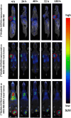
Results: In vivo biodistribution demonstrated strong contrast in HER2-positive tumors, particularly in orthotopic tumors, where uptake was significantly higher than in the blood pool and other organs from 24 h onwards and consistently higher than in ectopic HER2-positive tumors at all time points. Significantly higher tumor-to-blood and tumor-to-muscle ratios were observed in HER2-positive ectopic tumors compared to HER2-negative tumors but only at 4 and 24 h; the differences were likely due to non-specific binding of the tracer. The ratios for orthotopic HER2-positive tumors were significantly higher than those for ectopic HER2-negative tumors and melanoma at all time points. However, the differences between HER2-positive and HER2-negative tumors decreased at later time points.
Conclusion: These results suggest that [52Mn]Mn-DOTAGA(anhydride)-trastuzumab demonstrates efficient tumor-to-background contrast, emphasize the higher tumor uptake observed in orthotopic tumors, and highlight the influence of tumor environment characteristics on uptake.
Keywords: 52Mn; HER2; breast cancer; positron emission tomography; trastuzumab.
Copyright © 2025 Ngô, Vágner, Nagy, Ország, Nagy, Szoboszlai, Csikos, Váradi, Trencsényi, Tircsó and Garai.
Conflict of interest statement
AV, GN, GO, TN, ZS, GTr, and IG are employees of Scanomed Medical Diagnostic Training and Research Ltd. The remaining authors declare that the research was conducted in the absence of any commercial or financial relationships that could be construed as a potential conflict of interest.
Figures


Similar articles
-
Development of 52Mn Labeled Trastuzumab for Extended Time Point PET Imaging of HER2.Mol Imaging Biol. 2024 Oct;26(5):858-868. doi: 10.1007/s11307-024-01948-4. Epub 2024 Aug 27. Mol Imaging Biol. 2024. PMID: 39192059 Free PMC article.
-
52Mn-labelled Beta-cyclodextrin for Melanoma Imaging: A Proof-of-concept Preclinical Study.In Vivo. 2024 Nov-Dec;38(6):2591-2600. doi: 10.21873/invivo.13735. In Vivo. 2024. PMID: 39477386 Free PMC article.
-
Positron-Emission Tomography of HER2-Positive Breast Cancer Xenografts in Mice with 89Zr-Labeled Trastuzumab-DM1: A Comparison with 89Zr-Labeled Trastuzumab.Mol Pharm. 2018 Aug 6;15(8):3383-3393. doi: 10.1021/acs.molpharmaceut.8b00392. Epub 2018 Jul 16. Mol Pharm. 2018. PMID: 29957952
-
Tumor uptake and tumor/blood ratios for [89Zr]Zr-DFO-trastuzumab-DM1 on microPET/CT images in NOD/SCID mice with human breast cancer xenografts are directly correlated with HER2 expression and response to trastuzumab-DM1.Nucl Med Biol. 2018 Dec;67:43-51. doi: 10.1016/j.nucmedbio.2018.10.002. Epub 2018 Oct 16. Nucl Med Biol. 2018. PMID: 30390575
-
111In/68Ga-Labeled anti-epidermal growth factor receptor, native chemical ligation cyclized Affibody ZHER2:342min.2013 Apr 3 [updated 2013 May 23]. In: Molecular Imaging and Contrast Agent Database (MICAD) [Internet]. Bethesda (MD): National Center for Biotechnology Information (US); 2004–2013. 2013 Apr 3 [updated 2013 May 23]. In: Molecular Imaging and Contrast Agent Database (MICAD) [Internet]. Bethesda (MD): National Center for Biotechnology Information (US); 2004–2013. PMID: 23700641 Free Books & Documents. Review.
References
MeSH terms
Substances
LinkOut - more resources
Full Text Sources
Medical
Research Materials
Miscellaneous

