

Pathological Characterization of African Swine Fever Viruses With Genetic Deletions Detected in South Korea
- PMID: 40365486
- PMCID: PMC12074837
- DOI: 10.1155/tbed/9917280
Pathological Characterization of African Swine Fever Viruses With Genetic Deletions Detected in South Korea
Abstract
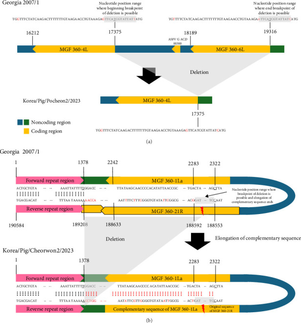
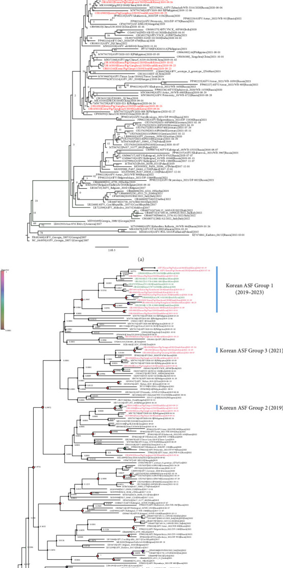
African swine fever virus (ASFV) genotype II has been circulating in South Korea, causing substantial economic losses to the Korean pig industry since 2019. Genetic epidemiological investigations using whole-genome sequencing have been conducted to track the genetic evolution of ASFV. Two ASFV strains were detected in domestic pig farms in South Korea, one with a large deletion in the MGF 360-6L gene and the other in the MGF 360-21R gene. Phylogenetic analysis indicated that all Korean isolates belonged to the Asian subgroup of ASFV genotype II and were further divided into distinct subclusters of Korean African swine fever (ASF) group I. To identify the pathological changes caused by the deletion of MGF 360-6L and MGF 360-21R genes, we evaluated their pathogenicity in experimentally infected domestic pigs. No significant changes in pathogenicity were observed compared to other viruses evaluated in our previous studies. All inoculated pigs died 7-10 days post-inoculation (dpi), showing acute forms of illness with common pathological lesions. These results highlight that large genetic deletions can occur naturally in ASFV, but the deletions in MGF 360-6L and MGF 360-21R genes did not alter pathogenicity in domestic pigs. Further research is needed to understand the roles of these genes, especially in viral replication and pathogenicity in wild boars and ticks.
Keywords: African swine fever; South Korea; animal experiment; genetic characterization; next-generation sequencing; pig farms; virulence.
Copyright © 2025 Seong-Keun Hong et al. Transboundary and Emerging Diseases published by John Wiley & Sons Ltd.
Conflict of interest statement
The authors declare no conflicts of interest.
Figures


Similar articles
-
The first complete genome sequence of the African swine fever virus genotype X and serogroup 7 isolated in domestic pigs from the Democratic Republic of Congo.Virol J. 2021 Jan 21;18(1):23. doi: 10.1186/s12985-021-01497-0. Virol J. 2021. PMID: 33478547 Free PMC article.
-
Comparison of the Virulence of Korean African Swine Fever Isolates from Pig Farms during 2019-2021.Viruses. 2022 Nov 13;14(11):2512. doi: 10.3390/v14112512. Viruses. 2022. PMID: 36423121 Free PMC article.
-
Genomic Epidemiology of African Swine Fever Virus Identified in Domestic Pig Farms in South Korea during 2019-2021.Transbound Emerg Dis. 2024 Jan 16;2024:9077791. doi: 10.1155/2024/9077791. eCollection 2024. Transbound Emerg Dis. 2024. PMID: 40303146 Free PMC article.
-
[African swine fever in Russian Federation].Vopr Virusol. 2012 Sep-Oct;57(5):4-10. Vopr Virusol. 2012. PMID: 23248852 Review. Russian.
-
Three Years of African Swine Fever in South Korea (2019-2021): A Scoping Review of Epidemiological Understanding.Transbound Emerg Dis. 2023 Feb 23;2023:4686980. doi: 10.1155/2023/4686980. eCollection 2023. Transbound Emerg Dis. 2023. PMID: 40303829 Free PMC article.
References
-
- Montgomery R. E. On A Form of Swine Fever Occurring in British East Africa (Kenya Colony) Journal of Comparative Pathology and Therapeutics . 1921;34:159–191. doi: 10.1016/S0368-1742(21)80031-4. - DOI
-
- Quembo C. J., Jori F., Vosloo W., Heath L. Genetic Characterization of African Swine Fever Virus Isolates From Soft Ticks at the Wildlife/Domestic Interface in Mozambique and Identification of a Novel Genotype. Transboundary and Emerging Diseases . 2018;65(2):420–431. doi: 10.1111/tbed.12700. - DOI - PMC - PubMed
MeSH terms
LinkOut - more resources
Full Text Sources
