. 2025 Apr 29:15:1585020.
doi: 10.3389/fcimb.2025.1585020. eCollection 2025.

Escherichia coli aggravates inflammatory response in mice oral mucositis through regulating Th17/Treg imbalance

Affiliations

Escherichia coli aggravates inflammatory response in mice oral mucositis through regulating Th17/Treg imbalance

Jia Liu et al. Front Cell Infect Microbiol. .

Abstract

Introduction: Microbial dysbiosis links to mucosal immune dysregulation, but the specific bacterial contributions to oral mucosal inflammation remain unclear. Escherichia coli (E. coli), a pathogen well-characterized in mucosal immunity and immune regulation studies, has been observed to be enriched in chronic oral inflammatory lesions and was reported to modulate T helper 17 cells (Th17)/T regulatory cells (Treg) homeostasis. Here, we developed an oral mucositis mouse model via tongue scratch and E. coli topical application to investigate its role in Th17/Treg imbalance.

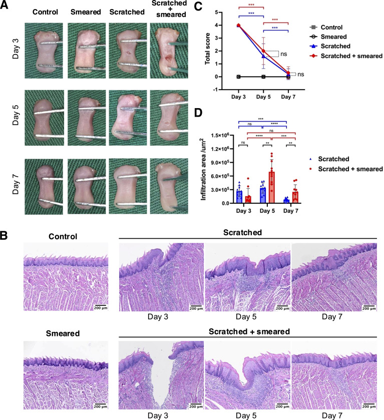
Methods: The inflammatory infiltration was evaluated by macroscopic photography and HE staining. The expression of inflammatory factors in tongue tissue and peripheral blood of mice were detected by immunohistochemical staining and enzyme-linked immunosorbent assay. The number of Th17 and Treg in mice spleen lymphocytes were evaluated with flow cytometry. Differential gene expression analysis, functional enrichment analysis and immune infiltration analysis were performed using RNA-seq data from oral lichen planus (OLP).

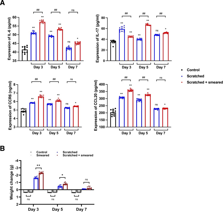
Results: E. coli stimulation aggravated inflammatory responses induced by scratching in lingual mucosa of mice, including increased local and systemic expression of interleukin 6 (IL6), interleukin 17 (IL17), chemokine receptor 6 (CCR6) and chemokine C-C motif ligand 20 (CCL20), increased proportions of Th17 cells and increased Th17/Treg ratio in spleen lymphocytes. Analysis of RNA-seq data from OLP revealed alterations in antimicrobial responses and inflammatory factors associated with upregulation of Th17/Treg balance.

Conclusion: This study supports the role of E. coli in promoting oral mucosal inflammation and provides an experimental basis for in vivo study of OLP from the perspective of microorganisms.

Keywords: Escherichia coli; Th17/Treg response; mice model; mucosal immunity; oral mucosal immune inflammation.


Conflict of interest statement

The authors declare that the research was conducted in the absence of any commercial or financial relationships that could be construed as a potential conflict of interest.

Figures

Figure 1
Figure 1
Macroscopic manifestations and HE staining of tongue tissues of mice. (A) Macrophotography of mice tongue tissues on days 3/5/7. (B) HE staining results of mice tongue tissues in different groups. Scale bar: 100 μm. (C) The total clinical symptom scores of the mice tongue tissue to assess the severity of the modeling lesions(n = 10/group/day). The asterisk (*) indicates statistical significance by Student's t test. *** p < 0.001, ns, no significance. (D) The infiltration area of inflammatory cells at the lesion sites from the mice tongue tissues based on HE staining results (n = 10/group/day). The asterisk (*) indicates statistical significance by Student's t test. ** p < 0.01, *** p < 0.001, **** p < 0.0001, ns, no significance.
Figure 2
Figure 2
Expression of IL-6, IL-17, CCR6 and CCL20 in the tongue mucosa of mice. (A) Representative immunohistochemical image of IL-6, IL-17, CCR6 and CCL20 expression in the tongue tissues of mice in the control group, the scratched group, and the scratched + smeared group. Scale bar: 50 μm. (B) Statistical analysis of expression of IL-6, IL-17, CCR6 and CCL20 in the tongue tissues of mice in the control group, the scratched group, and the scratched + smeared group (n = 10/group/day). Asterisks (*) represent significant differences versus the control group; hash symbols (#) represent significant differences between the scratched group and the scratched + smeared group (Student’s t test, * p < 0.05, ** p < 0.01, ## p < 0.01, ns, no significance).
Figure 3
Figure 3
Typical inflammatory factors were upregulated in mice treated with scratching and E. coli smearing. (A) ELISA was used to detect the expression of IL-6, IL-17, CCR6 and CCL20 in peripheral blood serum samples of mice (n = 10/group/day). Asterisks (*) represent significant differences versus the control group; hash symbols (#) represent significant differences between the scratched group and the scratched + smeared group (Student’s t test, * p < 0.05, ** p < 0.01, ## p < 0.01, ns, no significance). (B) The body weight changes of mice on days 3/5/7 during modeling. The body weight of the control group and the smeared group increased, while the scratched group and the scratched + smeared group decreased. Data present the individual values and mean with SD of each group. Student’s t tests were conducted between the control group and the smeared group, as well as between the scratched group and the scratched + smeared group (n = 10/group/day). * p < 0.05, ** p < 0.01, ns, no significance.
Figure 4
Figure 4
The ratio of Th17/Treg cells were upregulated in the scratched + E. coli smeared group. (A) Representative image of flow cytometry of Th17 cells from the spleen lymphocytes in the scratched group and the scratched + smeared group. (B) Representative image of flow cytometry of Treg cells from the spleen lymphocytes in the scratched group and the scratched + smeared group. (C, D) The proportion of Th17 and Treg cells from spleen lymphocytes of the scratched group and the scratched + smeared group (n=10/group/day). (E) The Th17/Treg ratio of the scratched group and the scratched + smeared group (n=10/group/day). Asterisks (*) in (C-E) represent significant differences versus the control group; hash symbols (#) in (C-E) represent significant differences between the scratched group and the scratched + smeared group (Student’s t test, * p < 0.05, ** p < 0.01, # p < 0.05, ## p < 0.01, ns, no significance).
Figure 5
Figure 5
RNA signature of EOLP characterized by upregulation of antimicrobial response and inflammatory factors. (A) Volcano plots showed differential expression genes between EOLP-RE group and NEOLP-S group. (B) Count number of IL6, IL17A, CCR6, and CCL20 between NEOLP-S (n = 24) and EOLP-RE groups (n = 7) in RNA-seq data. *p < 0.05, **p < 0.01, ****p < 0.0001. (C) Results of Gene Ontology (GO) enrichment analysis. (D) Results of KEGG pathway analysis. (E) Heat map showed immune infiltration scores between NEOLP-S and EOLP-RE samples based on CIBERSORT. (F-I) Results of Gene Set Enrichment Analysis. (EOLP-RE, erosive oral lichen planus with recurrent erosive oral lichen planus follow-up, NEOLP-S, non-erosive oral lichen planus with stable oral lichen planus follow-up).

Similar articles

References

    1. Al-Azri A. R., Gibson R. J., Bowen J. M., Stringer A. M., Keefe D. M., Logan R. M. (2015). Involvement of matrix metalloproteinases (MMP-3 and MMP-9) in the pathogenesis of irinotecan-induced oral mucositis. J. Pathol. Med. 44, 459–467. doi: 10.1111/jop.12255 - DOI - PubMed
    1. Altieri C., Speranza B., Corbo M. R., Sinigaglia M., Bevilacqua A. (2023). Gut-microbiota, and multiple sclerosis: background, evidence, and perspectives. Nutrients 15 (4), 942. doi: 10.3390/nu15040942 - DOI - PMC - PubMed
    1. Araújo R. P. N., Da Silva Freitas F. V., Nunes D. B., Da Silva Brito A. K., Da Costa D. S., De Sousa D. P., et al. . (2024). Investigating the pharmacological potential of phytol on experimental models of gastric ulcer in rats. Naunyn Schmiedebergs Arch. Pharmacol. 397, 7757–7766. doi: 10.1007/s00210-024-03085-9 - DOI - PubMed
    1. Baek K., Lee J., Lee A., Lee J., Yoon H. J., Park H. K., et al. . (2020). Characterization of intratissue bacterial communities and isolation of Escherichia coli from oral lichen planus lesions. Sci. Rep. 10, 3495. doi: 10.1038/s41598-020-60449-w - DOI - PMC - PubMed
    1. Baker J. L., Mark Welch J. L., Kauffman K. M., Mclean J. S., He X. (2024). The oral microbiome: diversity, biogeography and human health. Nat. Rev. Microbiol 22, 89–104. doi: 10.1038/s41579-023-00963-6 - DOI - PMC - PubMed

MeSH terms

LinkOut - more resources