. 2025 Jun 20;137(1):26-42.
doi: 10.1161/CIRCRESAHA.125.326230. Epub 2025 May 14.

Novel Aortic Dissection Model Links Endothelial Dysfunction and Immune Infiltration

Affiliations

Novel Aortic Dissection Model Links Endothelial Dysfunction and Immune Infiltration

Kenichi Kimura et al. Circ Res. .

Abstract

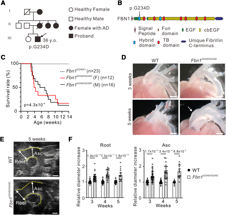
Background: Aortic dissection (AD) is the separation of medial layers of the aorta and is a major cause of death in patients with connective tissue disorders such as Marfan syndrome. However, molecular triggers instigating AD, its temporospatial progression, and how vascular cells in each vessel layer interact and participate in the pathological process remain incompletely understood. To unravel the underlying molecular mechanisms of AD, we generated a spontaneous AD mouse model.

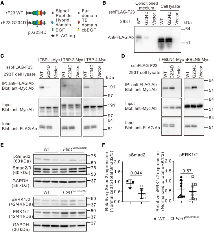
Methods: We incorporated a novel missense variant (p.G234D) in FBN1, the gene for fibrillin-1, identified in a patient with nonsyndromic familial AD into mice using the CRISPR/Cas9 (clustered regularly interspaced short palindromic repeats/CRISPR-associated protein 9) system. We performed molecular pathological analyses of the aortic lesions by histology, immunofluorescence staining, electron microscopy, synchrotron-based imaging, and single-cell RNA sequencing. Biochemical analysis was performed to examine the binding capacity of mutant human FBN1G234D (fibrillin 1 Gly234Asp) protein to LTBPs (latent TGFβ [transforming growth factor-beta] binding proteins), and signaling pathways in the mutant aortic wall were examined by the Western blot analysis.

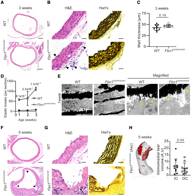
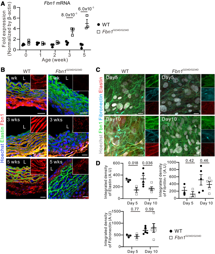
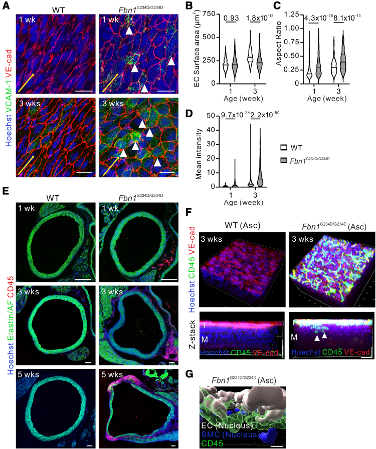
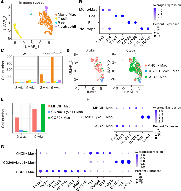
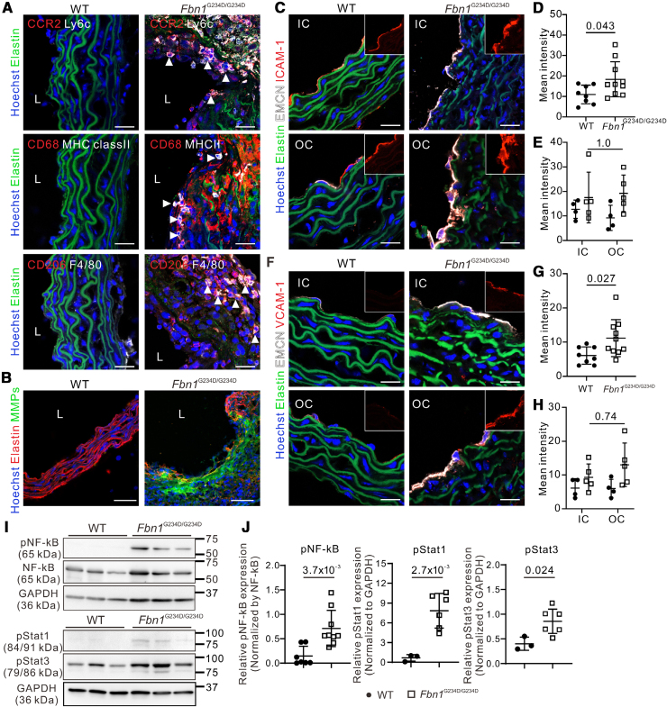
Results: Fifty percent of the Fbn1G234D/G234D mutant mice died within 5 weeks of age from multiple intimomedial tears that expanded longitudinally and progressed to aortic rupture accompanied by massive immune cell infiltration. Fbn1G234D/G234D endothelial cells exhibited altered mechanosensing with loss of parallel alignment to blood flow and upregulation of VCAM-1 (vascular cell adhesion molecule-1) and ICAM-1 (intercellular adhesion molecule-1) as early as 1 week of age. Single-cell RNA sequencing, validated by immunostaining, revealed a cluster of monocyte/macrophage predominantly in the intima at 3 weeks of age before the dissection, and the second cluster of macrophages increased during the progression of intimomedial tears, exhibiting strong CCR2+ (C-C motif chemokine receptor 2 positive) and both M1- and M2-like features. Consistently, upregulation of MMP2/9 (matrix metalloproteinase 2 and 9) was observed. Biochemically, FBN1G234D lost the ability to bind to LTBP-1, -2, and -4, resulting in the downregulation of TGFβ signaling in the aortic wall.

Conclusions: We show that interactions involving endothelial cells and macrophages/monocytes in the intima, where the extracellular matrix (ECM) microenvironment contains reduced TGFβ signaling, contribute to the initiation of AD. Our novel AD mouse model provides a unique opportunity to identify target molecules involved in the intimomedial tears that can be utilized for the development of therapeutic strategies.

Keywords: Marfan syndrome; aortic dissection; endothelial cells; fibrillin-1; macrophages.


Conflict of interest statement

None.

Figures

Figure 1.
Figure 1.
Familial aortic dissection with a novel FBN1 (fibrillin-1) missense variant. A, Family tree of the young male patient with a novel FBN1 missense variant. B, Schematic image of FBN1 domains. The missense variant (c.701 G>A, p.234 Gly>Asp, arrow) in the first hybrid domain was identified in the FBN1 gene. C, Kaplan-Meier survival curve of Fbn1 mutant mice, Fbn1G234D/G234D female (F) mice (n=12), and Fbn1G234D/G234D male (M) mice (n=16). Heterozygous (n=23) vs homozygous (n=28) mutant mice. D, Gross images of wild-type (WT) and Fbn1G234D/G234D aortas at 3 and 5 weeks of age. Note the dilated ascending aorta in a 5-week-old mutant mouse (arrow). E, Representative images of echocardiography of aortas from WT and mutant mice at 5 weeks of age. F, Relative diameter increase of the root and ascending aortas normalized with those of WT aortas by echocardiography. Increased diameters of the aortic root and ascending aorta starting at 3 weeks of age. Three weeks (root: 1.27±0.04; ascending aorta [Asc]: 1.38±0.05), 4 weeks (root: 1.48±0.05; Asc: 1.63±0.09), and 5 weeks (root: 1.43±0.08; Asc: 1.69±0.10). At 3-week WT: n=43 mice, Fbn1G234D/G234D: n=37 mice; 4-week WT: n=50 mice, Fbn1G234D/G234D: n=26 mice; and 5-week WT: n=43 mice, Fbn1G234D/G234D: n=18 mice. Data were presented as mean±SEM and analyzed by the log-rank test (C) and the Mann-Whitney U test or the Student t test (F). Bars are 1 mm (D and E). cbEGF indicates calcium binding epidermal growth factor-like domain; and EGF, epidermal growth factor-like domain.
Figure 2.
Figure 2.
Aortic dissection with intimomedial tears in the ascending aortas of Fbn1G234D/G234D mouse. A and B, Histological images of wild-type (WT) and Fbn1G234D/G234D aortas with hematoxylin and eosin (H&E) and Hart staining at 3 weeks of age. Elastin breaks and accumulation of immune cells (arrowheads) are observed in the aorta from Fbn1G234D/G234D mice. C, The morphometric analysis of wall thickness displays no significant differences between WT and Fbn1G234D/G234D aortas. WT: n=9 mice, Fbn1G234D/G234D: n=7 mice. D, The quantification of elastin breaks in the aortas from WT and Fbn1G234D/G234D mice at 1 to 3 weeks of age. At 1-week WT: n=3 mice, Fbn1G234D/G234D: n=3 mice; 2-week WT: n=6 mice, Fbn1G234D/G234D: n=5 mice; and 3-week WT: n=9 mice, Fbn1G234D/G234D: n=7 mice. E, Electron microscopic images of ascending aorta obtained from WT and Fbn1G234D/G234D mice at 1 week of age. In the WT aorta, continuous elastic laminae were being formed with the elastin-contractile unit (arrows), whereas elastic laminae of Fbn1G234D/G234D aortas were disrupted with loss of contacts with smooth muscle cells (arrowheads). F and G, Histological images of WT and Fbn1G234D/G234D aortas with H&E and Hart staining at 5 weeks of age. There were multiple intimomedial tears (arrowheads) in the aorta from Fbn1G234D/G234D mice (F). The continuity of medial layers was disrupted, and red blood cells were detected within the medial layers (arrows; G). H, 3D structure of intimomedial tears and quantification of its volume normalized by ascending (ASC) volume in the aorta from Fbn1G234D/G234D mice at 5 weeks. Multiple lesions (red) with disrupted medial layers expanded longitudinally, but no significant difference was observed between the inner curvature (IC) and outer curvature (OC). n=10 mice; the Mann-Whitney U test. An average number of disruptions per mouse is presented. Data were presented as mean±SEM and analyzed by the Student t test (C and D) or Mann-Whitney U test (H). Bars are 200 (A and F), 20 (B and G), 2 (E), and 1 (magnified in E).
Figure 3.
Figure 3.
Smooth muscle cells (SMCs) derived from Fbn1G234D/G234D aortas show defects in elastogenesis. A, Evaluation of Fbn1 (fibrillin-1) mRNA expression by quantitative PCR (qPCR) in primary aortic cells at passage 0, isolated from postnatal day 3 (0 week) to 5-week-old wild-type (WT) and Fbn1G234D/G234D mice. n=3 mice for each group. B, Representative immunofluorescence images showing markedly decreased FBN1 expression (red) in the media of aorta from Fbn1G234D/G234D mice at 1, 3, and 5 weeks of age. n=3 mice for each group. C, Representative immunofluorescence images of elastogenesis assay showing the delayed extracellular matrix (ECM) formation in primary SMCs from Fbn1G234D/G234D mice compared with WT. SMCs were analyzed at passage 2-3. D, Quantitative analysis of the integrated density of elastin, FN (fibronectin), and FBN1. n=3 mice for each group (day 5); n=5 mice per group (day 10). Data were presented as mean±SEM and analyzed by the Student t test (A and D). Bars are 20 (B) and 10 µm (C). L indicates lumen side.
Figure 4.
Figure 4.
Endothelial cell (EC) abnormality in the aorta of Fbn1 G234D/G234D mice. A, Representative immunofluorescence images of en face staining of the ascending aorta (Asc) show upregulated VCAM-1 (vascular cell adhesion molecule-1) expression (green) in ECs (red) that fail to align with blood flow (yellow arrows) in Fbn1G234D/G234D mice at 1 and 3 weeks of age. n=3 mice for each group. B through D, Quantitative analysis revealed that ECs from Fbn1G234D/G234D mice exhibit reduced cell surface area (B), increased cell roundness (C), and upregulated VCAM-1 expression (D). n=3 mice for each group. E, Representative immunofluorescence images showing the infiltration of CD45 (leukocyte common antigen) positive cells (red) in the intima (I) and the adventitia (A) of the aorta from Fbn1G234D/G234D mice at 1 (n=3 mice), 3 (n=8 mice), and 5 weeks (n=5 mice) of age. F, Representative 3D confocal images show that CD45 positive cells (green) spread out in the intima and infiltrate into the media (M; arrowheads) in Fbn1G234D/G234D mice at 3 weeks of age. n=3 mice for each group. G, 3D surface rendering by Imaris shows immune cells distributed underneath ECs. Data were presented as mean±SEM and analyzed by the Mann-Whitney U test (B–D). Bars are 100 (E), 20 (A and F), and 5 µm (G). VE-cad indicates vascular endothelial cadherin.
Figure 5.
Figure 5.
Identification of immune cell clusters in wild type (WT) and Fbn1G234D/G234D by single-cell RNA sequencing (scRNA-seq). A, Uniform manifold approximation and projection (UMAP) for immune cell clusters extracted from the major cell subclusters at 3 and 5 weeks of age. B, A dot plot indicates the normalized relative expression of marker genes in the distinct cell subcluster. C, The percentages of each subcluster in the total number of immune cells. D, UMAP for Fbn1G234D/G234D monocyte/macrophage subcluster extracted from the immune cell clusters shows an increase in CCR2+ (C-C chemokine receptor type 2 positive) macrophage (CCR2+ Mac, green) in the aorta of Fbn1G234D/G234D mice at 5 weeks of age. E, The percentages of each cluster in the total number of monocyte/macrophage subclusters in Fbn1G234D/G234D aortas. F, Dot plots showing the normalized average expression of marker genes in each cell cluster in Fbn1G234D/G234D aortas. G, Dot plots showing the normalized average expression of selective genes. In the dot plots, the dot size reflects the percentage of cells expressing the selected gene, and the dot color corresponds to the level of expression. Lyve1 indicates lymphatic vessel endothelial hyaluronan receptor 1; and MHCII, major histocompatibility complex class II.
Figure 6.
Figure 6.
Macrophages accumulate in the aortic wall of Fbn1 G234D/G234D mice. A, Representative immunofluorescence images showing the differential distribution of macrophages and monocytes (arrowheads) populations in the aorta from 5-week-old mice. n=3 mice for each group. B, Representative in situ zymography with Dye-quenched (DQ)-gelatin showing upregulation of MMP (matrix metalloprotease) activity (green) in the lesions in wild-type (WT) and Fbn1G234D/G234D aortas at 5 weeks of age. n=3 mice for each group. C through E, Representative immunofluorescence images at 5 weeks of age (C) and quantification of and ICAM-1 (intercellular adhesion molecule-1) abundance (D and E) showing the upregulation of ICAM-1 in the intima in Fbn1 G234D/G234D aortas, but no significant difference was observed between the inner curvature (IC) and the outer curvature (OC; E). WT: n=8 mice; Fbn1G234D/G234D: n=10 mice. F through H, Representative immunofluorescence images at 5 weeks of age (F) and quantification of VCAM-1 (vascular cell adhesion molecule-1) abundance (G and H) showing the upregulation of VCAM-1 in the intima, but no significant difference was observed between the IC and OC (H). WT: n=8 mice; Fbn1G234D/G234D: n=10 mice. I and J, Western blot of total protein extracts from 3-week-old WT and Fbn1G234D/G234D ascending aortas. WT: n=7 mice (nuclear factor-kappa B [NF-κB]), n=3 mice (stats); Fbn1G234D/G234D: n=9 mice (NF-κB), n=6 mice (stats). Data were presented as mean±SEM and analyzed by the Student t test (D, G, and J), the 2-way ANOVA and Šídák multiple comparisons test (E and F), or the Mann-Whitney U test (J). Bars are 50 (B) and 20 µm (A, C, and F). EMCN indicates endomucin.
Figure 7.
Figure 7.
Molecular and biochemical analysis displayed a significant impairment of rF23-G234D in the binding to LTBPs (latent TGFβ binding proteins) and affected canonical TGFβ (transforming growth factor-beta) pathways. A, Domain structure of peptide sequence DYKDDDDK (FLAG)-tagged partial fibrillin-1 (recombinat fragment of human fibrillin-1 23 [rF23]) and fibrillin-1 mutant (rF23 with G234D mutant [rF23-G234D]). B, Western blot analysis with anti-FLAG antibody using conditioned medium and cell lysates from human embryonic kidney 293T (HEK293T) cells transfected with plasmids encoding the rF23 wild type (WT) or rF23-G234D. C, In vitro binding assay using the cell lysates from transfected 293T cells. The lysates were mixed with c-myc (Myc)-tagged LTBPs and immunoprecipitated with anti-FLAG antibodies. D, In vitro binding assay using the cell lysates from transfected 293T cells. The lysates were mixed with Myc-tagged fibulins (FBLNs) and immunoprecipitated with anti-FLAG antibodies. E and F, Western blot of total protein extracts from 3-week-old WT and Fbn1G234D/G234D ascending aortas. WT: n=3 mice (suppressor of mothers against decapentaplegic [Smad]), n=8 mice (extracellular signal-regulated kinase [ERK]); Fbn1G234D/G234D: n=6 mice (Smads), n=8 mice (ERK). Data were presented as mean±SEM and analyzed by the Student t test (F).

Comment in

Similar articles

References

    1. Nienaber CA, Clough RE, Sakalihasan N, Suzuki T, Gibbs R, Mussa F, Jenkins MP, Thompson MM, Evangelista A, Yeh JS, et al. Aortic dissection. Nat Rev Dis Primers. 2016;2:16071. doi: 10.1038/nrdp.2016.71 - PubMed
    1. Tamori Y, Akutsu K, Kasai S, Sakamoto S, Okajima T, Yoshimuta T, Yokoyama N, Ogino H, Higashi M, Nonogi H, et al. Coexistent true aortic aneurysm as a cause of acute aortic dissection. Circ J. 2009;73:822–825. doi: 10.1253/circj.cj-08-0427 - PubMed
    1. Bossone E, Eagle KA. Epidemiology and management of aortic disease: aortic aneurysms and acute aortic syndromes. Nat Rev Cardiol. 2021;18:331–348. doi: 10.1038/s41569-020-00472-6 - PubMed
    1. Ostberg NP, Zafar MA, Ziganshin BA, Elefteriades JA. The genetics of thoracic aortic aneurysms and dissection: a clinical perspective. Biomolecules. 2020;10:182. doi: 10.3390/biom10020182 - PMC - PubMed
    1. Murray CA, Edwards JE. Spontaneous laceration of ascending aorta. Circulation. 1973;47:848–858. doi: 10.1161/01.cir.47.4.848 - PubMed

MeSH terms

LinkOut - more resources