. 2025 May 6;9(5):e0680.
doi: 10.1097/HC9.0000000000000680. eCollection 2025 May 1.

HBV core protein enhances WDR46 stabilization to upregulate NUSAP1 and promote HCC progression

Affiliations

HBV core protein enhances WDR46 stabilization to upregulate NUSAP1 and promote HCC progression

Fanyun Kong et al. Hepatol Commun. .

Abstract

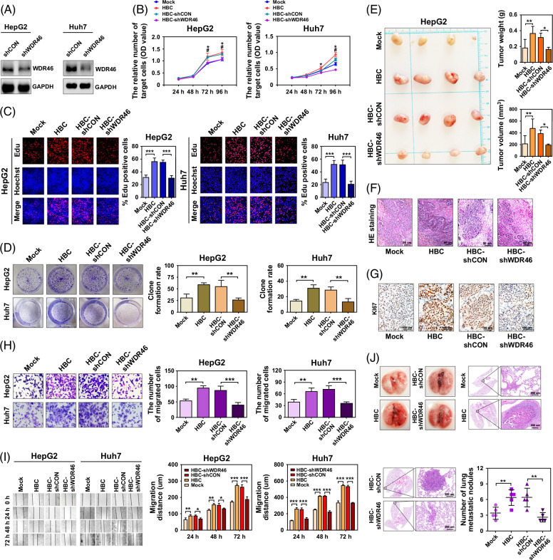
Background: The HBV core protein (HBC) is crucial for the progression of HCC. WD repeat-containing (WDR) 46 (WDR46) is implicated in the development of different tumors. Nevertheless, whether WDR46 is controlled by HBC to drive hepatocarcinogenesis remains unclear.

Methods: Different HCC cohorts, immunohistochemical staining, and bioinformatics analysis were utilized to estimate the clinical correlation between WDR46 and HBV-associated HCC. Western blotting, co-immunoprecipitation, chromatin immunoprecipitation, and oncology functional assays were performed to evaluate the effect of HBC on WDR46 in upregulating nucleolar spindle-associated protein 1 (NUSAP1), the influence of WDR46 on HBC-mediated HCC cell biological functions, and the mechanisms of WDR46 upregulation mediated by HBC to increase NUSAP1.

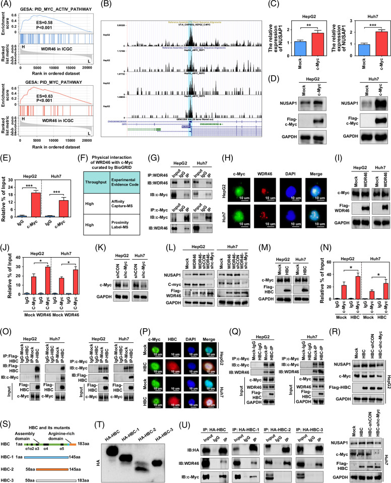
Results: WDR46 expression was elevated in HBV-related HCC in a HBC-dependent manner. Overexpression of WDR46 is closely linked to severe prognosis of tumors. Functionally, WDR46 contributes to HBC-induced cell growth and migration in vitro and in vivo. Furthermore, HBC enhanced WDR46 protein stabilization by hampering the interaction between WDR46 and TRIM25, thereby decreasing WDR46 ubiquitination. NUSAP1, a DNA replication-related molecule, is a vital downstream target of WDR46. Relying on WDR46, HBC promoted NUSAP1 upregulation to modulate the biological functions of HBC in HCC cells. Importantly, HBC enhanced the interaction between WDR46 and the transcription factor c-Myc to facilitate c-Myc recruitment to the NUSAP1 promoter, leading to the increase of NUSAP1 transcription.

Conclusions: Our comprehensive data provides new insights into the mechanisms responsible for HBC-induced hepatocarcinogenesis. WDR46 and its downstream molecule, NUSAP1, may act as novel therapeutic targets for HBV-related tumors.

Keywords: HBV core protein; NUSAP1; TRIM25; WDR46; c-Myc.


Conflict of interest statement

The authors have no conflicts to report.

Figures

FIGURE 1
FIGURE 1
The expression of WDR46 is increased in HCC, and it can be modulated by HBC. (A) The expression of WDR46 in HCC and adjacent tissues that detected by IHC. Scale bar, 100 μm. (B) The expression of WDR46 in the ICGC cohort. (C) WDR46 expression in 7 HCC cohorts from the HCCDB database. (D) The expression of WDR46 in HCC tissues in patients with TNM stage ≤ II or >II. (E) WDR46 expression in HCC tissues in patients with or without portal vein invasion. (F) The association of WDR46 with the OS of HCC patients in the ICGC database. (G) The relationship of WDR46 with significant pathways in the ARCHS4 database. (H) The expression of WDR46 in HBV-associated HCC tissues and adjacent tissues in the Gao cohort. (I) The expression of WDR46 in HBV-associated HCC tissues and adjacent tissues in the GSE94660. (J) The expression of WDR46 in HBV-associated HCC patients with age ≤ 50 or >50. (K) The expression of WDR46 in HBV-associated HCC patients with AFP ≤20 ng/mL or >20 ng/mL. (L) The association of WDR46 with the OS of HBV-associated HCC patients in the Gao cohort. (M) The association of WDR46 with the RFS of HBV-associated HCC patients in the Gao cohort. (N) The expression of WDR46 in HBV-positive HCC, HCV-positive HCC, and non-HBV/non-HCV HCC in the ICGC cohort. (O) The expression of the WDR46 gene in HCC cells induced by HBV. (P) The expression of WDR46 protein in HCC cells induced by HBV. (Q) The expression of WDR46 protein in HCC cells is induced by different HBV-encoded proteins. (R) The expression of the WDR46 gene in HCC cells induced by HBC. (S) The expression of WDR46 protein in HCC cells induced by HBC. (T) The interaction between WDR46 with HBC was detected by Co-IP assay. (U) The colocation of WDR46 with HBC in HCC cells that detected by immunofluorescence experiment. Scale bar, 10 μm; *p<0.05, **p<0.01, and ***p<0.001. Abbreviations: AFP, alpha-fetoprotein; Co-IP, co-immunoprecipitation; GAPDH, glyceraldehyde-3-phosphate dehydrogenase; HBC, HBV core protein; IHC, immunohistochemistry; NBNC, non-HBV/non-HCV; OS, overall survival; RFS, recurrence-free survival; WDR46, WD repeat-containing 46.
FIGURE 2
FIGURE 2
The effect of WDR46 on cell proliferation and migration mediated by HBC. (A) The inhibition of WDR46 protein expression is mediated by its specific shRNA. (B) The effect of WDR46 on cell viability mediated by HBC that detected by CCK-8. *p<0.05, Mock group compares to HBC group, #p<0.05, HBC-shCON group compares to HBC-shWDR46 group. (C) The effect of WDR46 on cell proliferation mediated by HBC that detected by EdU analysis. (D) The effect of WDR46 on colony formation mediated by HBC that detected by clone formation assay. (E) The effect of WDR46 on HCC cell proliferation mediated by HBC in nude mice. (F) H&E staining on the HCC tissues from nude mice in different groups. Scale bar, 50 μm. (G) KI67 expression was examined by IHC in tissues from nude mice in different groups. Scale bar, 100 μm. (H) The effect of WDR46 on cell migration mediated by HBC that detected by transwell assay. (I) The effect of WDR46 on cell migration mediated by HBC that detected by wound healing assay. (J) The effect of WDR46 on lung metastasis in HCC cells mediated by HBC in nude mice detected by H&E staining. Scale bar, 200 μm; *p<0.05 and **p<0.01. Abbreviations: CCK-8, cell counting kit-8; GAPDH, glyceraldehyde-3-phosphate dehydrogenase; H&E, hematoxylin–eosin; HBC, HBV core protein; IHC, immunohistochemistry; shRNA, short-hairpin RNA; WDR46, WD repeat-containing 46.
FIGURE 3
FIGURE 3
HBC disrupts the interaction between WDR46 with TRIM25 to stabilize WDR46 protein. (A) The significant pathways predicted by GSEA analysis with WDR46 in the ICGC HCC cohort. (B) The effect of BafA1 (200 nM) and MG132 (100 nM) on WDR46 expression in HCC cells. (C) The effect of HBC on the half-life of WDR46 protein in HCC tissues, when cells were treated with CHX (60 μg/mL). (D) The effect of HBC on stabilization of WDR46 protein in HCC tissues, when cells were treated with MG132 (100 nM). (E) The role of HBC on WDR46 ubiquitination in HCC cells. (F) The information on TRIM25 as a E3 ligase of WDR46 in the Ubibrowser database. (G) The predicted interaction between WDR46 and TRIM25 in the BioGRID database. (H) The expression of WDR46 in HCC cells with TRIM25 knockdown. (I) The interaction between WDR46 and TRIM25 in HCC cells was detected by Co-IP assays. (J) The colocation of WDR46 with TRIM25 in hepatoma cells was detected by immunofluorescence assay. Scale bar, 10 μm. (K) The effect of TRIM25 on half-life of WDR46 protein in HCC tissues, when cells were treated with CHX (60 μg/mL). (L) The effect of TRIM25 on stabilization of WDR46 protein in HCC tissues, when cells were treated with MG132 (100 nM). (M) The role of TRIM25 on WDR46 ubiquitination in HCC cells. (N) The effect of HBC on TRIM25 protein expression in HCC cells. (O) The effect of HBC on the interaction between WDR46 and TRIM25. (P) The effect of HBC on the expression of WDR46 mediated by TRIM25. (Q) The effect of TRIM25 on the expression of WDR46 mediated by HBC. (R) The effect of TRIM25 on the half-life of WDR46 protein mediated by HBC in HCC cells, when cells were treated with CHX (60 μg/mL). (S) The effect of TRIM25 on stabilization of WDR46 protein mediated by HBC in HCC cells, when cells were treated with MG132 (100 nM). (T) The role of TRIM25 on WDR46 ubiquitination mediated by HBC in HCC cells. (U) Pattern diagram of WDR46 mutants. (V) The expression of WDR46 mutants in HEK293T cells. (W) The interaction of WDR46 mutants with TRIM25 or HBC in HEK293T cells. Abbreviations: BafA1, bafilomycin A1; Co-IP, co-immunoprecipitation; CHX, cycloheximide; GAPDH, glyceraldehyde-3-phosphate dehydrogenase; GSEA, gene set enrichment analysis; HBC, HBV core protein; Ub, ubiquitin; WDR46, WD repeat-containing 46.
FIGURE 4
FIGURE 4
WDR46 promotes NUSAP1 transcription in HBC-positive HCC cells. (A) The heat map shows the effect of WDR46 on the expression of DEGs that were detected by RNA sequencing. (B) The pathway is enriched by GSEA analysis, based on the RNA-sequencing data. (C) The pathway is enriched by GSEA analysis, based on the ICGC HCC data. (D) The association of NUSAP1 with significant pathways in the ARCHS4 database. (E) The expression of NUSAP1 in HCC and adjacent tissues that detected by IHC. Scale bar, 50 μm. (F) The expression of NUSAP1 in the ICGC cohort. (G) NUSAP1 expression in 7 HCC cohorts in the HCCDB database. (H) The expression of NUSAP1 in HCC tissues in patients with or without vein invasion. (I) The expression of NUSAP1 in HCC tissues in patients with or without portal vein invasion. (J) The association of NUSAP1 with the OS of HCC patients in the ICGC database. (K) The association of WDR46 with NUSAP1 in HCC tissues in the ICGC cohort. (L) The expression of NUSAP1 in HBV-associated HCC tissues in the Gao cohort. (M) The expression of NUSAP1 in HBV-associated HCC tissues in GSE94660. (N) The expression of NUSAP1 in patients aged ≤50 years or >50 years in Gao cohort. (O) The expression of NUSAP1 in HBV-related HCC patients with AFP ≤20 ng/mL or >20 ng/mL in Gao cohort. (P) The association of NUSAP1 with the OS of HBV-related HCC patients in the Gao cohort. (Q) The association of NUSAP1 with the RFS of HBV-related HCC patients in the Gao cohort. (R) The association of WDR46 with NUSAP1 in HBV-associated HCC tissues in the ICGC cohort. (S) The expression of NUSAP1 in HBV-positive HCC, HCV-positive HCC, and non-HBV/non-HCV HCC in the ICGC cohort. (T) The effect of WDR46 on NUSAP1 gene expression in HCC cells. (U) The effect of WDR46 on NUSAP1 protein expression in HCC cells. (V) The expression of NUSAP1 gene in HCC cells induced by HBC. (W) The expression of NUSAP1 protein in HCC cells induced by HBC. (X) The expression of NUSAP1 protein in HCC cells induced by HBV. (Y) The effect of WDR46 on NUSAP1 protein expression in HCC cells mediated by HBC. *p<0.05, **p<0.01, and ***p<0.001. Abbreviations: DEGs, differentially expressed genes; GAPDH, glyceraldehyde-3-phosphate dehydrogenase; GSEA, gene set enrichment analysis; HBC, HBV core protein; IHC, immunohistochemistry; NBNC, non-HBV/non-HCV; NUSAP1, Nucleolar spindle-associated protein 1; OS, overall survival; RFS, recurrence-free survival; WDR46, WD repeat-containing 46.
FIGURE 5
FIGURE 5
The effect of NUSAP1 on cell proliferation and migration is mediated by WDR46. (A) The inhibition of NUSAP1 protein expression is mediated by its specific shRNA. (B) The effect of NUSAP1 on cell viability mediated by WDR46 that detected by CCK-8; *p<0.05, Mock group compares to WDR46 group, #p<0.05, WDR46-shCON group compares to WDR46-shNUSAP1 group. (C) The effect of NUSAP1 on cell proliferation mediated by WDR46 that detected by EdU analysis. (D) The effect of NUSAP1 on colony formation mediated by WDR46 that detected by clone formation assay. (E) The effect of NUSAP1 on HCC cell proliferation mediated by WDR46 in nude mice. (F) H&E staining on the HCC tissues from nude mice in different groups. Scale bar, 200 μm. (G) KI67 expression was examined by IHC in tissues from nude mice in different groups. Scale bar, 100 μm. (H) The effect of NUSAP1 on cell migration mediated by WDR46 that detected by transwell assay. (I) The effect of NUSAP1 on cell migration mediated by WDR46 that detected by wound healing assay. (J) The effect of NUSAP1 on lung metastasis in HCC cells mediated by WDR46 in nude mice that were detected by H&E staining. Scale bar, 400μm; *p<0.05 and **p<0.01. Abbreviations: CCK-8, cell counting kit-8; H&E, hematoxylin–eosin; IHC, immunohistochemistry; NUSAP1, nucleolar spindle-associated protein 1; shRNA, short-hairpin RNA; WDR46, WD repeat-containing 46.
FIGURE 6
FIGURE 6
The effect of c-Myc on the expression of NUSAP1 mediated by WDR46 and HBC. (A) The pathways associated with WDR46 in the ICGC HCC cohort that predicted by GSEA analysis. (B) The information of ChIP sequencing on the interaction of c-Myc with NUSAP1 promoter in HepG2 cells in the Cistrome Data Browser database. (C) The effect of ectopic c-Myc on the expression of NUSAP1 gene in HCC cells. (D) The effect of ectopic c-Myc on the expression of NUSAP1 protein in HCC cells. (E) The binding of c-Myc to NUSAP1 promoter in HCC cells that detected by ChIP assay. (F) The interaction between WDR46 with c-Myc in the BioGRID database. (G) The interaction between WDR46 and c-Myc detected by Co-IP assay. (H) The colocation of WDR46 with c-Myc in HCC cells detected by immunofluorescence assay. Scale bar, 10 μm. (I) The effect of WDR46 on c-Myc expression. (J) The effect of WDR46 on the binding of c-Myc to NUSAP1 promoter in HCC cells that detected by ChIP assay. (K) The inhibition of c-Myc mediated by its shRNA. (L) The effect of c-Myc on expression of NUSAP1 mediated by WDR46. (M) The effect of HBC on c-Myc expression. (N) The effect of HBC on the binding of c-Myc to NUSAP1 promoter in HCC cells that detected by ChIP assay. (O) The interaction between HBC and c-Myc detected by Co-IP assay. (P) The colocation of HBC with c-Myc in HCC cells detected by immunofluorescence assay. Scale bar, 10 μm. (Q) The effect of HBC on the interaction between WDR46 and c-Myc. (R) The effect of c-Myc on expression of NUSAP1 mediated by HBC. (S) Pattern diagram of HBC mutants. (T) The expression of HBC mutants in HEK293T cells. (U) The interaction of HBC mutants with c-Myc or WDR46 in HEK293T cells; *p<0.05, **p<0.01, and ***p<0.001. Abbreviations: ChIP, chromatin immunoprecipitation; Co-IP, co-immunoprecipitation; GAPDH, glyceraldehyde-3-phosphate dehydrogenase; GSEA, gene set enrichment analysis; HBC, HBV core protein; NUSAP1, nucleolar spindle-associated protein 1; WDR46, WD repeat-containing 46.
FIGURE 7
FIGURE 7
The role of NUSAP1 on cell growth and migration mediated by HBC. (A) The effect of NUSAP1 on cell viability mediated by HBC that detected by CCK-8; *p<0.05, Mock group compares to HBC group, #p<0.05, HBC-shCON group compares to HBC-shNUSAP1 group. (B) The effect of NUSAP1 on cell proliferation mediated by HBC that detected by EdU analysis. (C) The effect of NUSAP1 on colony formation mediated by HBC that detected by clone formation assay. (D) The effect of NUSAP1 on HCC cell proliferation mediated by HBC in nude mice. (E) H&E staining on the HCC tissues from nude mice in different groups. Scale bar, 100 μm. (F) KI67 expression examined by IHC in tissues from nude mice in different groups. Scale bar, 100 μm. (G) The effect of NUSAP1 on cell migration mediated by HBC that detected by transwell assay. (H) The effect of NUSAP1 on cell migration mediated by HBC that detected by wound healing assay. (I) The effect of NUSAP1 on the lung metastasis in HCC cells mediated by HBC in nude mice that detected by H&E staining. Scale bar, 200 μm; *p<0.05 and **p<0.01. Abbreviations: CCK-8, cell counting kit-8; HBC, HBV core protein; H&E, hematoxylin–eosin; IHC, immunohistochemistry; NUSAP1, Nucleolar spindle-associated protein 1.
FIGURE 8
FIGURE 8
A schematic diagram showing the mechanisms regarding the increase of WDR46 induced by HBC to upregulate NUSAP1 and facilitate hepatocarcinogenesis. In HCC cells, HBC strengthens WDR46 stabilization by disrupting the interaction between WDR46 and TRIM25 to reduce ubiquitination-dependent degradation of WDR46. Subsequently, HBC promotes the interaction of WDR46 with c-Myc to benefit the recruitment of c-Myc to the NUSAP1 promoter, therefore increasing NUSAP1 gene transcription and enhancing cell growth and migration. Abbreviations: HBC, HBV core protein; NUSAP1, Nucleolar spindle-associated protein 1; WDR46, WD repeat-containing 46.

Similar articles

References

    1. Zhang Y, Cao W, Wang S, Zhang L, Li X, Zhang Z, et al. . Epigenetic modification of hepatitis B virus infection and related hepatocellular carcinoma. Virulence. 2024;15:2421231. - PMC - PubMed
    1. Zhang M, Chen H, Liu H, Tang H. The impact of integrated hepatitis B virus DNA on oncogenesis and antiviral therapy. Biomark Res. 2024;12:84. - PMC - PubMed
    1. Lefeuvre C, Le Guillou-Guillemette H, Ducancelle A. A pleiotropic role of the hepatitis B virus core protein in hepatocarcinogenesis. Int J Mol Sci. 2021;22:13651. - PMC - PubMed
    1. Kong F, Tao Y, Yuan D, Zhang N, Li Q, Yu T, et al. . Hepatitis B virus core protein mediates the upregulation of C5alpha receptor 1 via NF-kappaB pathway to facilitate the growth and migration of hepatoma cells. Cancer Res Treat. 2021;53:506–527. - PMC - PubMed
    1. Kong F, Li N, Tu T, Tao Y, Bi Y, Yuan D, et al. . Hepatitis B virus core protein promotes the expression of neuraminidase 1 to facilitate hepatocarcinogenesis. Lab Invest. 2020;100:1602–1617. - PubMed

MeSH terms