. 2025 May 14;16(5):e0133824.
doi: 10.1128/mbio.01338-24. Epub 2025 Apr 9.

Dual oxic-anoxic co-culture enables direct study of anaerobe-host interactions at the airway epithelial interface

Affiliations

Dual oxic-anoxic co-culture enables direct study of anaerobe-host interactions at the airway epithelial interface

Patrick J Moore et al. mBio. .

Abstract

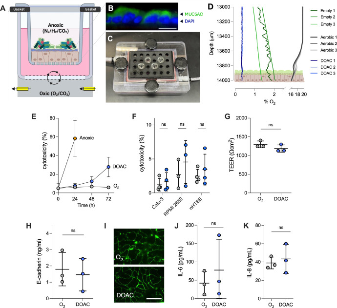
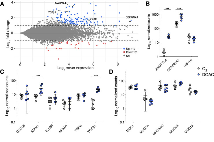
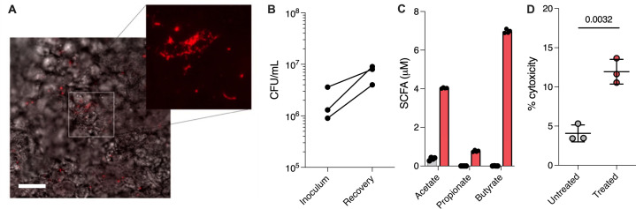
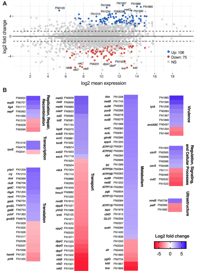
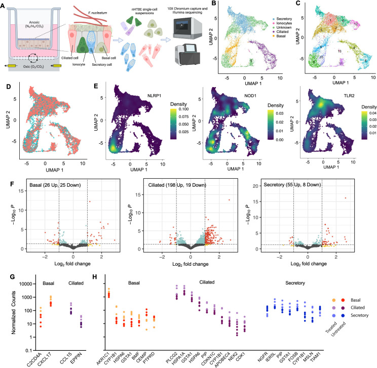
Strict and facultative anaerobic bacteria are widely associated with both acute and chronic airway diseases. However, their potential role(s) in disease pathophysiology remains poorly understood due to inherent limitations of existing laboratory models and conflicting oxygen demands between anaerobes and host cells. To address these limitations, here, we describe a dual oxic-anoxic culture (DOAC) approach that maintains an oxygen-limited microenvironment at the apical epithelial interface while host cells are oxygenated basolaterally. This platform enables epithelial-anaerobe co-culture for ~48 h, and we demonstrate its utility by evaluating reciprocal interactions between the oxygen-sensitive anaerobic bacterium, Fusobacterium nucleatum, and oxygen-demanding airway epithelial cells at the transcriptional level. Using bulk RNAseq, we demonstrate that epithelial colonization results in altered gene expression by F. nucleatum, highlighted by the differential expression of genes associated with virulence, ethanolamine and lysine metabolism, metal uptake, and other transport processes. We also combine DOAC with single-cell RNA sequencing to reveal a cell type-specific transcriptional response of the airway epithelium to F. nucleatum infection, including the increased expression of inflammatory marker genes and cancer-associated pathways. Together, these data illustrate the versatility of DOAC while revealing new insights into anaerobe-host interactions and their mechanistic contributions to airway disease pathophysiology.IMPORTANCEConflicting oxygen demands between anaerobes and host cells present a significant barrier to in vitro modeling of how these cell types interact. To this end, the significance of our dual oxic-anoxic culture (DOAC) approach lies in its ability to maintain anaerobe and epithelial viability during co-culture, paving the way for new insights into the role(s) of anaerobic microbiota in disease. We use DOAC to interrogate reciprocal interactions between the airway epithelium and Fusobacterium nucleatum-an anaerobic commensal with pathogenic potential. Given its link to a range of diseases, from localized infections to various cancers, these data showing how F. nucleatum bacterium re-shapes its metabolism and virulence upon epithelial colonization provide new mechanistic insight into F. nucleatum physiology and how the host responds. We use F. nucleatum as our model, but the DOAC platform motivates additional studies of the gut, lung, and oral cavity, where host-anaerobe interactions and the underlying mechanisms of pathogenesis are poorly understood.

Keywords: Fusobacterium nucleatum; RNAseq; airway epithelium; single-cell RNA sequencing.


Conflict of interest statement

The authors declare no conflict of interest.

Figures

Fig 1
Fig 1
Optimization of a dual oxic-anoxic epithelial culture platform. (A) Primary and immortalized (Calu-3, RPMI 2650) epithelial cells are cultured on Transwell inserts at ALI for 21–28 days, transferred to a gas-permeable multi-well plate manifold in an anaerobic chamber, and incubated under dual oxic-anoxic culture conditions where the apical compartment is exposed to the oxygen-limited environment of the chamber and mixed blood gas (21% O2, 5% CO2, 74% N2) is delivered to the basolateral compartment to oxygenate the epithelial cells. Figure created with BioRender.com. (B) Calu-3 cells produce a confluent mucus layer on the apical surface, mimicking chronic airway disease (blue, 4',6-diamidino-2-phenylindole (DAPI); green, anti-MUC5AC; bar = 25 µm). (C) Transwells are mounted in a custom 3D-printed thermoplastic polyurethane gasket mounted on a gas-permeable multi-well plate (see also Fig. S1). Blood gas is delivered and removed through cable glands mounted to the basolateral compartment of the Transwell-containing apparatus. (D) Representative oxygen microprofiles. Under normoxic growth conditions (grayscale), Calu-3 cells exhibit a slight oxycline at the epithelial surface. DOAC cells (blue) maintain an anoxic epithelial microenvironment below the level of detection (<0.3%). Empty Transwells in the DOAC system (green) show a gradual oxycline above the Transwell membrane. Profiles represent n = 3 biological replicates. (E) Cytotoxicity as measured by lactate dehydrogenase (LDH) in the DOAC medium over time relative to normoxic (O2+) and strict anoxic (O2) culture conditions. LDH activity is expressed as a percent relative to normoxic cultures at t = 0 and a lysed control. (F) LDH release by Calu-3, RPMI 2650, and primary nHTBE cells under normoxic culture (gray) and DOAC (blue) conditions after 24 h. (G) Transepithelial electrical resistance (TEER), (H) E-cadherin concentrations, (I) immunofluorescence of zonula occludens-1 (ZO-1) tight junction proteins (bar = 20 µm), and cytokines (J) IL-6 and (K) IL-8 showed no significant differences between normoxic culture and DOAC conditions after 24 h. All data shown for panels E–H, J, and K were derived from at least three independent experiments with three technical replicates each and were compared using a Mann-Whitney U-test. Bars in each panel represent the mean ± SD.
Fig 2
Fig 2
Dual oxic-anoxic culture of Calu-3 cells yields a similar transcriptomic profile to normoxic culture conditions (ALI). (A) Log ratio/mean average (MA plot) representation of Calu-3 gene expression under DOAC conditions relative to normoxic culture at ALI (117 upregulated [blue], 31 downregulated [red], log2fc > 1, Padj <0.001). (B) ANGPTL4 and SERPINA1 were differentially expressed, though HIF-1α was consistent between cultures. Few changes in (C) inflammatory biomarkers and (D) mucin gene expression were observed. Data shown in panels B–D are log10-normalized gene counts from five or six independent biological replicates and were compared using the Wald test, Benjamini-Hochberg adjusted (***, P < 0.001).
Fig 3
Fig 3
DOAC enables F. nucleatum colonization of and co-culture with Calu-3 epithelial cells. (A) After 24 h of DOAC co-culture, HCR-FISH imaging was used to confirm and visualize F. nucleatum colonization of Calu-3 cells and aggregation at the apical interface. Bar = 50 µm. (B) After 24 h of co-culture, F. nucleatum-treated Calu-3 cells were resuspended with PBS, and bacterial recovery relative to the inoculum was quantified by plating on selective agar. (C) SCFA quantification in spent culture media from challenged Calu-3 cells (blue) relative to untreated controls (gray). (D) F. nucleatum exhibited a slight cytotoxic effect after 24 h. Data shown in panel C represent the mean ± SD of four independent experiments. Data in panel D represent the mean ± SD of three independent experiments with three technical replicates for each condition and were compared using a Mann-Whitney U-test.
Fig 4
Fig 4
F. nucleatum alters its global transcriptional profile during anaerobic co-culture with Calu-3 cells. (A) Log ratio/mean average (MA plot) representation of F. nucleatum differential gene expression during DOAC co-culture with Calu-3 epithelial cells relative to planktonic culture in EMEM. (l2fc > 1; Padj < 0.001). (B) Heat maps depicting fold change gene expression in specific F. nucleatum genes by functional category (KEGG orthology). Genes encoding hypothetical proteins and those of unknown function are not shown (see Table S1).
Fig 5
Fig 5
Single-cell RNA sequencing of F. nucleatum-treated nHTBE cells reveals a cell type-specific transcriptional response. (A) Experimental schematic. Primary airway epithelial cells were cultured under DOAC conditions and challenged with F. nucleatum. Host cells were dissociated and collected via the 10X Genomics platform prior to scRNAseq. (B) UMAP projection of nHTBEs showing four major cell types (and unknown cells) from (C) 13 distinct clusters. (D) UMAP projection of nHTBE cell types by treatment condition. (E) Density plots (Nebulosa projections) of NLRP1, NOD1, and TLR2 showing heterogeneous expression of pattern recognition receptors between cell types. (F) Volcano plot representation of differential gene expression in basal, ciliated, and secretory cells between F. nucleatum-treated nHTBE cultures relative to untreated controls. (red, log10 expression >1 and l2fc > 1; blue, log10 expression >1 only; yellow, l2fc > 1 only). (G, H) Scatterplot representations of differentially expressed (G) inflammatory markers and (H) pathways associated with cell migration, proliferation, and migration, arranged by cell type. scRNAseq data are derived from at least three independent replicates for each condition (untreated, n = 6; treated n = 3).

Similar articles

Cited by

References

    1. Segal LN, Alekseyenko AV, Clemente JC, Kulkarni R, Wu B, Gao Z, Chen H, Berger KI, Goldring RM, Rom WN, Blaser MJ, Weiden MD. 2013. Enrichment of lung microbiome with supraglottic taxa is associated with increased pulmonary inflammation. Microbiome 1:19. doi:10.1186/2049-2618-1-19 - DOI - PMC - PubMed
    1. Dickson RP, Erb-Downward JR, Freeman CM, McCloskey L, Falkowski NR, Huffnagle GB, Curtis JL. 2017. Bacterial topography of the healthy human lower respiratory tract. MBio 8:e02287-16. doi:10.1128/mBio.02287-16 - DOI - PMC - PubMed
    1. Pragman AA, Kim HB, Reilly CS, Wendt C, Isaacson RE. 2012. The lung microbiome in moderate and severe chronic obstructive pulmonary disease. PLoS One 7:e47305. doi:10.1371/journal.pone.0047305 - DOI - PMC - PubMed
    1. Tunney MM, Field TR, Moriarty TF, Patrick S, Doering G, Muhlebach MS, Wolfgang MC, Boucher R, Gilpin DF, McDowell A, Elborn JS. 2008. Detection of anaerobic bacteria in high numbers in sputum from patients with cystic fibrosis. Am J Respir Crit Care Med 177:995–1001. doi:10.1164/rccm.200708-1151OC - DOI - PubMed
    1. Rogers GB, van der Gast CJ, Cuthbertson L, Thomson SK, Bruce KD, Martin ML, Serisier DJ. 2013. Clinical measures of disease in adult non-CF bronchiectasis correlate with airway microbiota composition. Thorax 68:731–737. doi:10.1136/thoraxjnl-2012-203105 - DOI - PubMed

MeSH terms

LinkOut - more resources