. 2025 Jun 17;99(6):e0221424.
doi: 10.1128/jvi.02214-24. Epub 2025 May 14.

Drosophila melanogaster Toll-9 elicits antiviral immunity against Drosophila C virus

Affiliations

Drosophila melanogaster Toll-9 elicits antiviral immunity against Drosophila C virus

Manish Chauhan et al. J Virol. .

Abstract

The Toll pathway plays a pivotal role in innate immune responses against pathogens. The evolutionarily conserved pattern recognition receptors (PRRs), including Toll-like receptors (TLRs), play a crucial role in recognition of pathogen-associated molecular patterns (PAMPs). The Drosophila genome encodes nine Toll receptors that are orthologous to mammalian TLRs. While mammalian TLRs directly recognize PAMPs, most Drosophila Tolls recognize the proteolytically cleaved ligand Spätzle to activate downstream signaling cascades. In this study, we demonstrated that Toll-9 is crucial for antiviral immunity against Drosophila C virus (DCV), a natural pathogen of Drosophila. A transposable element insertion in the Toll-9 gene renders the flies more susceptible to DCV. The stable expression of Toll-9 in Drosophila S2 cells results in increased Dicer2 induction and reduced AKT phosphorylation, collectively establishing an antiviral state that inhibits DCV replication. Toll-9 localizes to endosomes, where it binds viral double-stranded RNA (dsRNA), highlighting its role in detecting viral replication intermediates. Together, these findings identify Toll-9 as a key player in antiviral immunity against DCV infection, acting through its ability to recognize dsRNA and drive Dicer2 expression, along with other AKT-mediated antiviral responses.

Importance: Insects rely on innate immunity and RNA interference (RNAi) to combat viral infections. Our study underscores the pivotal role of Drosophila Toll-9 in antiviral immunity, aligning with findings in Bombyx mori, where Toll-9 activation upregulates the RNAi component Dicer2. We demonstrate that Drosophila Toll-9 functions as a pattern recognition receptor (PRR) for double-stranded RNA (dsRNA) during Drosophila C virus (DCV) infection, akin to mammalian Toll-like receptors (TLRs). Toll-9 activation during DCV infection leads to the upregulation of Dicer2 and Argonaute2 and dephosphorylation of AKT. This study also reveals that Toll-9 localizes in endosomal compartments where it interacts with dsRNA. These insights enhance our understanding of Drosophila innate immune mechanisms, reflecting the evolutionary conservation of immune responses across diverse species and providing impetus for further research into the conserved roles of TLRs across the animal kingdom.

Keywords: AKT; DCV; Dicer2; JAK/STAT; dsRNA; endosome.


Conflict of interest statement

The authors declare no conflict of interest.

Figures

Fig 1
Fig 1
Toll-9 mutant flies display increased susceptibility to DCV infection. (A) Survival analysis for isogenic control w1118 flies and Toll-90024-G4 mutant flies injected with phosphate-buffered saline (PBS) and DCV (N = 80 flies from three independent experiments). (B) qRT-PCR analysis of Toll-9 expression in w1118 flies and Toll-90024-G4 mutant flies. Data are representative of six biological replicates (groups of 3–5 flies) from three independent experiments. Error bars, SEM. Unpaired t test, ***P < 0.001. (C) qRT-PCR analysis of DCV-infected w1118 flies and Toll-90024-G4 mutant flies at 1 and 3 dpi for DCV mRNA. Data are representative of three biological replicates (groups of 3–5 flies), each from three independent experiments. Error bars, SEM. One-way analysis of variance (ANOVA), *P < 0.05. (D) Western blot analysis of DCV capsid protein with anti-DCV antibody from fly lysate (groups of 3–5 flies) of isogenic control w1118 flies and Toll-90024-G4 mutant flies injected with DCV at 1 and 3 dpi. Blots are representative of three independent experiments. (E) DCV titer in S2 cells infected with fly lysate from isogenic w1118 flies and Toll-90024-G4 mutant flies injected with DCV calculated as 50% tissue culture infectious dose (TCID50/mL). Data are representative of eight biological replicates (groups of 3–5 flies) from four independent experiments. Error bars, SEM. One-way ANOVA, *P < 0.05; ***P < 0.001.
Fig 2
Fig 2
Toll-9 regulates the expression of RNAi and JAK/STAT genes. qRT-PCR analysis of DCV-infected w1118 flies and Toll-90024-G4 mutant flies at 1 and 3 dpi for (A) Dicer2, (B) Argonaute2, (C) Upd2, (D) Upd3, and (E) Vir1. For the mock infection, both w1118 and Toll-90024-G4 mutant flies were injected with PBS and collected at indicated time points for normalization. Data are representative of six biological replicates (groups of 3–5 flies) from three independent experiments. Error bars, SEM. One-way ANOVA, *P < 0.05; ***P < 0.001.
Fig 3
Fig 3
Stable expression of Toll-9 reduces DCV infection in Drosophila S2 cells. qRT-PCR analysis of DCV-infected naïve S2 and Toll-9 OE cells in the presence and absence of CuSO4 at 1 dpi to detect expression of (A) Toll-9, (B) DCV replicase ORF 1, and (C) Dicer2 mRNA. Data are representative of three biological replicates (well of cells) from three independent experiments. Error bars, SEM. One-way ANOVA, **P < 0.01; ****P < 0.0001.
Fig 4
Fig 4
Toll-9 controls DCV infection via dephosphorylation of AKT. (A) Western blot analysis for indicated antibodies of cell lysate from DCV-infected S2 cells and Toll-9 OE cells in the presence and absence of CuSO4 at 1 dpi. Blots are representative of three independent experiments. (B and C) Densitometry quantitation of western blot showing expression for pAKT and DCV normalized to the expression of Actin. Data are representative of western blots from three independent experiments. Error bars, SEM. One-way ANOVA, *P < 0.05; ***P < 0.001. (D) Western blot analysis for indicated antibodies of cell lysate from DCV-infected S2 cells and Toll-9 OE cells in the presence and absence of CuSO4 at 1 dpi in the presence of AKT inhibitor (AKT VIII). Blots are representative of three independent experiments. (E and F) Micrographs showing the presence of DCV in naïve S2 cells and Toll-9 OE cells in the presence and absence of CuSO4 at 1 dpi in the presence and absence of AKT inhibitor. Data are representative of three independent experiments. (G) Viral titer of DCV-infected naïve S2 and Toll-9 OE cells in the presence and absence of AKT inhibitor calculated as TCID50/mL. Data are representative of eight biological replicates (well of cells) from four independent experiments. Error bars, SEM. One-way ANOVA, **P < 0.01.
Fig 5
Fig 5
Subcellular localization of Toll-9. (A) In silico prediction of signal peptide in Toll-9 protein sequence. Red solid line indicates the predicted N-terminal region, orange solid line indicates the predicted center hydrophobic region, and yellow solid line indicates the predicted C-terminal region of signal peptide. Black dotted line indicates the cleavage site (CS) of the signal peptide. Sec/SPI: Sec translocon transported secretory signal peptide/Signal Peptidase I; Tat/SPI: Tat translocon transported Tat signal peptides/Signal Peptidase I. (B) Western blot analysis demonstrating the presence of Toll-9/V5 in endosomes. Endosomal fractions were identified using Rab5 as a microsomal marker, while Actin served as a cytosolic marker. (C) Micrographs showing colocalization of Rab5-early endosome marker (anti-Rab5, green) and Toll-9 (anti-V5 tag, red) in Toll-9 OE cells and S2 cells treated with or without CuSO4 (500 µM). DAPI (blue) is used to stain the nucleus of the cell. Pearson’s correlation coefficient for localization overlap is shown on the merge panel of the micrograph. (D) Micrographs showing colocalization of Rab7-late endosome marker (anti-Rab7, green) and Toll-9 (anti-V5 tag, red) in Toll-9 OE cells and S2 cells treated with or without CuSO4 (500 µM). DAPI (blue) is used to stain the nucleus of the cell. Pearson’s correlation coefficient for localization overlap is shown on the merge panel of the micrograph. The results are representative of three independent experiments.
Fig 6
Fig 6
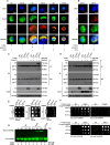
Toll-9 interacts with dsRNA in the endosomes. (A) Micrographs showing colocalization of Rab5 (anti-Rab5, green) and poly (I:C) or DCV dsRNA (J2 anti-dsRNA, red) in cells treated and untreated with CuSO4 (500 µM) in the presence and absence of poly (I:C) transfection or DCV infection. DAPI (blue) is used to stain the nucleus of the cell. Pearson’s correlation coefficient for localization overlap is shown on the merge panel of the micrograph. (B) Micrographs showing colocalization of DCV dsRNA (J2 anti-dsRNA, red) with Toll-9 (anti-V5 tag, green) in cells treated with CuSO4 (500 µM) in the presence and absence of DCV. DAPI (blue) is used to stain the nucleus of the cell. Pearson’s correlation coefficient for localization overlap is shown on the merge panel of the micrograph. (C) Western blot analysis using the indicated antibodies following immunoprecipitation of V5 tag (Toll-9) using anti-dsRNA (J2) antibody from naïve S2 cells and Toll-9 OE cells treated and untreated with poly (I:C) in the presence and absence of CuSO4 (500 µM). (D) Western blot analysis using the indicated antibodies following immunoprecipitation of V5 tag (Toll-9) using anti-dsRNA (J2) antibody of the lysate from uninfected and DCV-infected Toll-9 OE cells and S2 cells in the presence and absence of CuSO4 (500 µM). (E) dsRNA dot blot assay to detect dsRNA binding with purified Toll-9 protein. The dsRNA/Toll-9-V5 complex was blotted with an anti-V5 tag antibody. Control dsRNA-spotted blots were incubated with bovine serum albumin (BSA) and probed with J2 anti-dsRNA and anti-V5 tag antibodies. (F) Protein dot blot assay to detect purified Toll-9 protein in increasing concentration with dsRNA. Purified Toll-9 and S2 lysates were spotted onto membranes, incubated with or without dsRNA (2 µg/mL), and probed with J2 anti-dsRNA or anti-V5 antibodies. (G) RNA electrophoretic mobility shift assay (RNA EMSA) using purified Toll-9-V5 showing that Toll-9 binds to dsRNA in a concentration-dependent manner. The TBE gel represents the separation of bound and unbound dsRNA probes. The results are representative of three independent experiments.

Update of

Similar articles

References

    1. Agaisse H. 2007. An adaptive immune response in Drosophila? Cell Host Microbe 1:91–93. doi: 10.1016/j.chom.2007.04.003 - DOI - PubMed
    1. Lemaitre B, Nicolas E, Michaut L, Reichhart J-M, Hoffmann JA. 1996. The dorsoventral regulatory gene cassette spätzle/Toll/cactus controls the potent antifungal response in Drosophila adults. Cell 86:973–983. doi: 10.1016/s0092-8674(00)80172-5 - DOI - PubMed
    1. Litman GW, Rast JP, Fugmann SD. 2010. The origins of vertebrate adaptive immunity. Nat Rev Immunol 10:543–553. doi: 10.1038/nri2807 - DOI - PMC - PubMed
    1. Lin SJH, Cohen LB, Wasserman SA. 2020. Effector specificity and function in Drosophila innate immunity: getting AMPed and dropping Boms. PLOS Pathog 16:e1008480. doi: 10.1371/journal.ppat.1008480 - DOI - PMC - PubMed
    1. Lindsay SA, Wasserman SA. 2014. Conventional and non-conventional Drosophila Toll signaling. Dev Comp Immunol 42:16–24. doi: 10.1016/j.dci.2013.04.011 - DOI - PMC - PubMed

MeSH terms

Supplementary concepts

LinkOut - more resources