. 2025 Jun 17;99(6):e0132324.
doi: 10.1128/jvi.01323-24. Epub 2025 May 14.

Endothelial cell-released mitochondrial DNA promotes B cell differentiation and virus replication during severe fever with thrombocytopenia syndrome virus infection

Affiliations

Endothelial cell-released mitochondrial DNA promotes B cell differentiation and virus replication during severe fever with thrombocytopenia syndrome virus infection

Yun-Fa Zhang et al. J Virol. .

Abstract

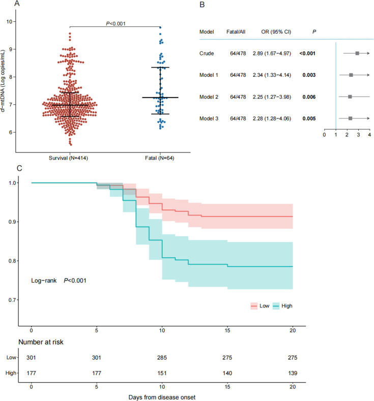
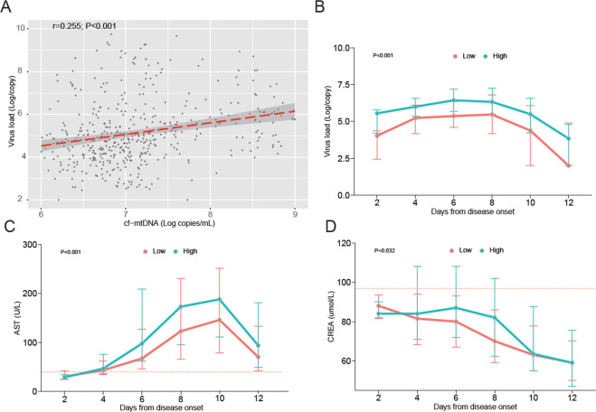
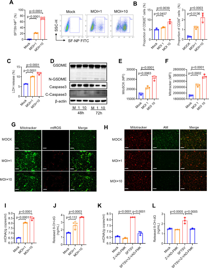
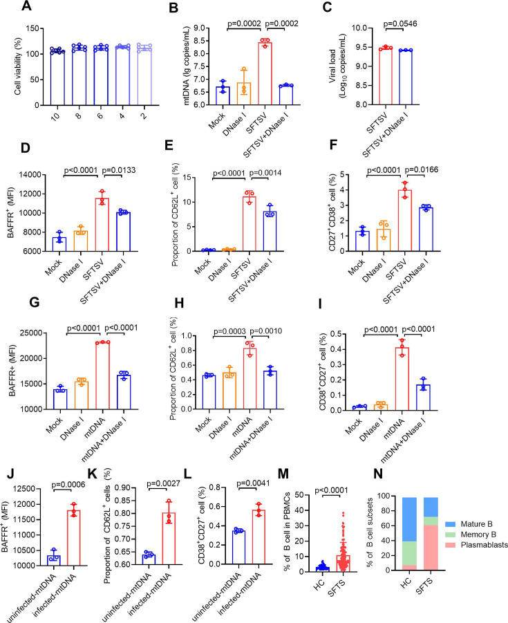
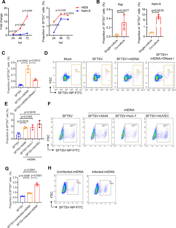
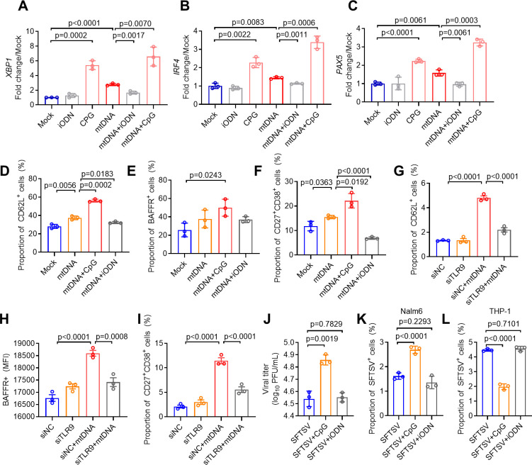
Severe fever with thrombocytopenia syndrome (SFTS) is an emerging infectious disease acquired through tick bites. We have previously demonstrated the correlation between SFTSV-induced mitochondrial dysfunction and inflammation induction, disease progression, and fatal outcome. In the current study, our clinical observation study establishes a strong correlation between elevated levels of circulating cell-free mtDNA and poor prognosis. In vivo studies further reveal endothelial cells as an important source responsible for releasing mtDNA into circulation, which promotes B cell activation, migration, and differentiation via Toll-like receptor 9 (TLR9). Notably, TLR9 activation enhances B-cell susceptibility to SFTSV infection. These findings suggest that mtDNA released by injured endothelial cells facilitates B cell differentiation and virus replication, emphasizing the significant role of mitochondrial damage within endothelial cells in contributing to the severity of SFTS outcomes.IMPORTANCESevere fever with thrombocytopenia syndrome (SFTS) is a new acute tick-borne infectious disease with a high fatality rate of 10%-50%. There is a strong correlation between SFTSV-induced mitochondrial dysfunction and inflammation induction, disease progression, and fatal outcome. Our research has revealed the crucial role of mtDNA in predicting the prognosis of SFTS and its impact on vascular endothelial injuries as well as B cell differentiation, two previously unexplored features of SFTSV infection. Moreover, mtDNA could activate the TLR9 signal to induce plasmablast differentiation in B cells and promote SFTSV infection. This study provides valuable mechanistic and clinical insights into the adverse outcomes associated with SFTSV infection.

Keywords: B cell; SFTS; TLR9; endothelial cell; mtDNA.


Conflict of interest statement

The authors declare no conflict of interest.

Figures

Fig 1
Fig 1
High levels of cf-mtDNA are associated with an elevated risk of death in patients with SFTS. (A) Quantification of cf-mtDNA showed a significant difference between survival and fatal groups. (B) The odds ratios and 95% CI were estimated from the crude model and three adjusted models. Model 1 adjusted for age; Model 2 adjusted for age and sex; and Model 3 adjusted for age, sex, and the presence of comorbidities. (C) Kaplan-Meier curves on the probability of survival in two groups with high and low cf-mtDNA levels. The numbers of at-risk patients at each time point were shown for each group. P values were calculated by log-rank test.
Fig 2
Fig 2
Measurement of cf-mtDNA in relation to virus load (A and B), AST (C), and CREA (D) in SFTS patients. (A) Spearman’s correlation coefficient was used to assess the correlation between cf-mtDNA copy number and viral loads. The red line represents the fitted regression line, and the gray area represents the 95% CI. (B–D) Kinetics of viral loads (B), AST (C), and CREA (D) compared between high and low cf-mtDNA groups with SFTSV infection. Data points are median values, and error bars show interquartile ranges (IQR). P values were calculated by the GEE model. The short red-dotted line represents the upper limit of the normal value of the indicator.
Fig 3
Fig 3
SFTSV induces endothelial cell damage and mitochondrial DNA release in HUVECs. (A) Flow cytometry analysis of SFTSV infection rate in HUVECs at 72 h post-infection (hpi). (B) The proportion of CD62E+ (left panel) and CD39+ (right panel) cells measured by flow cytometry in SFTSV-infected HUVECs at 72 hpi. (C) LDH release examined by the CytoTox 96 Non-Radioactive Cytotoxicity Assay Kit in SFTSV-infected HUVECs at 72 hpi. (D) Representative western blot images of HUVECs lysates post-SFTSV infection, processed for pyroptotic protein levels using GSDME, Caspase3, and C-Caspase3 specific antibody. β-Actin protein was tested as an internal loading control. (E and F) HUVECs were subject to flow cytometric analysis of mitochondrial reactive oxygen species (mtROS, E) and mitochondria mass (MitoTracker, F) at 72 hpi. (G) Immunofluorescence analysis of MitoTracker (Green) and mtROS (Red) in SFTSV-infected HUVECs at 72 hpi. Scale bar: 100 µm, Images shown are representative of three independent experiments. (H) HUVECs were infected with SFTSV for 72 h; then, immunofluorescence analysis of MitoTracker (Red) and AM (Green) in SFTSV-infected HUVECs at 72 hpi was performed to evaluate the opening of mitochondrial permeability. (I) The extracellular mtDNA released from HUVECs was detected by qPCR at 48 hpi. (J) The release of 8-OH-dG in the supernatant of HUVECs at indicated MOIs for 72 h. (K and L) HUVECs were infected with SFTSV and treated with Z-VAD-FMK for 72 h. The extracellular mtDNA (K) and 8-OH-dG (L) were analyzed. Data are present as mean ± s.d.
Fig 4
Fig 4
HUVECs release mtDNA that induces B cell activation and differentiation. (A) Viability of DNase I-treated HUVECs was measured using CCK-8 at 72 h post-infection (hpi). (B and C) SFTSV-infected HUVECs were treated with DNase I (10 U/mL) for 72 h, and mtDNA levels (B) and viral load (C) in the supernatant were measured with RT-qPCR. (D–F) HUVECs were infected with SFTSV at MOI = 5 or treated with DNase I (10 U/mL) for 24 h, followed by co-culture with Nalm-6 cells for 72 h. The proportion of BAFFR+ (D), CD62L+ (E), and plasmablasts (CD27+ CD38+ cells) (F) in Nalm-6 cells was detected by flow cytometry at 72 h post-co-culture. (G–I) B cells were isolated from PBMCs and treated with mtDNA purified from SFTSV-infected HUVECs (MOI = 5) rather than the cell culture supernatant or treated with DNase I (10 U/mL). The proportion of BAFFR+ (G), CD62L+ (H), and plasmablasts (CD27+ CD38+ cells) (I) was detected by flow cytometry at 72 h. (J–L) Nalm-6 cells were treated with mtDNA purified from SFTSV-infected HUVECs or non-infected HUVECs. The proportion of BAFFR+ (J), CD62L+ (K), and plasmablasts (CD27+ CD38+ cells) (L) was detected using flow cytometry at 24 h. (M) The proportion of B cells in PBMCs from healthy controls (HC) and SFTS patients was measured using flow cytometry. (N) Subpopulation of B cells in PBMCs from HC group and SFTS patients. Data represent the mean ± s.d.
Fig 5
Fig 5
HUVCE-derived mtDNA promotes the susceptibility of B cells to SFTSV. (A) The relative levels and proportion of SFTSV NP+ cells in plasmablasts cell lines (H929) and naive B cell lines (Nalm-6) at 72 h post-infection with SFTSV (MOI = 5). (B) The proportion of SFTSV NP+ cells in two B cell lines (Raji and Nalm-6) infected with SFTSV (MOI = 5) (single-culture group), or additionally co-cultured with HUVECs (co-culture group) at 72 h post-infection. (C and D) Effect of mtDNA, purified from SFTSV-infected HUVECs rather than the cell culture supernatant, treatment on the proportion of SFTSV NP+ cells in primary B cells isolated from human peripheral blood at 72 h post-infection (C). Representative images of flow cytometry analysis were shown (D). (E and F) Effect of different types of mtDNA, purified from SFTSV-infected A549, Huh-7, and HUVEC, treatment on the proportion of SFTSV NP+ cells in Nalm-6 (E), representative images of flow cytometry analysis were shown (F). (G and H) Effect of mtDNA from infected or non-infected HUVECs on the susceptibility of Nalm-6 to SFTSV (G). Representative images of flow cytometry analysis were shown (H). Data presented as mean ± s.d.
Fig 6
Fig 6
HUVEC-released mtDNA induces B cell activation and differentiation through the TLR9-signaling pathway. (A–C) B cells isolated from peripheral blood treated with 6 µg/mL CpG (a TLR9 agonist) and 0.5 µM iODN (a TLR9 inhibitor) were incubated with mtDNA for 72 h. The expression of differentiation-related genes XBP1 (A), IRF4 (B), and PAX5 (C) in B cells was measured with quantitative RT-PCR (qRT-PCR). (D–F) The proportion of CD62L+ (D), BAFFR+ (E), CD27 (F), and+ CD38+ (F) among B cells. (G–I) TLR9-knockdown Nalm-6 cells were treated with mtDNA purified from SFTSV-infected HUVECs. The proportion of CD62L+ (G), BAFFR+ (H), and CD27+ CD38+ (I) was examined. (J) SFTSV-infected B cells treated with CpG and iODN. The supernatant viral titer was measured with the immunological focus assay. (K and L) Effect of CpG (6 µg/mL) and iODN (0.5 µM) on the proportion of SFTSV NP+ cells in Nalm-6 (K) and THP-1 (L). Data presented as the mean ± s.d.

Similar articles

References

    1. Yu X-J, Liang M-F, Zhang S-Y, Liu Y, Li J-D, Sun Y-L, Zhang L, Zhang Q-F, Popov VL, Li C, et al. 2011. Fever with thrombocytopenia associated with a novel bunyavirus in China. N Engl J Med 364:1523–1532. doi: 10.1056/NEJMoa1010095 - DOI - PMC - PubMed
    1. Kuhn JH, Adkins S, Alioto D, Alkhovsky SV, Amarasinghe GK, Anthony SJ, Avšič-Županc T, Ayllón MA, Bahl J, Balkema-Buschmann A, et al. 2020. 2020 taxonomic update for phylum Negarnaviricota (Riboviria: Orthornavirae), including the large orders Bunyavirales and Mononegavirales. Arch Virol 165:3023–3072. doi: 10.1007/s00705-020-04731-2 - DOI - PMC - PubMed
    1. Takahashi T, Maeda K, Suzuki T, Ishido A, Shigeoka T, Tominaga T, Kamei T, Honda M, Ninomiya D, Sakai T, et al. 2014. The first identification and retrospective study of severe fever with thrombocytopenia syndrome in Japan. J Infect Dis 209:816–827. doi: 10.1093/infdis/jit603 - DOI - PMC - PubMed
    1. Kim M, Hong K-W, Kim SC, Kim RB, Cho M-C. 2022. Analysis of clinical characteristics and laboratory data related to the prognosis of Korean severe fever with thrombocytopenia syndrome patients: a single-center study. Vector Borne Zoonotic Dis 22:559–567. doi: 10.1089/vbz.2022.0035 - DOI - PubMed
    1. Tran XC, Kim SH, Lee J-E, Kim S-H, Kang SY, Binh ND, Duc PV, Phuong PTK, Thao NTP, Lee W, Bae J-Y, Park M-S, Kim M, Yoo JR, Heo ST, An KH, Kim JM, Cho N-H, Kee S-H, Lee KH. 2022. Serological evidence of severe fever with thrombocytopenia syndrome virus and IgM positivity were identified in healthy residents in Vietnam. Viruses 14:2280. doi: 10.3390/v14102280 - DOI - PMC - PubMed

MeSH terms

LinkOut - more resources