Review
. 2025 Jun 17;99(6):e0047125.
doi: 10.1128/jvi.00471-25. Epub 2025 May 14.

Clicking viruses-with chemistry toward mechanisms in infection

Affiliations
Review

Clicking viruses-with chemistry toward mechanisms in infection

Urs F Greber. J Virol. .

Abstract

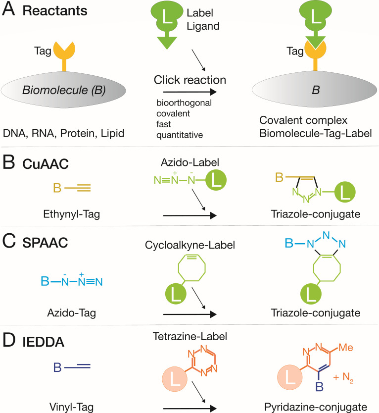
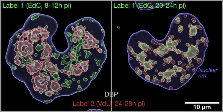
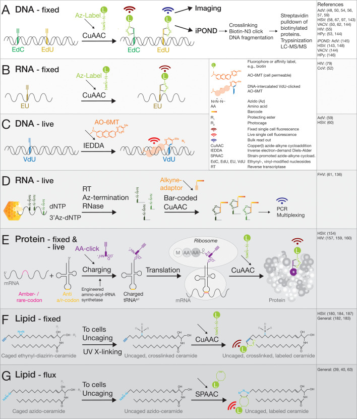
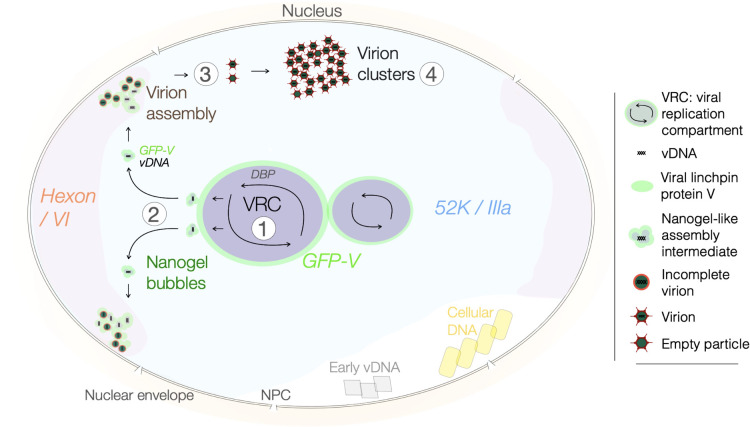
Viruses subvert cells and evade host defense. They emerge unpredictably and threaten humans and livestock through their genetic and phenotypic diversity. Despite more than 100 years since the discovery of viruses, the molecular underpinnings of virus infections are incompletely understood. The introduction of new methodologies into the field, such as that of click chemistry some 10 years ago, keeps uncovering new facets of viruses. Click chemistry uses bio-orthogonal reactions on chemical probes and couples nucleic acids, proteins, and lipids with tractable labels, such as fluorophores for single-cell and single-molecule imaging, or biotin for biochemical profiling of infections. Its applications in single cells often achieve single-molecule resolution and provide important insights into the widely known phenomenon of cell-to-cell infection variability. This review describes click chemistry advances to unravel infection mechanisms of a select set of enveloped and nonenveloped DNA and RNA viruses, including adenovirus, herpesvirus, and human immunodeficiency virus. It highlights recent click chemistry breakthroughs with viral DNA, viral RNA, protein, as well as host-derived lipid functions in both live and chemically fixed cells. It discusses new insights on specific processes including virus entry, uncoating, transcription, replication, packaging, and assembly and provides a perspective for click chemistry to explore viral cell biology, infection variability, and genome organization in the particle.

Keywords: DNA; RNA; adenovirus; click chemistry; herpesvirus; human immunodeficiency virus (HIV); lipid; protein; virus entry; virus genome packaging and assembly.


Conflict of interest statement

The author declares no conflict of interest.

Figures

Fig 1
Fig 1
Overview of the most common click chemistry reactions in virus research. (A) Principal reactants. Chemically modified biomolecules (B), such as DNA, RNA, proteins, or lipids, contain chemical tags, such as alkynes, that react in situ with bio-orthogonally modified ligands, for example, azido-fluorophores or -biotin. This reaction yields covalent products containing tractable labels. (B) CuAAC couples ethynyl-tagged biomolecules to azido-labels yielding stable triazole conjugates. Suitable for chemically fixed cells. (C) SPAAC couples azido-tags to strained alkynes in live cells, yielding stable triazole conjugates. (D) IEDDA used, for example, to couple vinyl-tagged biomolecules to tetrazine-containing labels yields stable pyridazine conjugates suitable for live-cell studies.
Fig 2
Fig 2
Imaging of vDNA replication dynamics by combined CuAAC and IEDDA pulse-chase click chemistry. Pulse-chase protocol with two bio-orthogonal click reactions, CuAAC and IEDDA. The former coupled ethynyl-modified deoxy-cytidine (EdC) to an azido-fluorophore, and the latter vinyl-modified deoxy-uridine (VdU) to a tetrazine fluorophore. The protocol allows for in situ distinction of vDNA synthesized early in AdV infection (early-replicated vDNA) compared to late-synthesized vDNA (late-replicated vDNA). The latter defines the viral replication center as indicated by immunofluorescence staining of the vDNA-binding protein (DBP, 72K). Note that labelling reagent 1 (EdC, green) was added to infected A549 cells 8–12 or 20–24 hpi, and label 2 (VdU, red) 24–28 hpi, followed by specimen fixation, DBP staining (gray), clicking with azide-AlexaFluor 647 (EdC) and AO-6MT (VdU), imaging in a Leica SP8 FALCON CLSM, and 3D rendering using Imaris10 (Oxford Instruments, Oxon, UK). The position of the nuclear rim was defined by staining with 4´,6-diamidino-2-phenylindole (blue). The images show the rendered volumes of infected A549 cell nuclei. Detailed procedures are described in reference . The images are courtesy of Dr. Alfonso Gomez-Gonzalez.
Fig 3
Fig 3
Schematic representation of a select set of click chemistry reactions with vDNA, vRNA, proteins, and lipids. (A) CuAAC-based detection of alkyne-tagged deoxynucleotides EdC or EdU incorporated into vDNA during viral replication. Azido-fluorophores or azido-biotin label the vDNA upon chemical fixation for analyses by fluorescence microscopy and iPOND (isolating proteins on nascent DNA) with mass spectrometry, respectively. (B) CuAAC-based detection of the alkyne-tagged nucleotide EU incorporated into vRNA, allowing for fluorescence microscopy analyses of newly synthesized vRNA. (C) IEDDA-based labeling of alkene-tagged deoxynucleotides (in this case, VdU) incorporated into vDNA, visualized with the DNA intercalating dye AO-6MT, which covalently links to the vinyl group by inverse Diels-Alder ligation. This allows for live-cell tracking of newly synthesized vDNA over several hours until the onset of cell toxicity. The labeling protocol developed for AdV is available in reference . An alternative protocol using IEDDA and strained trans-cyclooctene was developed for HSV-1 (60). (D) Population assay measuring stepwise heat-triggered vRNA exposure from flock house virus (FHV) particles. Exposed vRNA is random-primed with oligo-deoxynucleotides, reverse transcribed in the presence of chain-terminating 3’azido-tagged deoxynucleotides, which are stochastically incorporated by reverse transcriptase in the reaction mix, digested with RNase, tagged with an alkyne-containing barcode adaptor, and analyzed by ClickSeq and multiplexed PCR. This procedure mapped vRNA uncoating intermediates of FHV (61). (E) A minimal approach to click-tag proteins using genetically engineered bio-orthogonal tRNA and aminoacyl-tRNA synthetase to incorporate a noncanonical amino acid (ncAA) at a position of interest using an amber stop codon or a rare codon of choice. An alkyne group in the ncAA is covalently linked by CuAAC to an azido-fluorophore, or another reactive group, such as a tetrazine reacting with a strained functional group on the ncAA by IEDDA. (F, G) Multifunctional ceramide lipid probes for fixed as well as live-cell analyses of lipid localization and flux. Ultraviolet light irradiation of the diazirine group leads to photo-crosslinking by carbene insertion into nearby hydrogen-bonded carbon atoms. Esterified hydroxy groups masking their hydrophilic functionality enhance cell delivery of the probe and are removed by cytosolic esterases. Photo-destructible cages, such as coumarins or nitrobenzyl groups, prevent probe metabolization and can be released by blue light. The terminal alkyne tags on aliphatic chains can be coupled to azido- or strained-ligands by CuAAC and SPAAC reactions, respectively (62, 63).
Fig 4
Fig 4
Model derived from click chemistry experiments depicting stepwise assembly of AdV with concurrent vDNA packaging and capsid assembly in the cell nucleus. Dual-label, pulse-chase click chemistry and live-cell confocal, as well as electron microscopy, inform a stepwise concurrent model for AdV assembly in the cell nucleus (see reference 59). The model additionally builds on recent evidence involving the 52K biomolecular condensate in viral assembly (125) and viral assembly intermediates identified by transmission electron microscopy (126). Step 1: the viral replication center constitutes three essential viral proteins, DBP, DNA-Pol, and terminal protein, to amplify vDNA. Step 2: newly replicated vDNA is sequestered from the periphery of the VRC into nanoscale bubbles containing the viral capsid-vDNA linchpin protein V (GFP-V). The bubbles released into the viral 52K domain (containing the viral inner capsid protein IIIa) located around the VRC. Step 3: the GFP-V-vDNA nanogels diffuse in the 52K compartment and cluster in the nuclear periphery, where the major viral capsid protein hexon and its binding partner protein VI are enriched. These loci termed virion formation sites release virus particles that migrate by unrestricted diffusion and eventually form paracrystalline clusters in the nucleus (step 4).

Similar articles

References

    1. Lederberg J. 2000. Infectious history. Science 288:287–293. doi: 10.1126/science.288.5464.287 - DOI - PubMed
    1. Lustig A, Levine AJ. 1992. One hundred years of virology. J Virol 66:4629–4631. doi: 10.1128/JVI.66.8.4629-4631.1992 - DOI - PMC - PubMed
    1. Koonin EV, Dolja VV, Krupovic M. 2022. The logic of virus evolution. Cell Host Microbe 30:917–929. doi: 10.1016/j.chom.2022.06.008 - DOI - PubMed
    1. Ludmir EB, Enquist LW. 2009. Viral genomes are part of the phylogenetic tree of life. Nat Rev Microbiol 7:615. doi: 10.1038/nrmicro2108-c4 - DOI - PubMed
    1. Enard D, Cai L, Gwennap C, Petrov DA. 2016. Viruses are a dominant driver of protein adaptation in mammals. Elife 5:e12469. doi: 10.7554/eLife.12469 - DOI - PMC - PubMed

LinkOut - more resources