. 2025 Aug;48(8):6373-6396.
doi: 10.1111/pce.15611. Epub 2025 May 14.

A Holistic Investigation of Arabidopsis Proteomes Altered in Chloroplast Biogenesis and Retrograde Signalling Identifies PsbO as a Key Regulator of Chloroplast Quality Control

Affiliations

A Holistic Investigation of Arabidopsis Proteomes Altered in Chloroplast Biogenesis and Retrograde Signalling Identifies PsbO as a Key Regulator of Chloroplast Quality Control

Dario Di Silvestre et al. Plant Cell Environ. 2025 Aug.

Abstract

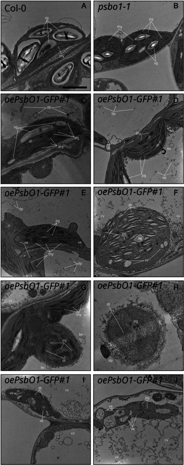
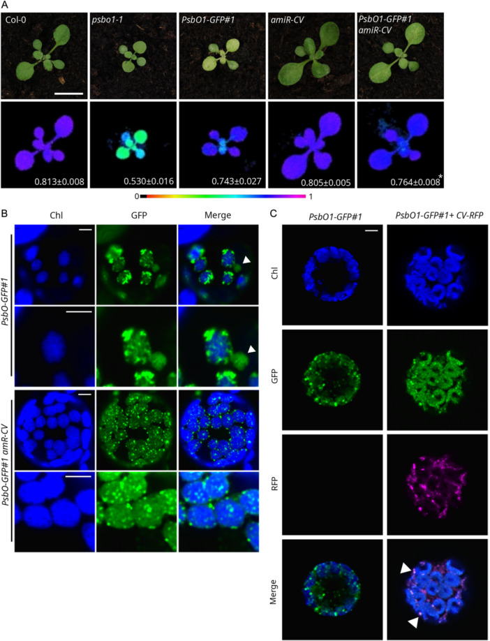
Communication between the diverse compartments of plant cells relies on an intricate network of molecular interactions that orchestrate organellar development and adaptation to environmental conditions. Plastid-to-nucleus signalling pathways play a key role in relaying information from developing, mature, and damaged or disintegrating chloroplasts to the nucleus, which serves to coordinate gene expression between the two genomes. To shed light on these mechanisms, we performed a comprehensive analysis of the response of the Arabidopsis thaliana proteomes to perturbation of chloroplast biogenesis by the antibiotic lincomycin (Lin) in the absence of GENOMES UNCOUPLED 1 (GUN1), a key player in plastid-to-nucleus signalling. The topological analysis of protein-protein interactions (PPIs) and co-expression networks enabled the identification of protein hubs in each genotype and condition tested, and highlighted whole-cell adaptive responses to the disruption of chloroplast biogenesis. Our findings reveal a novel role for PsbO, a subunit of the oxygen-evolving complex (OEC), which behaves as an atypical photosynthetic protein upon inhibition of plastid protein synthesis. Notably, and unlike all other subunits of the thylakoid electron transport chain, PsbO accumulates in non-photosynthetic plastids, and is crucial for the breakdown of damaged chloroplasts.

Keywords: chloroplast biology; chloroplast degradation; intracellular signalling; oxidative stress; proteome; proteomics.


Figures

Figure 1
Figure 1
Proteomes of A. thaliana Col‐0 and gun1 seedlings grown on MS medium in the absence or presence (±) of Lin. (A) Schematic overview of the experimental set‐up adopted in this study, which depicts the relevant conditions, data sets and replicates used. Each circle represents one replicate. Empty circles indicate control conditions and filled circles refer to Lin‐grown samples. Col‐0 samples are shown in red, gun1 in black. (B) Venn diagram of proteins identified in Sets 1, 2 and 3. The data in Set1 were generated in our laboratory from total protein extracts obtained from Col‐0 ± Lin and gun1‐102 ± Lin seedlings harvested 6 days after sowing (DAS). Set2 was derived from Col‐0 ± Lin and gun1‐102 ± Lin seedlings, 6 DAS, and is enriched in soluble proteins (Tadini, Peracchio, et al. 2020), while Set3 was obtained from total protein extracts from Col‐0 ± Lin; gun1‐101 ± Lin seedlings, 5 DAS (Wu, Meyer, Wu, et al. 2019). (C) Venn diagram showing the proteins identified in the three data sets [refer to the shape/colour code shown in (A)] and grouped according to the four conditions: Col‐0 and the gun1 mutant grown in the presence or absence of Lin (±Lin). (D) Hierarchical clustering of differentially abundant proteins (DAPs), p ≤ 0.001.
Figure 2
Figure 2
Selection of DAPs upregulated in the presence of Lin. These proteins are functionally involved in protein folding, oxidative stress response, vesicle trafficking (ER–Golgi–Vacuole), cell death regulation, proteolysis and ubiquitination. For each protein, the normalized spectral count value (nSpC) in the range 0–100 is shown. Highlighted comparisons have a statistical significance of p ≤ 0.05, while asterisks indicate a significance of p ≤ 0.01, calculated according to LDA. [Color figure can be viewed at wileyonlinelibrary.com]
Figure 3
Figure 3
Hubs derived from PPI and co‐expression network models. (A) PPI network hubs in Col‐0 and gun1 seedlings, grown in the absence or presence of Lin (±Lin). PPI hubs were selected on the basis of betweenness, centroid and bridging centralities. Larger and underlined nodes indicate proteins that were defined as hubs and were differentially abundant (p ≤ 0.01). (B) Hub proteins based on co‐expression network models and genetic background or growth condition. (C) Hub proteins exclusively found in Col‐0, gun1, Col‐0 + Lin or gun1 + Lin co‐expression network models. [Color figure can be viewed at wileyonlinelibrary.com]
Figure 4
Figure 4
Validation of extra‐plastid protein hubs. (A) Cytosolic folding stress and ubiquitination in total protein extracts obtained from Col‐0 and gun1‐102 samples grown in the absence or presence of Lin (±Lin). The immunoblots were obtained using antibodies specific for UBQ11 and cytosolic AtHSP90‐1, respectively. Coomassie Brilliant Blue (C.B.B.) staining of SDS–PAGE is shown as loading control. (B) Proteasome activity assay based on the level of fluorescence released by amino‐methyl‐coumarin. Asterisks indicate significant differences (*p < 0.05; **p < 0.01 according to ANOVA with Tukey's post hoc test). (C) Real‐time quantitative PCR of cytosolic ROS and the stress‐related marker genes HSFA2, CAT2 and APX2. Asterisks indicate significant differences (*p < 0.05; **p < 0.01 according to Student's t‐test). (D) NR enzyme activity. Asterisks indicate significant differences (*p < 0.05; **p < 0.01; *** p < 0.001 according to ANOVA with Tukey's post hoc test). (E) Measurement of mitochondrial oxygen consumption (nmol O2/min/mg protein) in Col‐0 and gun1‐102 ± Lin samples, together with (F) mitochondrial alternative oxidase (AltOX) activity. Asterisks indicate significant differences (*p < 0.05; **p < 0.01; ***p < 0.001; **** p < 0.0001 according to ANOVA with Tukey's post hoc test). (G) Real‐time quantitative PCR of AltOX1a, mtHSC70‐1 and mtHSC70‐5 transcripts, used as molecular marker genes for the investigation of mitochondrion‐to‐nucleus retrograde signalling and mtUPR. Asterisks indicate significant differences (*p < 0.05; **p < 0.01 according to Student's t‐test). [Color figure can be viewed at wileyonlinelibrary.com]
Figure 5
Figure 5
PsbO plays a role in the increased sensitivity of gun1 seedlings to lincomycin. (A) Immunoblot analyses based on total protein extracts from seedlings grown in absence or presence of 550 µM Lin (±Lin). Extracts obtained from Col‐0, gun1‐102, psbo1‐1 and gun1‐102 psbo1‐1 genotypes were probed with antibodies specific for PsbO, PsbR and PsbQ. Coomassie Brilliant Blue (C.B.B.) staining of SDS–PAGE is shown as loading control. Note that the PsbO antibody reacts with both PsbO1 and PsbO2, and PsbO2 is detectable in the psbo1 mutant genetic background. The arrowhead indicates the mature PsbO protein, the plus symbol (+) indicates the precursor protein and the band indicated by the asterisk (*) may refer to a post‐translational modification of PsbO precursor protein. (B) Visible phenotypes of 6 DAS wild‐type (Col‐0), single (gun1‐102, psbo1‐1) and double (gun1‐102 psbo1‐1) mutant seedlings grown on MS medium in the absence and presence of different concentrations of Lin (5.5, 55, 550 µM) in the growth medium. The Fv/Fm parameter (bottom right) is reported as an indicator of photosynthesis efficiency and the functional state of chloroplasts (average ± SD; n ≥ 15). (C) Transmission electron micrographs of mesophyll cells from 6 DAS seedlings of the indicated genotypes grown on MS medium with 55 µM Lin. thy, thylakoids; pg, plastoglobuli; ve, vesicle; bv, budding vesicles; dp, degradation products; cv, collapsing vacuole. The scale bar represents 1 µm. [Color figure can be viewed at wileyonlinelibrary.com]
Figure 6
Figure 6
PsbO is involved in both chloroplast quality control and chloroplast degradation. (A) Visible phenotypes displayed by 12 DAS plantlets of the indicated genotypes and fluorometric data depicting levels of the Fv/Fm parameter (shown in false colours) and the average ± SD values (n ≥ 4). Scale bar: 1 cm. (B) Fluorescence signals from chlorophylls (Chl, blue) and GFP (green) detected in protoplasts obtained from leaves of the indicated genotypes were observed with a confocal microscope. Solid arrowheads indicate budding, vesicle‐like structures, while empty arrowheads show detached vesicles. Scale bar: 10 µm. (C) Fluorescence signals from chlorophylls (Chl, blue) and GFP (green) detected in mesophyll tissue obtained from the indPsbO1‐GFP#2 line incubated for the indicated hours (HAI, hours after induction) in presence of DEX. Scale bar: 10 µm. (D) Immunoblot analyses of total lysates and pellet obtained by ultracentrifugation collected from leaves of the indicated genotypes and, where indicated, incubated in presence of DEX for the indicated hours. Filters were incubated with antibodies specific for PsbO and GFP. Asterisk (*) and arrowhead (<) indicate the GFP constructs and the endogenous PsbO, respectively. The comparable amount of endogenous PsbO in the total lysate indicates the equal loading in the ultracentrifuge tubes. [Color figure can be viewed at wileyonlinelibrary.com]
Figure 7
Figure 7
Transmission electron micrographs of mesophyll cells of Col‐0 (A), psbo1‐1 (B) and PsbO1‐GFP (C–J) lines from 12 DAS plantlets grown on soil. thy, thylakoids; dt, degrading thylakoids; sg, starch granule; pg, plastoglobuli; ve, vesicles; bv, budding vesicles, dp, degradation products; cv, collapsing vacuole. Scale bar: 1 µm.
Figure 8
Figure 8
Functional interaction of PsbO with CV. (A) Visible phenotypes of 12 DAS plantlets of the indicated genotypes and fluorometric data representing the Fv/Fm parameter (in false colours) and the average ± SD values (n ≥ 4). Scale bar: 1 cm. (B) Fluorescence signals from chlorophylls (Chl, blue) and GFP (green) detected in protoplasts obtained from leaves of the indicated genotypes observed at different magnifications with a confocal microscope. Solid arrowheads indicate budding vesicle‐like structures. Scale bar: 5 µm. (C) Fluorescence signals from chlorophylls (Chl, blue), GFP (green) and RFP (magenta) detected in protoplasts obtained from the PsbO1‐GFP#1 line and transformed with the CV‐RFP construct observed with a confocal microscope. Scale bar: 5 µm. [Color figure can be viewed at wileyonlinelibrary.com]

Similar articles

Cited by

References

    1. Allahverdiyeva, Y. , Suorsa M., Rossi F., et al. 2013. “ Arabidopsis Plants Lacking PsbQ and PsbR Subunits of the Oxygen‐Evolving Complex Show Altered PSII Super‐Complex Organization and Short‐Term Adaptive Mechanisms.” Plant Journal 75: 671–684. - PubMed
    1. Anjum, N. A. , Sharma P., Gill S. S., et al. 2016. “Catalase and Ascorbate Peroxidase—Representative H2O2‐detoxifying Heme Enzymes in Plants.” Environmental Science and Pollution Research 23: 19002–19029. - PubMed
    1. Assenov, Y. , Ramírez F., Schelhorn S.‐E., Lengauer T., and Albrecht M.. 2008. “Computing Topological Parameters of Biological Networks.” Bioinformatics 24: 282–284. - PubMed
    1. Bari, E. , Ferrera F., Altosole T., et al. 2023. “Trojan‐Horse Silk Fibroin Nanocarriers Loaded With a Re‐Call Antigen to Redirect Immunity Against Cancer.” Journal for Immunotherapy of Cancer 11: e005916. - PMC - PubMed
    1. Berardini, T. Z. , Reiser L., Li D., et al. 2015. “The Arabidopsis Information Resource: Making and Mining the ‘Gold Standard’ Annotated Reference Plant Genome.” Genesis 53: 474–485. - PMC - PubMed

MeSH terms