. 2025 Jul 2;24(7):976-992.
doi: 10.1158/1535-7163.MCT-24-0379.

A Novel B7-H4xCD3 Bispecific T-cell Engager (PF-07260437) Synergizes with Breast Cancer Standard of Care and Immune Checkpoint Therapies

Affiliations

A Novel B7-H4xCD3 Bispecific T-cell Engager (PF-07260437) Synergizes with Breast Cancer Standard of Care and Immune Checkpoint Therapies

Keith Abayasiriwardana et al. Mol Cancer Ther. .

Abstract

Immune checkpoint inhibitors have shown limited success in breast cancer, the most common and deadly cancer in women worldwide. Novel immune therapies, such as CD3-engaging bispecific antibodies, have shown clinical promise in hematologic malignancies. However, developing CD3 bispecifics for solid tumors has been challenging due to the difficulty in identifying tumor-specific antigens. B7-H4 is proposed as an attractive tumor-associated antigen for breast cancer therapeutics with comprehensive coverage regardless of breast cancer molecular subtype. We designed a B7-H4-targeting CD3 bispecific molecule, PF-07260437, and demonstrated B7-H4-dependent pharmacology in vitro by directing cytotoxic T-cell killing to breast cancer cell lines. Treatment of cell line- and patient-derived xenograft in vivo models of human breast cancer with PF-07260437 induced substantial tumoricidal activity, often resulting in complete responses. Mechanistically, PF-07260437 increased T-cell number and activation, leading to efficient tumor killing. Additionally, combining PF-07260437 with standard of care (palbociclib plus fulvestrant) and a checkpoint inhibitor (anti-PD-1) showed combinatorial benefits in an immune-competent in vivo model. Clinically relevant noninvasive PET/CT imaging with a CD8-targeting tracer demonstrated PF-07260437-mediated increases in intratumoral CD8 T cells, highlighting the utility of CD8-PET technology to potentially assess biomarker changes in the clinic. Finally, the manageable toxicity profile of PF-07260437 was highlighted in an exploratory toxicology study in cynomolgus monkeys. These data support the clinical testing of PF-07260437 for treating B7-H4-expressing solid tumors, including breast cancer.


Conflict of interest statement

L. Wu reports personal fees from Pfizer during the conduct of the study and has a patent for WO2022013775A1 pending. M. Nocula-Lugowska reports other support from Pfizer, Inc. during the conduct of the study and has a patent for WO_2022_013775_A1 pending. E. Rosfjord reports employment with AstraZeneca and ownership of Pfizer and AstraZeneca stock. D. Mathur reports a patent for US11434292 (B2) issued. K. Maresca reports personal fees from Pfizer, Inc. outside the submitted work. A. Giddabasappa reports employment with Pfizer. C. Lees reports other support from Pfizer outside the submitted work and employment with Pfizer and ownership of Pfizer shares. A.T. Hooper reports other support and personal fees from Pfizer, Inc. during the conduct of the study as well as personal fees from Regeneron Pharmaceuticals outside the submitted work and has a patent for WO2022/013775 A1 pending. No disclosures were reported by the other authors.

Figures

None
Graphical abstract
Figure 1.
Figure 1.
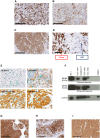
B7-H4 is highly expressed in primary human breast cancers and tumor models. A–C, B7-H4 expression in primary human breast cancer was measured by B7-H4 IHC (DAB) with hematoxylin counterstain. Representative images from (A) HR+HER2, (B) HER2+, and (C) TNBC molecular subtypes are shown. D, Comparison of expression in tumor tissue and normal adjacent mammary glands (NAT). E, B7-H4 (DAB) and CD68 (Vina Green, a macrophage marker) co-staining of primary human breast cancers with varying levels of B7-H4 expression: no, low, moderate, and high. F, Western blot of B7-H4 in primary human PBMCs, with human breast cancer cell lines for comparison. GAPDH is used as a loading control. G–I, B7-H4 expression (DAB, brown stain) with hematoxylin counterstain in human breast cancer PDXs grown in NSG mice: (G) PDX-BRX-26305 (H-score = 219), (H) PDX-BRX-11380 (H-score = 235), and (I) PDX-BRX-24301 (H-score = 205). All images are shown at 20× objective. Scale bar, 200 µm.
Figure 2.
Figure 2.
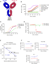
Design, binding, and in vitro activity of the B7-H4 bispecific molecule PF-07260437. A, Schematic illustration of the B7-H4 CD3 bispecific molecule PF-07260437. EE depicts two mutations in the hinge (C223E and P228E), and E depicts a mutation in CH3 (L368E) in the anti-B7-H4 arm. The RRR depicts three mutations in the hinge (C223R, E225R, and P228R), and R depicts a mutation in CH3 (K409R) in the anti-CD3 arm. B, Cell-based binding of PF-07260437 on the B7-H4-expressing human tumor cell line MX-1, HEK-293 cells transfected to express human B7-H4, and the human tumor cell line HCC-1806 that does not express B7-H4, parental HEK-293 cells, and CD3-mediated binding to primary human pan T cells and primary cynomolgus pan T cells. Data representative of three independent assessments are shown. C, Lactate dehydrogenase release-based in vitro cytotoxicity assay measuring T-cell killing of MX-1, MDA-MB-468, MDA-MB-453, and T-47D cells induced by PF-07260437. Representative data shown from a single T-cell donor from three separate T-cell donors tested. D, Luciferase-based cell viability assay measuring T-cell killing induced by PF-07260437 in HCC-1954-luc (cell line expressing B7-H4) and no T-cell killing induced by PF-07260437 in HCC-1806-luc (cell line that does not express B7-H4). E, Linear regression of log (EC50 of PF-07260437) and log (B7-H4 copy number measured by antibody bound per cell shown in Fig. 1K) in the five B7-H4–expressing cell lines tested in cytotoxicity assays. Goodness-of-fit R2 = 0.8045. Best fit and 95% confidence band are shown. F, Cell proliferation at days 2 and 6 after treatment with PF-07260437 without T cells measured by a CellTiter-Glo kit (Promega) in HR+HER2 cell line T-47D, HER2+ cell line HCC-1954, and TNBC cell line MX-1. Data in the figure are shown as nonlinear regression curves of mean ± SEM of two replicates. RLU, relative light units.
Figure 3.
Figure 3.
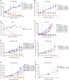
PF-07260437 induced significant and durable TGI in human CDX and PDX models of breast cancer in vivo. A, TNBC tumor cell line MDA-MB-468 and (B) HER2+ tumor cell line HCC-1954 were inoculated subcutaneously, and untreated human PBMCs were injected 6 days prior to drug dosing via an intravenous route. Once tumors were established, mice were dosed with PF-07260437, negative control bispecific, or vehicle either intravenously or subcutaneously on days 0, 7, and 14. C, HR+HER2 tumor cell line T-47D and (D) MX-1-Luc TNBC tumor cells were inoculated subcutaneously. Once tumors were established, mice were dosed with PF-07260437 or vehicle (T-47D) or with negative control bispecific (MX-1-Luc), either intravenously or subcutaneously. Twenty-four hours after the first dose, ex vivo expanded human pan T cells were injected intravenously, and mice received two subsequent doses of PF-07260437 on days 7 and 14. Efficacy in PDX models: PDX-BRX-11380 (E), PDX-BRX-24301 (F), PDX-BRX-26305 (G), and PDX-CRX-11201 (H) fragments were inoculated subcutaneously, and untreated human PBMCs were injected intravenously 6 days prior to treatment initiation. Once tumors were established, mice were dosed with PF-07260437, a negative control bispecific, or vehicle either intravenously or subcutaneously on days 0, 7, and 14. A–H, Mean ± SEM of each group of 10 mice is shown. Dashed vertical lines represent the days mice were dosed with the drug or vehicle. NC bispecific = negative control bispecific. Note in D that several treatment groups are superimposed on top of each other on the graph because of similar activity (intravenous 0.05 mg/kg green line triangle marker, intravenous 0.5 mg/kg red line circle marker, subcutaneous 0.05 mg/kg blue line square marker, and subcutaneous 0.5 mg/kg black line x marker).
Figure 4.
Figure 4.
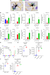
Immunomodulatory effects of PF-07260437 in vivo. MDA-MB-468 cells were inoculated subcutaneously, and human PBMCs were intravenously injected 6 days prior to treatment initiation. Once tumors were established, mice were dosed intravenously with PF-07260437 at 0.5 and 0.05 mg/kg or vehicle on days 0 and 7, and tumor samples were taken at different time points for PD evaluation. A, Magnified representative IHC images from day 5 samples to illustrate tumor cell and T-cell interaction: human IgG (DAB, brown arrows), CD3 (Vina Green, green arrows), and GzymB (purple stain, purple arrows) co-staining. B, Dose- and (C) time-dependent induction of human CD3E, IL2, IFNG, CXCL9, and CXCL10 genes was assessed by qRT-PCR analysis and standardized to the housekeeping gene GAPDH. Mean ± SEM of each group of four mice is shown. D–F, Flow cytometric analysis of tumor samples for T-cell number and activation at different doses and time points after PF-07260437 treatment. D, CD3+ T-cell density normalized to tumor weight. E, Time- and dose-dependent induction of GzymB in CD8+ T cells. F, Time- and dose-dependent upregulation of CD25 expression in CD8+ T cells. Mean ± SEM of each group of four mice is shown. Statistical analysis was done by unpaired t test for two-group comparisons or one-way ANOVA for multiple-group comparisons. *, P < 0.05; **, P < 0.01; ***, P < 0.001; ****, P < 0.0001.
Figure 5.
Figure 5.
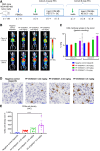
PET imaging with an anti-CD8 minibody detected kinetic and dose-dependent increases in CD8 T-cell infiltration following PF-07260437 treatment. A, Schematics showing the experimental design, dosing regimen (bsAb, bispecific antibody treatment), and imaging time points. B, Representative whole-body coronal maximum intensity projection images of mCT and PET overlay in the indicated treatment groups and time points. GI, gastrointestinal region; Kid, kidney; Liv, liver; Tu, tumor. C,Ex vivo quantitation of tumor uptake measured by gamma counting; N = 8/group. Statistical analysis was done by one-way ANOVA followed by Tukey multiple comparison test. *, P < 0.05; ****, P < 0.0001. D, Representative IHC images showing CD8a staining across different groups in cohort B. All images are shown at 20× objective. Scale bar, 50 µm. E, Quantitation of CD8a-positive T cells from the IHC images. N = 5–8. Statistical analysis was done by one-way ANOVA followed by the Kruskal–Wallis multiple comparisons test. ****, P < 0.001. C and E Data are mean ± SEM.
Figure 6.
Figure 6.
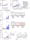
PF-07260437 induced significant TGI as a monotherapy and enhanced TGI in combination with standard-of-care and ICI therapies. AF, TGI and PD of various treatments in HR+ ZR-75-1 human cancer cell line xenografts. A, HR+ ZR-75-1 human cancer cells were inoculated subcutaneously, and once tumors were in the 200–250 mm3 range (day 53 after tumor implant), mice were treated with vehicle or predosed with fulvestrant (subcutaneously; every 3 days loading dose followed by once weekly for 4 weeks), palbociclib [twice a day (BID) orally], or both agents for 21 days (pretreatment phase prior to PF-07260437 dosing). B, Subsequently, each of the predosed groups was injected intravenously with human PBMCs 7 days prior to being subdivided and receiving either PF-07260437 or negative control bispecific subcutaneously at weekly intervals in addition to continuing the same predosing treatment regimen. Mean ± SEM of each group of 10 mice is shown; tumor means are plotted until the first animal is lost from each group independently. Dashed vertical lines represent the days mice were dosed. C, To perform expression analyses, tumors were harvested on day 28 after fulvestrant + palbociclib initiation and processed into formalin-fixed, paraffin-embedded blocks, and a NanoString IO360 panel assay was performed. CD3 gene expression across various treatment groups is shown. D, CD3 IHC (DAB) with hematoxylin counterstain of selected groups. Representative images from the fulvestrant + palbociclib + negative control bispecific, vehicle + PF-07260437, and fulvestrant + palbociclib + PF-07260437 groups are shown at 20× objective. Scale bar, 200 µm. E, NanoString IO360 panel PD-L1 gene expression across various treatment groups. F, PD-L1 IHC (DAB) with hematoxylin counterstain of selected groups. Representative images from the fulvestrant + palbociclib + negative control bispecific, vehicle + PF-07260437, and fulvestrant + palbociclib + PF-07260437 groups are shown at 20× objective. Scale bar, 200 µm. G and H, Combination benefit of PF-07260437 and anti–PD-1 therapy in an immunocompetent humanized model. G, The TNBC syngeneic cell line E0771-hB7-H4 was inoculated subcutaneously into huCD3e mice, and tumors were allowed to establish. Once tumor volume was in the 100–150 mm3 range, mice were dosed once with PF-07260437 subcutaneously or vehicle control; a mouse anti–PD-1 blocking antibody was administered intraperitoneally every 3 days for six doses, or a combination of PF-07260437 and anti–PD-1 was given according to their respective schedules. Mean ± SEM of each group of 15 mice is shown; tumor means are plotted until the first animal is lost from each group independently. Dashed vertical lines represent days mice were dosed. H, Tumors were harvested 9 days after treatment start with PF-07260437/anti–PD-1, dissociated into single-cell suspensions, and analyzed by flow cytometry for CD3 and CD8 expression. Statistical analysis was done using one-way ANOVA. *, P < 0.05; **, P < 0.01; ***, P < 0.001; ****, P < 0.0001.

References

    1. WHO . Breast cancer. Cancer 2020. Available from:https://www.who.int/news-room/fact-sheets/detail/breast-cancer.
    1. Colditz GA, Bohlke K. Priorities for the primary prevention of breast cancer. CA Cancer J Clin 2014;64:186–94. - PubMed
    1. Kimbung S, Loman N, Hedenfalk I. Clinical and molecular complexity of breast cancer metastases. Semin Cancer Biol 2015;35:85–95. - PubMed
    1. Choi IH, Zhu G, Sica GL, Strome SE, Cheville JC, Lau JS, et al. Genomic organization and expression analysis of B7-H4, an immune inhibitory molecule of the B7 family. J Immunol 2003;171:4650–4. - PubMed
    1. Wescott EC, Sun X, Gonzalez-Ericsson P, Hanna A, Taylor BC, Sanchez V, et al. Epithelial expressed B7-H4 drives differential immunotherapy response in murine and human breast cancer. Cancer Res Commun 2024;4:1120–34. - PMC - PubMed

MeSH terms

Substances