. 2025 May 14;20(5):e0322147.
doi: 10.1371/journal.pone.0322147. eCollection 2025.

Efficacy and immunogenicity of rKVAC85B in a BCG prime-boost regimen against H37Rv and HN878 Mycobacterium tuberculosis strains

Affiliations

Efficacy and immunogenicity of rKVAC85B in a BCG prime-boost regimen against H37Rv and HN878 Mycobacterium tuberculosis strains

Eunkyung Shin et al. PLoS One. .

Abstract

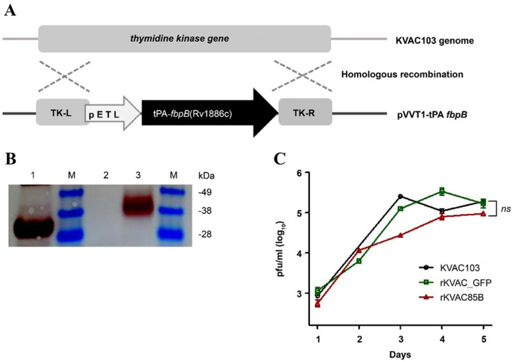
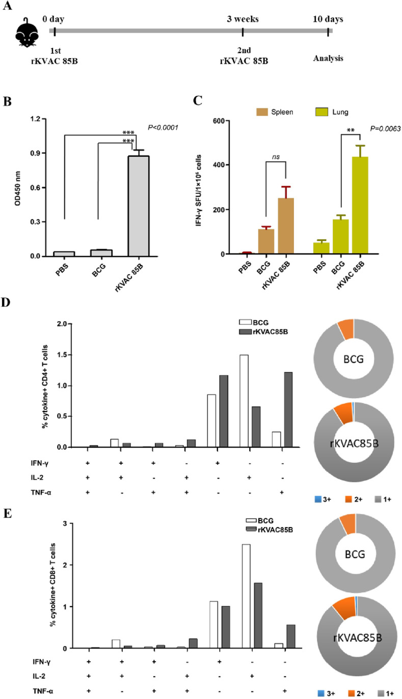
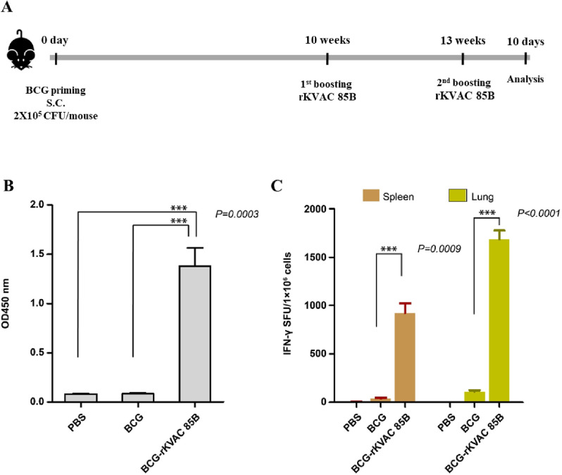
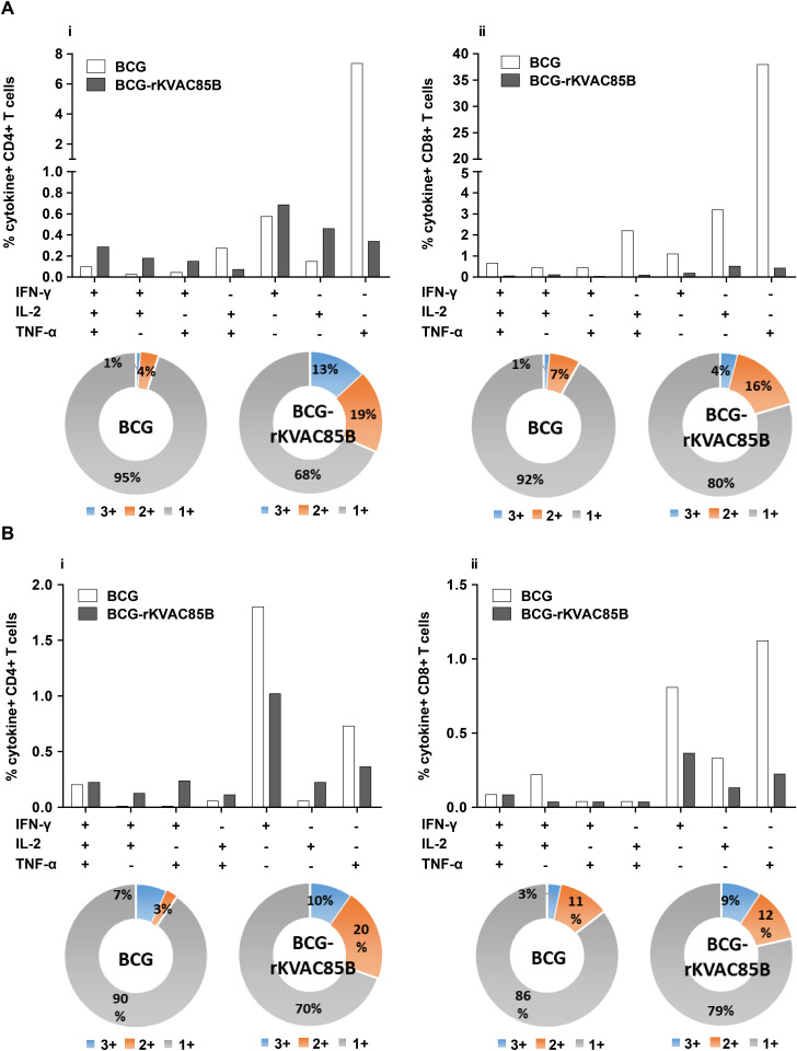
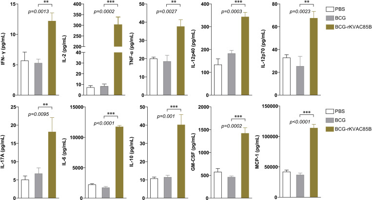
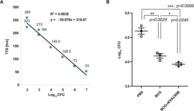
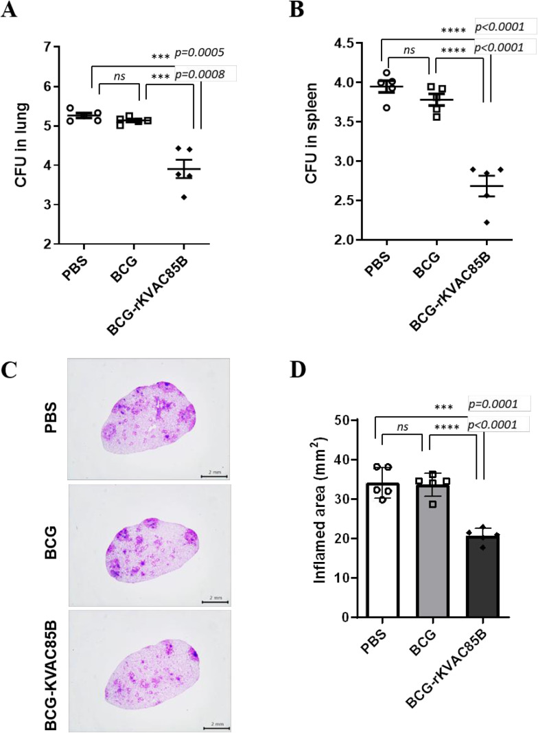
Mycobacterium tuberculosis infection accounted for 1.3 million deaths worldwide in 2022. Bacillus Calmette-Guérin (BCG) is the only licensed vaccine against tuberculosis (TB); however, it has limited protective efficacy in adults. In this study, we constructed a recombinant vaccinia virus expressing Ag85B from M. tuberculosis using a novel attenuated vaccinia virus (KVAC103). We then analyzed the immunogenicity of prime-boost inoculation strategies using recombinant KVAC103 expressing Ag85B (rKVAC85B) compared to BCG. In both rKVAC85B prime-boost and BCG prime-rKVAC85B boost inoculation regimens, rKVAC85B induced the generation of specific immunoglobulin G (IgG) and secretion of interferon-γ by immune cells. In vitro analysis of Mycobacterium growth inhibition revealed a comparable immune-mediated pattern of outcomes. Furthermore, bacterial loads in the lungs were significantly lower in mice inoculated with the BCG prime-rKVAC85B boost than in the BCG-only group following a rechallenge infection with both H37Rv and HN878 strains of M. tuberculosis. These findings collectively suggest that KVAC103, incorporated into a viral vector, is a promising candidate for the development of a novel TB vaccine platform that is effective against multiple M. tuberculosis strains, including H37Rv and HN878, and that rKVAC85B effectively stimulates immune responses against M. tuberculosis infection.


Conflict of interest statement

The authors have declared that no competing interests exist.

Figures

Fig 1
Fig 1. Construction and expression of rKVAC85B in Vero cells.
(A) Schema illustrating the generation of recombinant KVAC virus expressing the M. tuberculosis Ag85B protein. To create rKVAC85B, a codon-optimized Rv1886c was inserted into the KVAC genome, containing TK-L and TK-R regions, through homologous recombination. TK: thymidine kinase. (B) Confirmation of Ag85B protein expression was conducted through western blot analysis using a rabbit polyclonal antibody against Ag85B (Abcam, Cambridge, UK). The purchased Ag85B protein (Abcam, Cambridge, UK) was used as the positive control, whereas the lysate obtained from the rKVAC-GFP infected group was used as the negative control. M: protein marker; Line 1: positive control (purified Ag85B protein), Line 2: negative control (Vero cell lysate), Line 3: rKVAC85B (Vero cell-infected lysate). (C) Comparative growth analysis was performed for the rKVAC85B and rKVAC-GFP groups. The growth titrations of these two viruses were assessed in Vero cells at a multiplicity of infection of 0.1, and the supernatants of the virus culture media were collected and harvested at 24 h intervals. Virus titrations were determined using a plaque assay.
Fig 2
Fig 2. Immune responses of rKVAC85B prime-boost immunized mice.
(A) Scheme of rKVAC85B prime-boost immunazation. rKVAC85B (5 × 10⁷ pfu/mouse) was subcutaneously inoculated 2 dose 3 weeks intervals and then immunized mice were sacrificed after 10-14 days from 2nd inoculation of rKVAC85B. (B) IgG antibody response to M. tuberculosis Ag85B antigen. Serum levels of antigen-specific IgG were quantified using ELISA, with microtiter plates coated with recombinant Ag85B protein (Abcam, 100 ng/well) to assess humoral immunity post-vaccination. (C) Frequency of IFN-γ-secreting T-cells in the lungs and spleens as measured using ELISPOT. Lymphocytes isolated from the lungs and spleens of vaccinated mice were stimulated ex vivo with M. tuberculosis Ag85B peptide mixture (JPT, 100 ng/well). Spot-forming units per 1 × 10⁶ cells were enumerated to determine the Ag-specific T-cell responses. (D) Polyfunctionality of CD4+ T-cells. The chart represents the percentage of CD4+ T-cells secreting different combinations of IFN-γ, TNF-α, and IL-2 upon stimulation with Ag85B protein. The pie charts illustrate the distribution of T-cells based on cytokine-secretion profiles: single (gray), double (orange), and triple (blue) cytokine producers. (E) Polyfunctionality of CD8+ T-cells. Similar to panel (D), this chart depicts the percentage of CD8+ T-cells secreting cytokines IFN-γ, TNF-α, and IL-2, with the accompanying pie charts showing the proportions of mono-, bi-, and tri-cytokine-secreting cells.
Fig 3
Fig 3. IgG titers and IFN-γ ELISPOT responses were assessed via BCG prime-rKVAC85B boost immunization in mice.
(A) Scheme of BCG prime-rKVAC85B boost immunization. To induce BCG immune responses, mice were inoculated BCG 2 × 10⁵ CFU/mouse and then maintained for 10 weeks. The mice were subcutaneously inoculated rKVAC85B (5x 10⁷ pfu/mouse) 2 dose 3 weeks intervals and then immunized mice were sacrificed after 10-14 days from 2nd inoculation of rKVAC85B. (B) Determination of Ag85B-specific IgG titers. Sera from the immunized mice were diluted at 1:200 and applied to ELISA plates coated with the Ag85B antigen (Abcam, 100 ng/well) to measure specific IgG titers. The optical density was recorded at 450 nm (OD_450 nm). (C) IFN-γ ELISPOT assay to identify activated T-cells. The ELISPOT method was used to quantify IFN-γ-secreting T-cells in the spleen and lung tissues. Cells were stimulated with Ag85B peptides (JPT, 100 ng/well) for 36 h, and the frequency of IFN-γ-producing cells was measured. Notably, the lung tissue from the rKVAC85B-boosted mice showed a significant elevation in IFN-γ-secreting cells (***p < 0.0001) compared to the spleen, which displayed a less marked increase (***p = 0.0009).
Fig 4
Fig 4. Polyfunctional T-cell responses via BCG prime-rKVAC85B boost immunization in mice.
Polyfunctional T-cell profiling was conducted to assess the capacity of CD4+ and CD8+ T-cells to secrete the cytokines IFN-γ, TNF-α, and IL-2 following antigenic stimulation. (A) Frequency of polyfunctional CD4+ (i) and CD8+ T-cells (ii) in the spleen. Data are presented as the percentage of cells secreting any combination of the three cytokines, with the pie charts reflecting the cell proportion secreting mono-, bi-, or tri-cytokines. (B) Frequency of polyfunctional CD4+ T-cells (i) and CD8+ T-cells (ii) in the lungs. The pie charts show the distribution of T-cells based on their cytokine-secretion profiles, categorized by their ability to secrete mono-, bi-, or tri-cytokines.
Fig 5
Fig 5. Augmented pulmonary cytokine response following BCG prime-rKVAC85B boost.
Cytokine responses in the pulmonary compartment were quantified following a prime-boost vaccination regimen using bead-based ELISA. Lung cells were harvested and stimulated with antigen Ag85B peptides for 36 h to assess post-vaccination cytokine levels. This comparative analysis delineated cytokine induction across three groups: PBS control, BCG priming alone, and BCG priming followed by rKVAC85B booster. The cytokines measured include IFN-γ, IL-2, TNF-α, IL-12p40, IL-12p70, IL-17A, IL-6, IL-10, GM-CSF, and MCP-1. Results are expressed as mean ± standard deviation (SD), and levels of statistical significance are marked with asterisks: **p < 0.01, ***p < 0.001, indicating a significant increase in the cytokine levels in the BCG and rKVAC85B co-immunized groups.
Fig 6
Fig 6. In vitro growth inhibition of M. bovis BCG with splenocytes isolated from BCG prime-rKVAC85B boost-immunized mice.
(A) MGIA standard curve, depicting the relationship between the logarithm of colony-forming units (log10 CFUs) and time to detection (TTD) in days. The standard curve demonstrates a high coefficient of determination (R2 = 0.9638), indicating a strong inverse correlation between Log_10 CFUs and TTD, as described by the linear regression equation y = −39.979x + 318.67. (B) One week after the final immunization, 1 × 10⁶ splenocytes from immunized mice groups were co-cultured with 50 CFUs of M. bovis BCG. Splenocytes were obtained from six control animals in each group, represented by an individual data point. The p-values of the differences were determined using a one-way ANOVA with Tukey’s multiple comparison tests.
Fig 7
Fig 7. Comparative efficacy of BCG prime-rKVAC85B boost immunization against M. tuberculosis HN878.
(A) Bacterial-load quantification in the lungs and (B) spleen of mice, following an immunization schedule of BCG priming and subsequent rKVAC85B boosts, was performed to determine the immunoprotective effects against two distinct strains of M. tuberculosis HN878 and H37Rv (S2 Fig). Mice were challenged with M. tuberculosis HN878 3 weeks after the last immunization dose. Eight weeks post-infection, CFUs in the lung tissue were enumerated to quantify the bacterial burden and infer the level of protection conferred by immunization. (C) and (D) display histological sections of the lung tissues stained with hematoxylin and eosin (H&E) post-infection with HN878 strains and compared to inflamed area, respectively. The scale bar represents 2 μm. Statistical significance of differences in CFUs between the groups was determined using the one-way ANOVA, with p-values indicating the level of significance, where *p ≤ 0.05, **p ≤ 0.01, and ***p ≤ 0.001. “ns” denotes not significant.

Similar articles

References

    1. Global tuberculosis report 2023. Licence: CC BY-NC-SA 3.0 IGO. Geneva: World Health Organization; 2023.
    1. Colditz GA. Efficacy of BCG vaccine in the prevention of tuberculosis. Meta-analysis of the published literature. JAMA: The Journal of the American Medical Association. JAMA. 1994;271(9):698-702. doi: 10.1001/jama.1994.03510330076038 - DOI - PubMed
    1. Talbot EA, Silva SFM, Frothingham R. Disseminated Bacille Calmette-Guérin Disease After Vaccination: Case Report and Review. Clinical Infectious Diseases. 1997;24:1139–46. - PubMed
    1. Darrah PA, Patel DT, De Luca PM, Lindsay RW, Davey DF, Flynn BJ, et al.. Multifunctional TH1 cells define a correlate of vaccine-mediated protection against Leishmania major. Nat Med. 2007;13(7):843–50. doi: 10.1038/nm1592 - DOI - PubMed
    1. Du S, Li C, Wang Y, Liu C, Ren D, Li Y, et al.. Construction and evaluation of a new triple-gene expression cassette vaccinia virus shuttle vector. J Virol Methods. 2012;185(2):175–83. doi: 10.1016/j.jviromet.2012.06.022 - DOI - PubMed

MeSH terms