. 2025 May 16;11(20):eadq4623.
doi: 10.1126/sciadv.adq4623. Epub 2025 May 14.

Identification and characterization of the de novo methyltransferases for eukaryotic N6-methyladenine (6mA)

Affiliations

Identification and characterization of the de novo methyltransferases for eukaryotic N6-methyladenine (6mA)

Ting Cheng et al. Sci Adv. .

Abstract

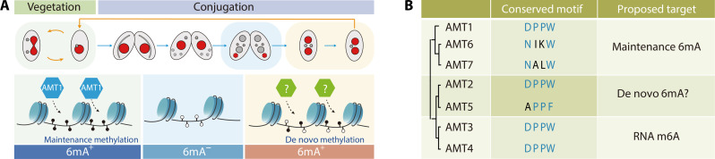
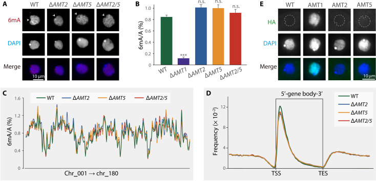
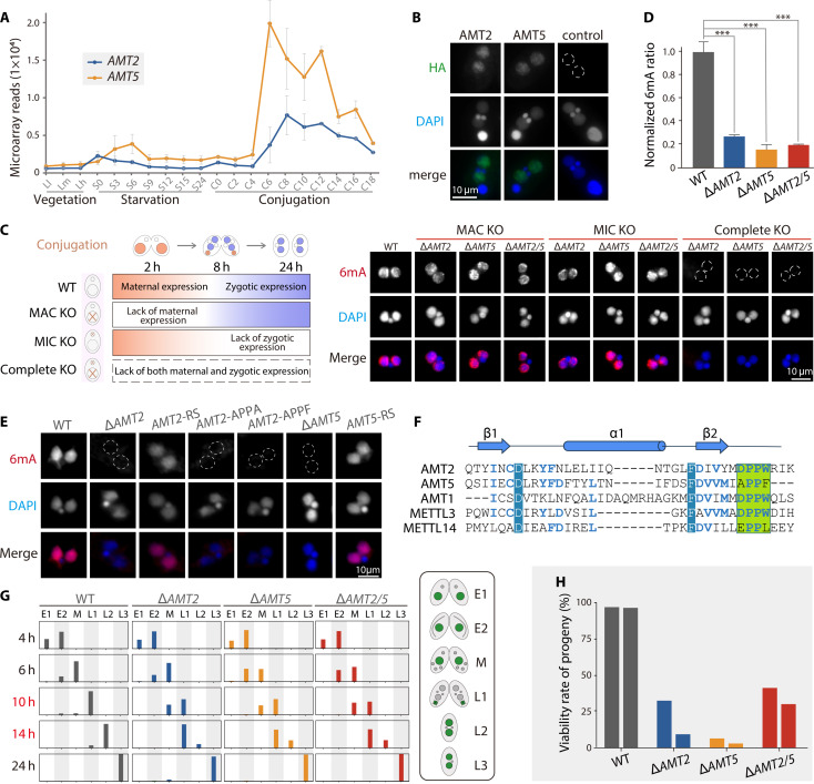
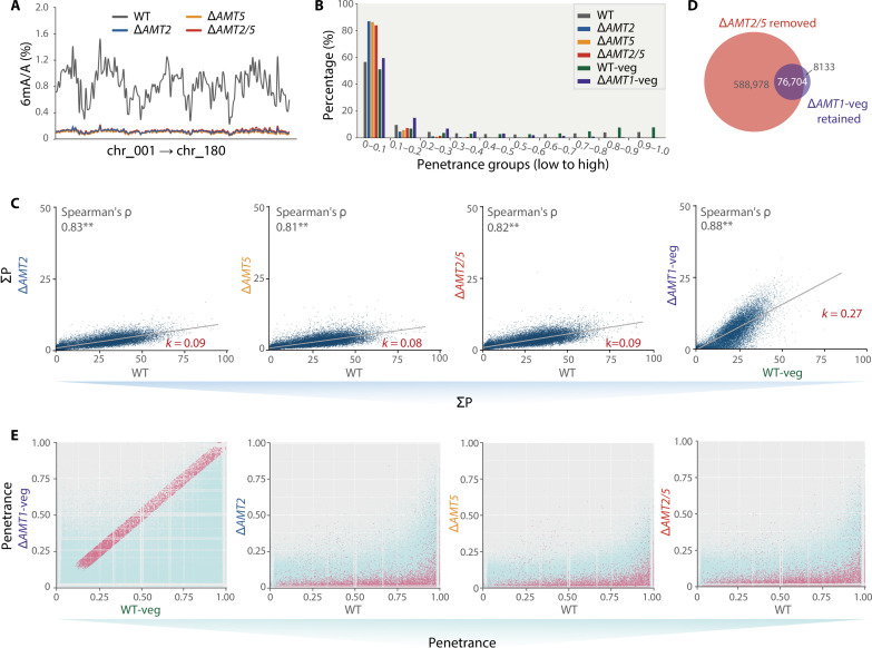
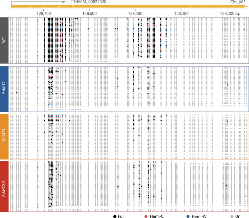
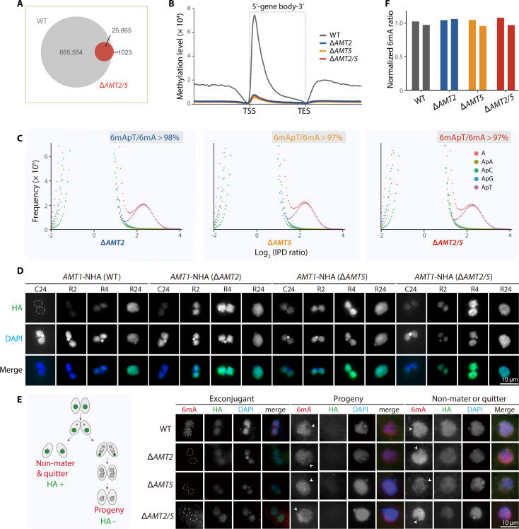
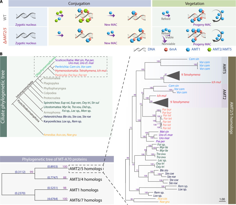
N6-methyladenine (6mA) is an intensively investigated epigenetic modification in eukaryotes. 6mA is maintained through semiconservative transmission during DNA replication, but the identity of de novo methyltransferase (MTase) catalyzing its establishment remains unknown. Here, we identified MT-A70 family proteins AMT2 and AMT5 as the de novo MTases responsible for 6mA establishment, using the unique sexual reproduction process of the unicellular eukaryote Tetrahymena thermophila. Deletion of AMT2 and AMT5 led to a substantial decrease in 6mA levels in the progeny macronucleus, resulting in an altered gene expression pattern and a substantial decline in the survival rate of sexual progenies. Additionally, the maintenance MTase AMT1 could exhibit a much diminished de novo methylation activity in cells lacking AMT2 and AMT5. Our study delineated the establishment-maintenance pathway of 6mA and underscored the biological importance of de novo methylation, revealing a notable parallel between 6mA and the classical 5-methylcytosine in eukaryotes.


Figures

Fig. 1.
Fig. 1.. 6mA dynamics and MT-A70 family MTases in Tetrahymena.
(A) Top: Schematic drawing of nuclear events and 6mA distribution during vegetation and conjugation stages (red, 6mA positive; gray, 6mA negative). Bottom: Graphics of 6mA deposition by de novo and maintenance MTases. (B) Simplified phylogenetic tree of Tetrahymena MT-A70 family MTases, marked with their catalytic motifs and proposed functions.
Fig. 2.
Fig. 2.. AMT2 and AMT5 were not required for maintenance 6mA during the vegetative stage.
(A) 6mA levels were not affected in the KO cells during the vegetative stage, as demonstrated by immunofluorescence (IF) staining. Arrowheads indicated the absence of 6mA signals in the MIC. (B) Mass spectrometry (MS) analysis revealed that 6mA levels in KO cells were not substantially changed during the vegetative growth. ΔAMT1 strain was used as a control, showing a marked reduction in 6mA levels. Three biological replicates were conducted for each strain. ***P < 0.001; n.s., not significant (P > 0.05). (C) 6mA ratios, defined as the percentage of methylated adenines among all adenines (6mA/A), were comparable on 180 non-rDNA (nonribosomal DNA) MAC chromosomes between KO and WT cells during the vegetative stage. MAC chromosomes were arranged from left to right in accordance with their positions on the MIC chromosomes. (D) 6mApT dinucleotides were enriched toward the 5′ end of gene bodies in both KO and WT cells. Genes were scaled to unit length and were extended to each side by one unit length. One unit length was divided into 30 bins, and the distribution frequency was calculated as the ratio of the 6mA amount at each specific position to the total 6mA amount. TSS, transcription start site; TES, transcription end site. (E) IF staining showed that AMT2 and AMT5 were not detectable in the somatic MAC (dashed circles) of vegetative cells. Hemagglutinin (HA)–tagged AMT1 cells and WT (SB210) cells without the HA tag were used as positive and negative controls, respectively.
Fig. 3.
Fig. 3.. AMT2 and AMT5 were essential for de novo 6mA during the conjugation stage.
(A) AMT2 and AMT5 are highly expressed during new MAC formation of conjugation stage (30). (B) AMT2 and AMT5 were localized in the newly formed MAC. Note the absence of the HA signal in the new MAC (dashed circle) of WT control cells. (C) Both somatically and zygotically expressed AMT2 and AMT5 contributed to de novo 6mA deposition. Left: Expression pattern of maternal and zygotic AMT2 and AMT5 in WT and KO cells. Right: 6mA signals in the new MAC of MAC KO (no maternal expression), MIC KO (no zygotic expression), and complete KO cells. Note the absence of 6mA signals in the new MAC (dashed circles) of complete KO cells. (D) MS analysis of 6mA levels in the flow cytometry–purified new MAC. Three biological replicates for each strain. ***P < 0.001. (E) IF staining analysis of 6mA levels in the new MAC. The outline of the nuclei without 6mA signals was delineated with dashed circles. (F) Alignment of sequences surrounding the catalytic motif in AMT1, AMT2, and AMT5 of Tetrahymena and in human METTL3 and METTL14. Identical sequences were indicated by white letters with blue background, while similar sequences were indicated by bold blue letters. The DPPW catalytic motif was depicted with a green background. (G) Conjugation progress of KO and WT cells. The schematic on the right illustrated the corresponding nuclear morphologies at each stage. E1, pre-meiosis; E2, meiosis, M, mitosis; L1, new MAC development, L2, pair separation; L3, one of the new MIC degraded. n > 200 for each strain. n represents the number of single cells. h, hours. (H) The viability rate of KO and WT progenies. Two biological replicates for each strain.
Fig. 4.
Fig. 4.. 6mA establishment was severely impaired in the new MAC of AMT2 and/or AMT5 KO cells.
(A) 6mA ratios exhibited a global decrease across all 180 non-rDNA chromosomes in KO cells at 24 hours post-mixing. Chromosome numbering followed the assignment outlined by Sheng et al. (65). (B) 6mApT positions with high penetrance in the new MAC of WT cells were depleted in KO cells. 6mApT positions were divided into 10 groups according to their penetrance, from low to high. 6mA SMRT-CCS data from the somatic MAC of ΔAMT1AMT1-veg) and WT (WT-veg) cells were included for comparison. (C) 6mA levels of individual genes were markedly reduced in the new MAC of KO cells, as well as in ΔAMT1-veg. 6mA level was calculated as “the sum of penetrance of all 6mApT positions (ΣP) on a specific gene.” (D) Venn diagram illustrated that the vast majority of the retained 6mA sites in ΔAMT1 cells were unmethylated in the new MAC of ΔAMT2/5 cells. ΔAMT2/5 removed sites were as defined as “positions with less than 10× coverage of 6mApT in the new MAC of ΔAMT2/5 cells.” ΔAMT1-veg retained sites were defined as “positions with more than 10× coverage of 6mApT in the somatic MAC of ΔAMT1 cells.” (E) The penetrance of AMT1-independent 6mApT positions was more substantially reduced in ΔAMT2/5 cells. AMT1-independent 6mApT positions, depicted as red points, were defined as having a penetrance difference ≤ ±0.05 between WT-veg and ΔAMT1-veg, with 6mApT coverage ≥ 5× in both strains. The reduction in penetrance of AMT1-independent 6mApT positions was more pronounced than the rest majority of 6mApT positions in the new MAC of KO cells.
Fig. 5.
Fig. 5.. Typical DNA single molecules revealed that a substantial proportion of highly methylated 6mApT sites in WT cells were either unmethylated or lowly methylated in KO cells.
The methylation states of a self-complementary ApT duplex were divided into four categories: full (methylation on both Watson and Crick strands, black dots), hemi-C (methylation only on the Crick strand, red dots), hemi-W (methylation only on the Watson strand, blue dots), and un (no methylation, gray dots). bp, base pairs.
Fig. 6.
Fig. 6.. The methylation pattern of the remaining 6mA sites in the new MAC of KO cells was attributed to AMT1.
(A) Venn diagram illustrated the overlap between methylated 6mA sites in the new MAC of WT and ΔAMT2/5 cells. The same cutoff (6mApT coverage ≥ 10×) was applied to both samples. (B) 6mApT exhibited an enrichment toward the 5′ end of gene bodies in both KO and WT cells. Genes were scaled to a unit length and were extended by one unit length on each side. One unit length was divided into 30 bins, and the sum of penetrance within each bin represented the methylation level. (C) 6mA displayed a preference for ApT dinucleotides in KO cells. Note that the bimodal distributions of both adenines (A) and ApT dinucleotides (ApT) were less obvious in KO cells than in WT cells (fig. S11A), due to the highly reduced number of 6mApT sites. (D) AMT1 levels were up-regulated in KO cells both at late conjugation and after refeeding. AMT1 was N-terminally HA-tagged in both MAC and MIC endogenous loci. New MACs without the HA signal were delineated with dashed circles. C24: 24 hours post-mixing. R2, R4, and R24: 2, 4, and 24 hours after refeeding. (E) 6mA signal intensity was restored in KO progenies. Left: Schematic diagram showing that the RPB3-CHA construct was transformed into somatic MACs to distinguish progeny (HA negative) from non-mater or quitter (HA positive). Right: 6mA occurrence in KO exconjugants before refeeding was abolished but was restored after refeeding (progeny). New MACs of the exconjugants were circled with white dashed lines, and the arrowheads indicated the absence of 6mA signals in MICs. (F) MS analysis of 6mA levels in KO and WT progenies. Two biological replicates for each strain.
Fig. 7.
Fig. 7.. Proposed regulatory model for 6mA and phylogenetic analysis of AMT2 and AMT5 homologs in ciliates.
(A) A proposed model for 6mA establishment and maintenance in WT and KO cells. In the new MAC of WT cells, AMT2 and AMT5 initiated the de novo 6mA deposition on unmethylated ApT dinucleotides, possibly helped by AMT1, generating hemi-6mApT. Subsequently, these hemi-6mApT sites were converted into full-6mApT by the maintenance activity of AMT1. In the somatic MAC of progenies during the vegetative stage, established full-6mApT sites were faithfully maintained by AMT1. In AMT2 and/or AMT5 KO cells, however, the deposition of de novo 6mA dependent on AMT2 and AMT5 was abolished, while AMT1 could exhibit both de novo and maintenance activities and generate both hemi- and full-6mA on a limited number of sites. After refeeding, 6mA levels in KO cells could be restored to that of WT cells, probably owing to the up-regulated level of AMT1. (B) Phylogenetic analysis showed the divergence of AMT2 and/or AMT5 homologs in ciliates. Proteins were labeled by species abbreviations and colored on the basis of their phylogenetic positions in the ciliate phylogenetic tree (inset). Phylogenetic analysis of MT-A70 family proteins (homologs of AMT1, AMT2/AMT5, AMT3/AMT4, and AMT6/AMT7) was shown as the evolutionary tree in the lower left corner. The homologs of AMT2 and AMT5 were shown in detail on the right, and homologs of nine Tetrahymena species were represented by black triangles. The scale bar corresponds to 1 expected amino acid substitution per site. Species abbreviations and NCBI GI numbers were listed in file S1.

Similar articles

References

    1. Fu Y., Luo G., Chen K., Deng X., Yu M., Han D., Hao Z., Liu J., Lu X., Dore L. C., Weng X., Ji Q., Mets L., He C., N6-methyldeoxyadenosine marks active transcription start sites in Chlamydomonas. Cell 161, 879–892 (2015). - PMC - PubMed
    1. Greer E. L., Blanco M. A., Gu L., Sendinc E., Liu J., Aristizábal-Corrales D., Hsu C., Aravind L., He C., Shi Y., DNA methylation on N6-adenine in C. elegans. Cell 161, 868–878 (2015). - PMC - PubMed
    1. Zhang G., Huang H., Liu D., Cheng Y., Liu X., Zhang W., Yin R., Zhang D., Zhang P., Liu J., Li C., Liu B., Luo Y., Zhu Y., Zhang N., He S., He C., Wang H., Chen D., N6-methyladenine DNA modification in Drosophila. Cell 161, 893–906 (2015). - PubMed
    1. Zhang M., Yang S., Nelakanti R., Zhao W., Liu G., Li Z., Liu X., Wu T., Xiao A., Li H., Mammalian ALKBH1 serves as an N6-mA demethylase of unpairing DNA. Cell Res. 30, 197–210 (2020). - PMC - PubMed
    1. Wu T. P., Wang T., Seetin M. G., Lai Y., Zhu S., Lin K., Liu Y., Byrum S. D., Mackintosh S. G., Zhong M., Tackett A., Wang G., Hon L. S., Fang G., Swenberg J. A., Xiao A. Z., DNA methylation on N6-adenine in mammalian embryonic stem cells. Nature 532, 329–333 (2016). - PMC - PubMed

MeSH terms

LinkOut - more resources