. 2025 Dec;17(1):2498164.
doi: 10.1080/19420862.2025.2498164. Epub 2025 May 14.

Development of potent humanized TNFα inhibitory nanobodies for therapeutic applications in TNFα-mediated diseases

Affiliations

Development of potent humanized TNFα inhibitory nanobodies for therapeutic applications in TNFα-mediated diseases

Tao Yin et al. MAbs. 2025 Dec.

Abstract

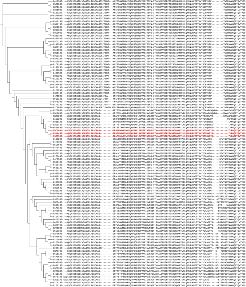
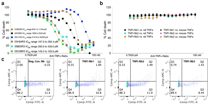
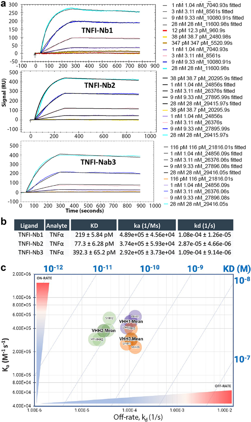
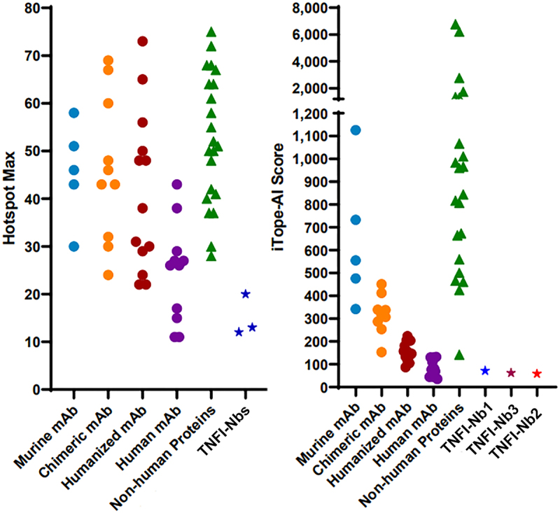
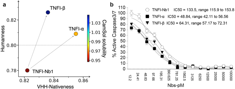
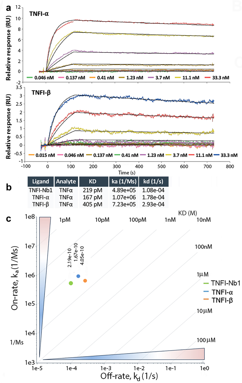
Tumor necrosis factor-alpha (TNFα) is a key pro-inflammatory cytokine implicated in the pathogenesis of numerous inflammatory and autoimmune diseases, including rheumatoid arthritis, inflammatory bowel disease, and neurodegenerative disorders such as Alzheimer's disease. Effective inhibition of TNFα is essential for mitigating disease progression and improving patient outcomes. In this study, we present the development and comprehensive characterization of potent humanized TNFα inhibitory nanobodies (TNFI-Nbs) derived from camelid single-domain antibodies. In silico analysis of the original camelid nanobodies revealed low immunogenicity, which was further reduced through machine learning-guided humanization and developability optimization. The two humanized TNFI-Nb variants we developed demonstrated high anti-TNFα activity, achieving IC₅₀ values in the picomolar range. Binding assays confirmed their high affinity for TNFα, underscoring robust neutralization capabilities. These TNFI-Nbs present valid alternatives to conventional monoclonal antibodies currently used in human therapy, offering potential advantages in potency, specificity, and reduced immunogenicity. Our findings establish a solid foundation for further preclinical development and clinical translation of TNFα-targeted nanobody therapies in TNFα-mediated diseases.

Keywords: Alzheimer disease; TNFα; TNFα inhibition; nanobody; nanobody humanization; single-domain antibody.


Conflict of interest statement

Dr. Luciano D’Adamio is a Professor at Rutgers University and also the founder of NanoNewron, a biotechnology company focused on developing innovative therapeutics for central nervous system diseases. The contribution to this study of Pietro Sormanni, Aubin Ramon and Matthew Greenig was conducted in a consultancy capacity and was remunerated by NanoNewron LLC.

Figures

Shown here are 100 unique α-TNFα nanobodies derived from an alpaca and a llama immunized with active, trimeric human TNFα. Amino acid sequences were aligned and grouped based on CDR similarity and overall sequence identity. A phylogenetic tree was constructed using BioPython and visualized through iTOL to illustrate sequence divergence and relatedness. The three nanobodies with the strongest TNFα inhibitory activity, as determined in functional assays, are indicated in red.
Figure 1.
Phylogenetic analysis and amino acid sequences of 100 unique α-TNFα nanobodies (nbs) isolated from immunized alpaca and Llama.
A multi-panel figure illustrating the inhibitory activity, species specificity, and membrane-bound TNFα binding of TNFI-Nb1, TNFI-Nb2, and TNFI-Nb3. (a) A dose–response inhibition curve displaying the percentage inhibition of human TNFα by each nanobody across increasing concentrations, analyzed using a non-linear regression model. (b) A separate inhibition curve demonstrating that TNFI-Nb1, TNFI-Nb2, and TNFI-Nb3 do not inhibit rat or mouse TNFα, as indicated by the absence of dose-dependent activity. (c) Flow cytometry data showing TNFI-Nb1, TNFI-Nb2, and TNFI-Nb3 binding to membrane-bound TNFα in HEK293 cells expressing human TNFα and EGFP. Cells were stained with an anti-His-APC antibody to detect nanobody binding, with a negative control nanobody included for comparison.
Figure 2.
TNFI-Nb1, TNFI-Nb2, and TNFI-Nb3 exhibit potent, human-specific TNFα inhibition and bind membrane-bound TNFα.
A multi-panel figure presenting the kinetics of TNFα binding to three nanobodies (TNFI-Nb1, TNFI-Nb2, and TNFI-Nb3) immobilized on Carboxyl sensors. (a) A sensor gram with overlaid fitted curves illustrating TNFα binding responses across different concentrations, showing specific and dose-dependent interactions. Colored lines represent raw data, while black curves indicate fitted 1:1 binding model result. (b) A bar graph summarizing the average kinetic parameters (association and dissociation rates, affinity constants) from three or four independent experiments, with error bars representing the standard error of the mean. (c) A two-dimensional iso-affinity kinetic plot where individual data points (circles) represent kinetic values from independent experiments. Diagonal lines indicate equilibrium binding constants to facilitate visualization of affinity distribution, with mean values labeled in bold.
Figure 3.
Kinetics of TNFα binding to immobilized TNFI-Nb1, TNFI-Nb2 and TNFI-Nb3.
Bar graphs comparing the predicted immunogenicity of TNFI-Nb1, TNFI-Nb2, and TNFI-Nb3 to a range of therapeutic antibodies and non-human proteins. The graph displays Total Scores and Hotspot Max values, with lower scores indicating a reduced risk of immunogenicity. TNFI-Nbs show lower immunogenicity scores compared to non-human proteins and are within the range of established therapeutic antibodies, suggesting minimal immunogenicity risk.
Figure 4.
TNFI-Nb1, TNFI-Nb2 and TNFI-Nb3 have low predicted immunogenicity.
Figure 5.
Figure 5.
In silico engineering of TNFI-α and TNFI-β nanobodies: optimized mutants of TNFI-Nb1 with enhanced TNFI activity.
Figure 6.
Figure 6.
Kinetics of TNFα analytes binding to immobilized nanobodies.
Figure 7.
Figure 7.
Humanized TNFI-α binds to membrane-bound human TNFα in a transfection model.

Similar articles

References

    1. Zouris G, Evangelopoulos DS, Benetos IS, Vlamis J.. The use of TNF-α inhibitors in active ankylosing spondylitis treatment. Cureus. 2024;16:e61500. doi: 10.7759/cureus.61500. - DOI - PMC - PubMed
    1. Lopetuso LR, Cuomo C, Mignini I, Gasbarrini A, Papa A. Focus on anti-tumour necrosis factor (TNF)-α-related autoimmune diseases. IJMS. 2023;24(9):8187. doi: 10.3390/ijms24098187. - DOI - PMC - PubMed
    1. Leone GM, Mangano K, Petralia MC, Nicoletti F, Fagone P. Past, present and (foreseeable) future of biological anti-TNF alpha therapy. J Clin Med. 2023;12(4):1630. doi: 10.3390/jcm12041630. - DOI - PMC - PubMed
    1. Kawanishi M, Fujii Y. Pharmacological profiles and clinical efficacy of ozoralizumab (Nanozora((r)) 30 mg syringes for S.C. Injection), the first Nanobody((R)) compound in Japan. Nihon Yakurigaku Zasshi. 2023;158:490–19. doi: 10.1254/fpj.23051. - DOI - PubMed
    1. Crommelin HA, Vorselaars AD, van Moorsel CH, Korenromp IH, Deneer VH, Grutters JC. Anti-TNF therapeutics for the treatment of sarcoidosis. Immunotherapy. 2014;6(10):1127–1143. doi: 10.2217/imt.14.65. - DOI - PubMed

MeSH terms

Substances

LinkOut - more resources