Review
. 2025 May 14:271678X251338971.
doi: 10.1177/0271678X251338971. Online ahead of print.

Therapeutic and diagnostic potential of extracellular vesicle (EV)-mediated intercellular transfer of mitochondria and mitochondrial components

Affiliations
Review

Therapeutic and diagnostic potential of extracellular vesicle (EV)-mediated intercellular transfer of mitochondria and mitochondrial components

Mingjin Wang et al. J Cereb Blood Flow Metab. .

Abstract

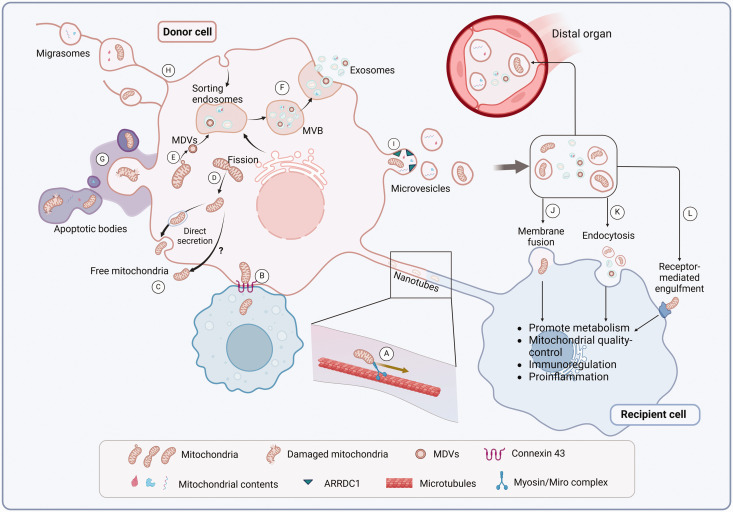
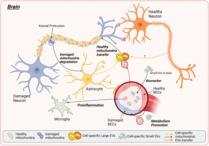
Extracellular vesicles (EVs) facilitate the transfer of biological materials between cells throughout the body. Mitochondria, membrane-bound organelles present in the cytoplasm of nearly all eukaryotic cells, are vital for energy production and cellular homeostasis. Recent studies highlight the critical role of the transport of diverse mitochondrial content, such as mitochondrial DNA (mt-DNA), mitochondrial RNA (mt-RNA), mitochondrial proteins (mt-Prots), and intact mitochondria by small EVs (<200 nm) and large EVs (>200 nm) to recipient cells, where these cargos contribute to cellular and mitochondrial homeostasis. The interplay between EVs and mitochondrial components has significant implications for health, metabolic regulation, and potential as biomarkers. Despite advancements, the mechanisms governing EV-mitochondria crosstalk and the regulatory effect of mitochondrial EVs remain poorly understood. This review explores the roles of EVs and their mitochondrial cargos in health and disease, examines potential mechanisms underlying their interactions, and emphasizes the therapeutic potential of EVs for neurological and systemic conditions associated with mitochondrial dysfunction.

Keywords: Extracellular vesicles; energy metabolism; mitochondria; mitochondrial components.


Conflict of interest statement

The author(s) declared no potential conflicts of interest with respect to the research, authorship, and/or publication of this article.

Figures

Figure 1.
Figure 1.
Mechanisms of mitochondrial transfer. (a) Tunnel-nanotubes transfer mitochondria mediated by microtubules and Myosin/Miro1 complex. (b) Mitochondrial Gap-junction-channel transfer mediated by Connexin 43. (c) Free mitochondria are released directly through the secretory autophagy pathway or an unclarified mechanism. (d) Mitochondrial fission promoted by PGC-1α signaling has been suggested to mediate extracellular mitochondrial secretion. (e) MDVs’ generation is mediated by the PINK1/Parkin, also regulated by Snx9 and Rab9. MDVs are delivered to endosomes, leading to the formation of MDV-containing MVBs (f). (g) Various pathological stimuli, such as hypoxia, induce mitochondria-containing apoptotic bodies releasing. (h) Kif5b and Myosin19 induce migrasome-mediated mitochondria transfer. (i) ARRDC1 mediates the transport of mitochondria and mitochondrial contents into microvesicles. (j–l) Mitochondria secreted extracellularly can be taken up by recipient cells through membrane fusion (j) or endocytosis (k). Extracellular free mitochondria may also interact with the recipient cell surface for uptake (l). Then these internalized mitochondria can elicit biological effects on recipient cells. (Created in BioRender.).
Figure 2.
Figure 2.
The EV-mediated cell-type-specific mitochondrial transfer and its potential role in neurological disease. EVs, carrying cell-specific surface markers, transfer mitochondrial components such as DNA, RNA, proteins, and intact mitochondria between neurons, astrocytes, and microglia (color indicated), supporting axonal maintenance, localized metabolic regulation in EV-targeting cells, and brain remodeling. These EVs can also cross the BBB and deliver their mitochondrial cargo to distant organs, contributing to the pathological biomarker and systemic metabolic homeostasis factor. (Created in BioRender.).

Similar articles

References

    1. Hayakawa K, Esposito E, Wang X, et al.. Transfer of mitochondria from astrocytes to neurons after stroke. Nature 2016; 535: 551–555. - PMC - PubMed
    1. Rangaraju V, Lewis TL, Hirabayashi Y, et al.. Pleiotropic mitochondria: the influence of mitochondria on neuronal development and disease. J Neurosci 2019; 39: 8200–8208. - PMC - PubMed
    1. Trigo D, Avelar C, Fernandes M, et al.. Mitochondria, energy, and metabolism in neuronal health and disease. FEBS Lett 2022; 596: 1095–1110. - PubMed
    1. Islam MT. Oxidative stress and mitochondrial dysfunction-linked neurodegenerative disorders. Neurol Res 2017; 39: 73–82. - PubMed
    1. Wang Y, Xu E, Musich PR, et al.. Mitochondrial dysfunction in neurodegenerative diseases and the potential countermeasure. CNS Neurosci Ther 2019; 25: 816–824. - PMC - PubMed

LinkOut - more resources