. 2025 May 14;16(1):4475.
doi: 10.1038/s41467-025-59639-9.

Macrophage-augmented intestinal organoids model virus-host interactions in enteric viral diseases and facilitate therapeutic development

Affiliations

Macrophage-augmented intestinal organoids model virus-host interactions in enteric viral diseases and facilitate therapeutic development

Guige Xu et al. Nat Commun. .

Abstract

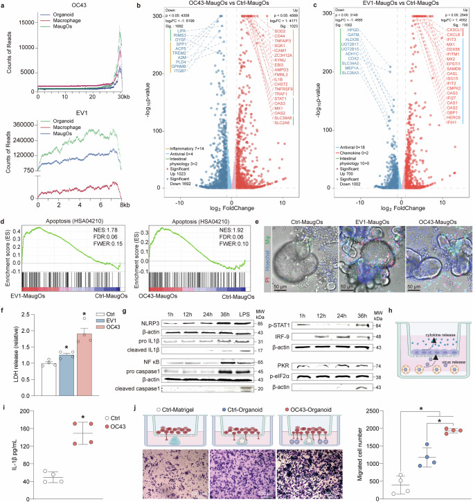
The pathogenesis of enteric viral infections is attributed to both viral replication and the resultant immune-inflammatory response. To recapitulate this complex pathophysiology, we engineer macrophage-augmented organoids (MaugOs) by integrating human macrophages into primary intestinal organoids. Echovirus 1, echovirus 6, rotavirus, seasonal coronavirus OC43 and SARS-CoV-2- known to directly invade the intestine- are used as disease modalities. We demonstrate that these viruses efficiently propagate in MaugOs and stimulate the host antiviral response. However, rotavirus, coronavirus OC43 and SARS-CoV-2, but not the two echoviruses, trigger inflammatory responses. Acetate, a microbial metabolite abundantly present in the intestine, potently inhibits virus-induced inflammatory responses in MaugOs, while differentially affecting viral replication in macrophages and organoids. Furthermore, we provide a proof-of-concept of combining antiviral agent with either anti-inflammatory regimen or acetate to simultaneously inhibit viral infection and inflammatory response in MaugOs. Collectively, these findings demonstrate that MaugOs are innovative tools for studying the complex virus-host interactions and advancing therapeutic development.


Conflict of interest statement

Competing interests: The authors declare no competing interests.

Figures

Fig. 1
Fig. 1. Establishment and characterization of macrophage-augmented intestinal organoids.
a Schematic overview of constructing three different types of macrophage-augmented intestinal organoids (MaugOs). Created in BioRender.com. https://BioRender.com/cegs5ln. b Representative images of time-lapse confocal microscopy during MaugOs formation. Unstained intestinal organoid fragments and CFSE pre-labelled macrophages differentiated from THP-1 monocytes were integrated for 18 hours. Images were taken every 30 minutes (also see Supplementary Movie 1). c Immunofluorescence staining images of MaugOs incorporating organoids from three donors. Epithelial membrane of organoids was stained by EpCAM (red); THP-1 monocytes-derived macrophages stained by CFSE (green). Scale bar, 150 μm. d Bright field and immunofluorescence staining images depicting organoids, THP-1 macrophages and MaugOs. e Venn diagram of overlapped differentially expressed genes in macrophages (Mφ), intestinal organoids and MaugOs. f Significantly enriched pathways by KEGG analysis of MaugOs. (p < 0.05). g The expression of inflammatory genes quantified by qRT-PCR upon stimulation of 1 μg/mL LPS for 1 hour and 12 hours. Each group was compared to its respective control without LPS treatment (n = 3). h Quantification of IL-1β protein production by ELISA in MaugOs integrated THP-1 monocytes-derived macrophages (n = 4). i Immunofluorescence staining images of MaugOs integrated PBMC monocytes-derived macrophages and iPSCs-derived macrophages. Epithelial membrane of organoids was stained by EpCAM (red); PBMC monocytes-derived macrophages were stained by CFSE, and iPSCs-derived macrophages expressed H2B-mEGFP protein (green). Scale bar, 50 μm. j The expression of inflammatory genes quantified by qRT-PCR upon stimulation of LPS in MaugOs integrated iPSCs-derived or PBMC monocytes-derived macrophages (n = 2). k Quantification of IL-1β protein production by ELISA in MaugOs integrated PBMC monocytes-derived macrophages and iPSCs-derived macrophages (n = 4). Data were presented as means of biological replicates ± SEM. Statistical analysis was performed using the two-tailed Mann–Whitney test. *p < 0.05.
Fig. 2
Fig. 2. MaugOs recapitulate enteric viral infections and virus-host interactions.
ab Viral RNA level in EV1 (a) or EV6 (b) infected MaugOs from 1 to 36 hours post-infection (hpi). Data was normalized to viral RNA level at 1 hpi (n = 2). c–d Quantification of infectious EV1 (c) and EV6 (d) titers in culture supernatants from 1 to 36 hpi (n = 4). Dotted line indicates detection limit. e–f Confocal imaging of viral replicating dsRNA (red) in EV1 (e) or EV6 (f) infected MaugOs at 36 hpi. Scale bar, 50 μm. g RNA expression of ISGs and inflammatory genes in EV1- or EV6-infected MaugOs from 1 to 36 hpi (n = 2). h Viral RNA level in rotavirus-infected MaugOs from 1 to 36 hpi (n = 2). Data was normalized to viral RNA level at 1 hpi. i Quantification of infectious rotavirus titers in culture supernatants from 1 to 36 hpi (n = 4). j Confocal imaging of rotavirus VP6-protein (red) in MaugOs at 36 hpi. Scale bar, 50 μm. k ISGs expression in rotavirus-infected MaugOs from 1 to 36 hpi (n = 2). l Quantification of IL-1β that secreted from uninfected- and rotavirus-infected MaugOs (n = 4). m Viral RNA expression in OC43-infected MaugOs from 1 to 36 hpi (n = 3). Data was normalized to viral RNA level at 1 hpi. n Quantification of infectious OC43 titres in culture supernatants from 1 to 36 hpi (n = 4). o Confocal imaging of OC43 N-protein (red) in MaugOs at 36 hpi. Scale bar, 50 μm. p ISGs expression in OC43-infected MaugOs from 1 to 36 hpi (n = 3). q Quantification of IL-1β that secreted from uninfected- and OC43-infected MaugOs (n = 4). r Viral RNA expression in OC43-infected MaugOs incorporating organoids from two additional donors from 1 to 36 hpi (n = 4). Data was normalized to RNA level at 1 hpi. s ISGs and inflammatory genes expression in OC43-infected MaugOs incorporating organoids from two additional donors at 36 hpi (n = 2). All data were presented as means of biological replicates ± SEM. Statistical analysis was performed using the two-tailed Mann–Whitney test. *p < 0.05.
Fig. 3
Fig. 3. Characterizing the complex virus-host interactions in MaugOs.
a Whole genome view of mapped transcripts by RNA-seq across the OC43 or EV1 genome in infected organoid, macrophage and MaugOs. b–c Differential gene expression analysis of OC43 infection (b) and EV1 infection (c) in MaugOs compared to uninfected MaugOs (n = 3). d Gene set enrichment analysis (GSEA) of KEGG apoptosis pathways (HSA04210) in MaugOs infected with OC43 or EV1. e Fluorescence staining of dead cells (PI, red), cell nuclei (Hoechst, blue), and bright field images at 36 hours post-inoculation following OC43 or EV1 infection in MaugOs. CFSE-labeled THP-1-derived macrophages are shown in green. Scale bar, 50 μm. f Quantification of lactate dehydrogenase (LDH) release in MaugOs upon OC43 or EV1 infection at 36 hours post-inoculation (n = 4). MaugOs without virus infection served as control (normalized as 1). g OC43-infected organoids were integrated with THP1 macrophages and samples were collected at 1 hour, 12 hour, 24 hour and 36 hour after MaugOs assembly. The protein level of NLRP3, pro IL-1β, NF-κB and pro caspase 1 in lysates, and cleaved IL-1β mature and cleaved caspase-1 in supernatant were determined by western blotting. LPS treatment for 36 hours in MaugOs was used as positive control. The protein level of p-STAT1, IRF-9, p-eIF2α in MaugOs lysates were determined by western blotting at 1 hour, 12 hour, 24 hour, and 36 hour post=-OC43 infection. h Schematic representation of OC43-infected organoids culturing in the down compartment and THP-1 macrophages culturing in the transwell insert. Created in BioRender.com. https://BioRender.com/3vjrdll. i ELISA quantification of IL-1β released into the insert supernatant by macrophages (n = 4). j Cell migration assay determining the macrophage number attracted by Matrigel (as control), uninfected and OC43-infected organoids cultured in Matrigel (n = 4). Scale bar, 200 μm. Created in BioRender.com. https://BioRender.com/jv8nl58. All data were presented as means of biological replicates ± SEM. Statistical analysis was performed using the two-tailed Mann–Whitney test. *p < 0.05.
Fig. 4
Fig. 4. Establishment of MaugOs using differentiated intestinal organoids.
a Morphology of organoids differentiating towards enterocytes-, goblet- and enteroendocrine (EEC)-phenotypes in different time points. b Gene expression level of Villin (enterocytes marker), Muc2 (goblet cell marker) and CHGA (enteroendocrine cell marker) upon different differentiation culture (n = 4). UD: undifferentiated organoids; EEC: enteroendocrine-differentiated organoids. c Characterizing cell types of differentiated organoids by immunofluorescence staining of Villin (enterocytes), Muc2 (goblet cells) and CHGA (enteroendocrine cells). EpCAM is the marker of epithelial cell membrane (red). Scale bar, 35 μm and 50 μm. d Morphology of MaugOs integrated with enterocytes- (Villin, red), goblet cells- (Muc2, red) and enteroendocrine cells (EEC)- (CHGA, red) differentiated organoids. THP1 macrophages were stained with CFSE (green). Scale bar, 50 μm. e QRT-PCR quantification of inflammatory and antiviral associated genes expression in MaugOs integrated differentiated or undifferentiated organoids upon OC43 infection (n = 2). f Quantification of IL-1β gene expression in MaugOs integrated undifferentiated or enterocytes-differentiated organoids (n = 4). g ELISA quantification of IL-1β protein production in MaugOs integrated undifferentiated or enterocytes-differentiated organoids (n = 4). h Quantification of OC43 viral RNA level in MaugOs integrated undifferentiated or differentiated organoids at 36 hours postinfection (n = 4). i Confocal imaging of viral replicating double-stranded RNA (dsRNA, red color) in enterocytes-differentiated organoids, at 48 hours post OC43 infection. Scale bar, 20 μm. j QRT-PCR quantification of released OC43 virus from polarized intestinal monolayers cultured in transwell system (n = 3). Created in BioRender.com. https://BioRender.com/yz1fjns. All data were presented as means of biological replicates ± SEM. Statistical analysis was performed using the two-tailed Mann–Whitney test. *p < 0.05.
Fig. 5
Fig. 5. Characterizing the function of acetate on enteric viral infection and inflammatory response in MaugOs.
a Profiling the influence of three metabolites on inflammatory gene expression triggered by LPS in MaugOs (n = 2). b–d ELISA quantification of IL-1β (b), IL-6 (c) and IL-8 (d) in the supernatant of MaugOs following LPS stimulation with or without 50 mM acetate treatment for 24 hours (n = 4). e–h ELISA quantification of IL-1β (e), IL-6 (f), IL-8 (g) and TNF-α (h) in the supernatant of MaugOs following OC43 infection with or without 50 mM acetate treatment for 36 hours (n = 4). i Quantification of IL-1β production in the supernatant of rotavirus-infected MaugOs with or without 50 mM acetate treatment for 36 hours (n = 4). jk Quantification of IL-1β production in the supernatant of MaugOs integrated PBMC- or iPSCs-derived macrophages after LPS stimulation (j) or OC43 infection (k), with or without 50 mM acetate treatment for 24 hours (n = 4). l The inhibitory effect of 50 mM acetate treatment for 12 hours on the IL-1β gene expression and protein production in MaugOs simultaneously elicited by LPS and OC43 infection (n = 4). m–o Quantification of viral RNA level in OC43-infected THP1 macrophages (m), organoids (n) and MaugOs (o) with or without 50 mM acetate treatment for 36 hours (n = 4). p–q Quantification of viral RNA level in rotavirus-infected (p), EV1 or EV6-infected (q) organoids with or without 50 mM acetate treatment for 36 hours (n = 4). r Top 20 significantly regulated genes upon OC43 infection in MaugOs (n = 3). s Significantly regulated pathways by KEGG analysis at 36 hours in OC43-infected MaugOs with 50 mM acetate treatment (p < 0.05), compared with the non-treatment group. Red: upregulated; blue: downregulated (n = 3). t–u The inhibitory effect of 50 mM acetate treatment on the protein level of NF-κB and NLRP3 signaling cascade in LPS-treated (t) or OC43-infected (u) MaugOs. All data were presented as means of biological replicates ± SEM. Statistical analysis was performed using the two-tailed Mann–Whitney test. *p < 0.05.
Fig. 6
Fig. 6. Dissecting the anti-inflammatory effects of acetate in enteric infections: mechanism-of-action.
a Schematic illustration of the key receptor and signaling pathways that targeted by acetate. Created in BioRender.com. https://BioRender.com/0vfnx9c. b The effect of GLPG0974 on the expression of IL-1β in OC43 virus-infected MaugOs quantified by qRT-PCR (n = 4). c The effect of GLPG0974 on the production of IL-1β in the supernatant of OC43-infected MaugOs quantified by ELISA (n = 4). d The effect of GLPG0974 on the expression of IL-1β in LPS-treated MaugOs quantified by qRT-PCR (n = 4). e The effect of GLPG0974 on the production of IL-1β in the supernatant of LPS-treated MaugOs quantified by ELISA (n = 4). f and g The effect of GLPG0974 on the protein level of NLRP3 signaling cascade in OC43-infected (f) or LPS-treated (g) MaugOs at 36 hours determined by western blotting. NLRP3 and NF-κB were detected from cell lysates; Cleaved caspase-1 and cleaved IL-1β were detected from culture supernatant. h The effect of AC-CoA inhibitor 1 on the expression of IL-1β in OC43-infected MaugOs quantified by qRT-PCR (n = 4). i The effect of AC-CoA inhibitor 1 on the production of IL-1β in the supernatant of OC43-infected MaugOs quantified by ELISA (n = 4). j The effect of AC-CoA inhibitor 1 on the expression of IL-1β in LPS-treated MaugOs quantified by qRT-PCR (n = 4). k The effect of AC-CoA inhibitor 1 on the production of IL-1β in the supernatant of LPS-treated MaugOs quantified by ELISA (n = 4). l and m The effect of AC-CoA inhibitor 1 on the protein level of NLRP3, pro IL-1β and cleaved caspase 1 in OC43-infected (l) or LPS-treated (m) MaugOs at 36 hours determined by western blotting. All data were presented as means of biological replicates ± SEM. Statistical analysis was performed using the two-tailed Mann–Whitney test. *p < 0.05.
Fig. 7
Fig. 7. Devising combination treatment in MaugOs against enteric viral infections.
a Profiling the effect of a group of clinically used immunosuppressants on the expression of IL-1β and viral RNA in OC43-infected MaugOs at 36 hours post-treatment (n = 2). 6-TG: 6-Thioguanine; 6-MP: 6-mercaptopurine; CsA: Cyclosporin A; MTX: Methotrexate; Tof: Tofacitinib; AZA: Azathioprine; 5-ASA: 5-aminosalicylic acid; Pred: Prednisolone; Dex: Dexamethasone; Bud: Budesonide. b IL-1β RNA expression in OC43-infected MaugOs following 36-hours treatment of budesonide, dexamethasone, and prednisolone, respectively, (n = 4). c OC43 viral RNA level in MaugOs treated with 1 μM nirmatrelvir (Nir) for 36 hours (n = 4). d The effect of nirmatrelvir, budesonide and MCC950 (NLRP3 inhibitor) on the protein level of pro IL-1β and NF-κB at 36 hours post-treatment in OC43-infected MaugOs. ef QRT-PCR quantification of OC43 viral RNA and IL- gene expression (e), and ELISA quantification of IL-1β production (f) in MaugOs following 36-hours combination treatment with 1 μM nirmatrelvir and 1 μM budesonide (n = 4). g PCA analysis of different MaugOs groups (n = 3). Control: uninfected MaugOs; OC43: OC43-infected MaugOs; OC43+Nir: 1 μM nirmatrelvir treatment in OC43-infected MaugOs; OC43+Acetate: 50 mM acetate treatment in OC43-infected MaugOs; OC43+Nir+Acetate: combination treatment of 1 μM nirmatrelvir and 50 mM acetate in OC43-infected MaugOs. h–i OC43 viral RNA level (h), IL-1β gene expression and protein production (I) in MaugOs following 36-hours combination treatment with 1 μM nirmatrelvir and 50 mM acetate (ace) (n = 4). j The percentages of mapped OC43 viral transcripts in different groups of MaugOs (n = 3). k Whole genome view of mapped transcripts by RNA-seq across the OC43 genome in different groups of MaugOs (n = 3). l Top 20 significantly regulated KEGG pathways in OC43-infected MaugOs upon combination treatment of nirmatrelvir and acetate, compared to nirmatrelvir treatment alone (n = 3). All data were presented as means of biological replicates ± SEM. Panel b, c, e, f, h and i use Mann–Whitney test with two tailed; J uses One-way ANOVA followed by Dunn’s multiple comparison test (two-sided). *p < 0.05; **p < 0.01; ***p < 0.001; ****p < 0.0001.

References

    1. Amimo, J. O. et al. Rotavirus Interactions With Host Intestinal Epithelial Cells. Front Immunol.12, 793841 (2021). - PMC - PubMed
    1. Kirk, M. D. et al. World Health Organization Estimates of the Global and Regional Disease Burden of 22 Foodborne Bacterial, Protozoal, and Viral Diseases, 2010: A Data Synthesis. PLoS Med.12, e1001921 (2015). - PMC - PubMed
    1. Banyai, K., Estes, M. K., Martella, V. & Parashar, U. D. Viral gastroenteritis. Lancet392, 175–186 (2018). - PMC - PubMed
    1. Zhang, M. et al. Clinical characteristics of severe neonatal enterovirus infection: a systematic review. BMC Pediatr.21, 127 (2021). - PMC - PubMed
    1. Li, C. et al. Human airway and nasal organoids reveal escalating replicative fitness of SARS-CoV-2 emerging variants. Proc. Natl Acad. Sci. USA120, e2300376120 (2023). - PMC - PubMed

MeSH terms

Substances