. 2025 May 14;25(1):176.
doi: 10.1186/s12935-025-03805-y.

BCR::ABL1 expression in chronic myeloid leukemia cells in low oxygen is regulated by glutamine via CD36-mediated fatty acid uptake

Affiliations

BCR::ABL1 expression in chronic myeloid leukemia cells in low oxygen is regulated by glutamine via CD36-mediated fatty acid uptake

Caterina Mancini et al. Cancer Cell Int. .

Abstract

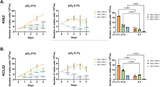
Background: Chronic myeloid leukemia (CML) is influenced by microenvironmental nutrients, glucose (Glc), and glutamine (Gln) which regulate cell proliferation, viability, and the expression of the driver oncoprotein (BCR::ABL1).

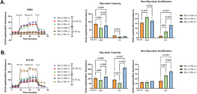
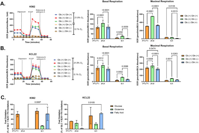
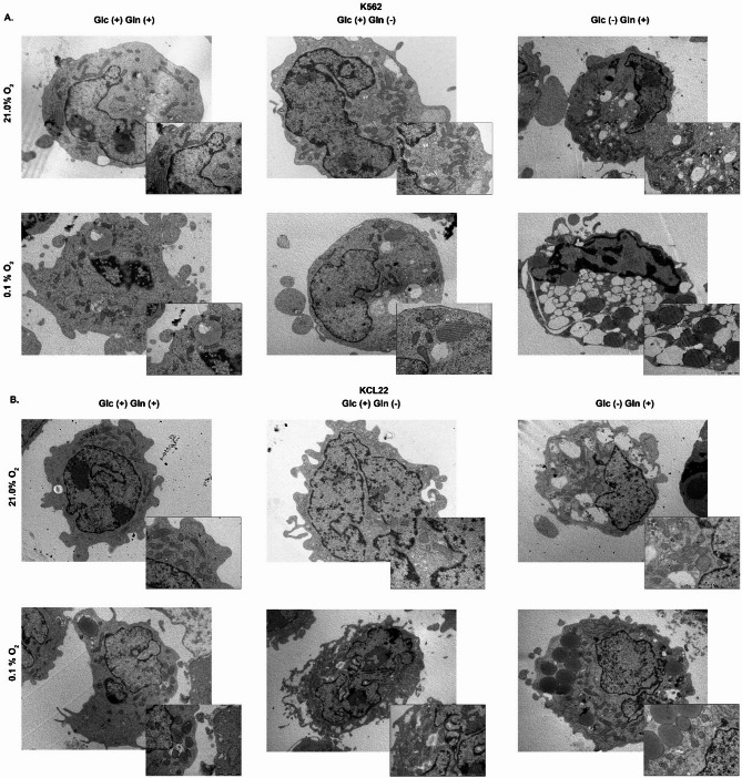
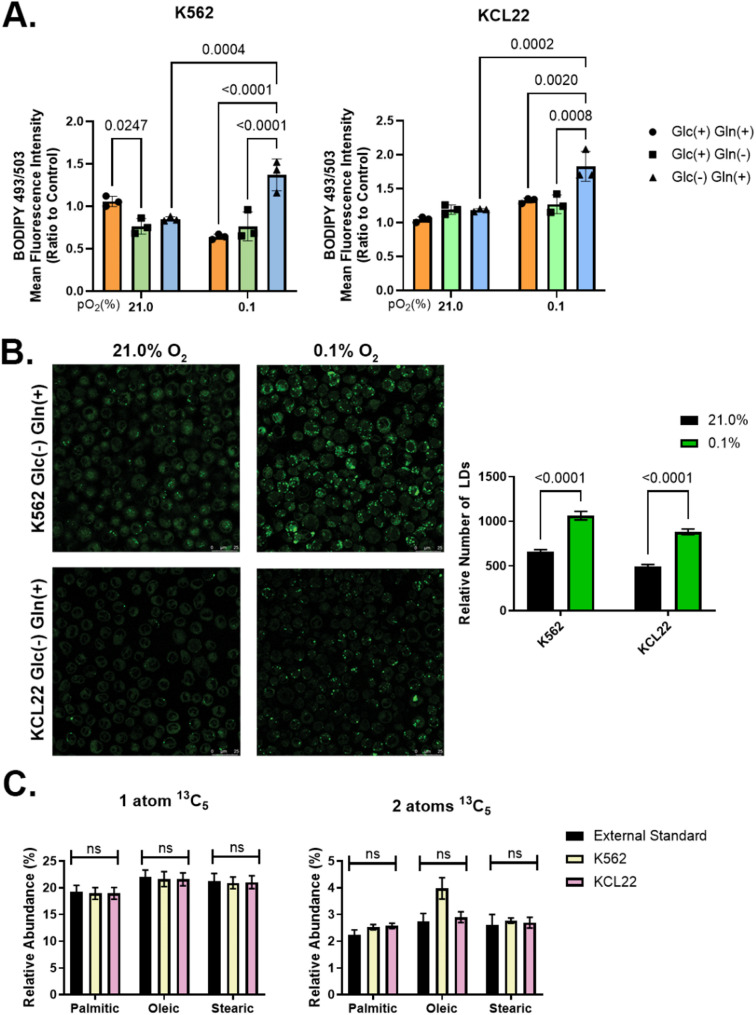
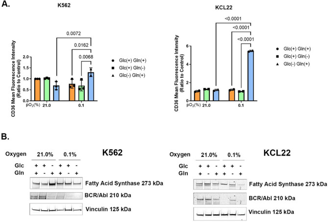
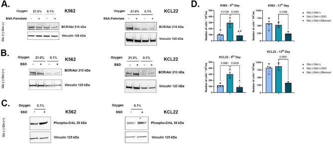
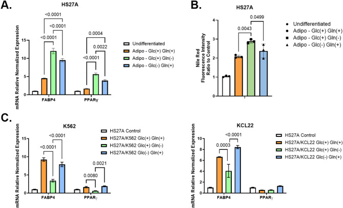
Results: Our study revealed that Glc, while partially supporting alone cell growth in normoxia, is essential in low oxygen conditions, whereas Gln is ineffective. Under low oxygen, Gln reduced oxidative respiratory activity while enhancing glycolysis. In these conditions, fatty acid (FA) metabolism becomes crucial, as evidenced by increased lipid droplets (LD) accumulation when Glc was absent. Gln, in particular, drives CD36-mediated FA uptake, suppressing the BCR::ABL1 oncoprotein and facilitating cell survival. By co-culturing leukemia cells with adipocytes, one of the main bone marrow (BM) cell components, we observed an enhanced FA release, suggesting a link between FA, microenvironmental BM cells, and the maintenance of leukemic stem cells (LSC).

Methods: K562 and KCL22 cell lines were subjected to Glc and/or Gln deprivation under hypoxic conditions (96 h at 0.1% O2). Metabolic profiling was conducted through the Seahorse XFe96 analyzer, and the contribution of L-Glutamine-13C5 to FA de novo synthesis was determined via GC/MS. Intracellular neutral LD were measured using BODIPY 493/503 in confocal microscopy and flow cytometry, with their presence and morphology further examined via transmission electron microscopy. BCR::ABL1 as well as several FA-related markers were evaluated via Western Blotting, whilst CD36 was determined through flow cytometry. LC2 assay was used for measuring leukemia stem cell potential by inhibiting FA uptake via the usage of the Sulfo-N-Succinimidyl Oleate, a CD36 inhibitor. qPCR was exploited to detect markers of FA secretion in CML-adipocytes co-culture together with Nile Red staining to assess free FA in the media.

Conclusions: These findings underscore the central role of FA in the regulation of the LSC compartment of CML, highlighting the importance of Gln in facilitating CML cell survival under restrictive metabolic conditions and preparing the cell population for expansion upon the release of these restrictions.

Keywords: BCR:ABL1; Chronic myeloid leukemia; Fatty acids; Hypoxia.


Conflict of interest statement

Declarations. Ethics approval and consent to participate: Not applicable. Consent for publication: Not applicable. Competing interests: The authors declare no competing interests.

Figures

Fig. 1
Fig. 1
Effects of glucose and/or glutamine on CML cell survival and proliferation. K562 (A) and KCL22 (B) cells were subjected to Glc and/or Gln deprivation for 4 days under low oxygen (0.1% O2) or standard conditions (21.0% O2), whereas cells in complete medium (Glc+/Gln+) were used as controls. Trypan blue-negative cells were counted at the indicated incubation times (left and middle panels), and the final results (day 4) are shown in the right panels. Data are expressed as the mean ± SD of the data from three independent experiments (n = 3). The differences for which statistical significance is indicated in the left and middle panels are with respect to Glc+/Gln + while on the right panels, the statistical significance was calculated for Normoxia Vs Hypoxia samples
Fig. 2
Fig. 2
Glutamine enhances glycolytic capacity. Cells were incubated as indicated for four days and then subjected to the GlycoStress assay. The ExtraCellular Acidification Rate (ECAR) measured (A, K562; B, KCL22) is shown as complete patterns obtained from representative Seahorse runs (left panels) or as the average of independent measures (middle and right panels). The correlation between EACR and intracellular and extracellular lactate production is shown in Supplementary Fig. 1E. The latter data are expressed as the mean ± SEM of the results from at least three independent experiments, each consisting of cell plating into six Seahorse microplate wells (n = 3)
Fig. 3
Fig. 3
The lack of oxygen affects cell metabolic demand. Cells were incubated as indicated for 4 days and then subjected to MitoStress or Mito Fuel Flex assays. The Oxygen Consumption Rate (OCR) measured by the mitostress test (A, K562; B, KCL22) is shown as complete patterns obtained from representative Seahorse runs (left panels) or as the average of independent measures (middle and right panels). The Fuel Oxidation Dependency (C) was calculated using the following equation: formula image. Only the oxidation dependence of FA was statistically significant. Averaged data are expressed as the mean ± SEM of results from at least three independent experiments, each consisting of cell plating into six Seahorse microplate wells (n = 3)
Fig. 4
Fig. 4
Glutamine stimulates lipid storage in the absence of glucose. K562 (A) and KCL22 (B) cells were incubated as indicated for four days and then fixed and prepared for TEM analysis. Images were captured using a JEM 1010 transmission electron microscope equipped with a MegaView III high-resolution digital camera (n = 3)
Fig. 5
Fig. 5
Glutamine induces FA accumulation. K562 and KCL22 cells were incubated as indicated for 4 days, stained with BODIPY 493/503, and analyzed by flow cytometry (A) and confocal analysis (B). For the latter, an algorithm to count green positive pixels was exploited on 60 z-stack slices to include the entire cell volume. The graph in (B) shows the total number of identifiable LDs in the examined slides. (C) Cell content of palmitic, oleic, and stearic acids, as determined by GC/MS using 13C5-labeled Gln and tracing 1 or 2 Gln-derived carbon atoms. Data are expressed as the mean ± SD of the data from three independent experiments (n = 3)
Fig. 6
Fig. 6
CD36-mediated FA uptake induces a block of de novo FA synthesis. K562 and KCL22 cells were incubated for 4 days as indicated. (A) CD36 expression was measured by flow cytometry. Glc(-) Gln(+) in hypoxia was compared only to Glc(+) Gln(+) and Glc(+) Gln(-) in the same oxygen condition, and versus Glc(-) Gln(+) in normoxia (B) Total cell lysates were subjected to immunoblotting to determine the expression levels of FA-synthase and BCR::ABL1. The loading of equal amounts of vinculin protein was confirmed by immunoblotting and the average densitometric values of bands from three independent experiments, obtained using ImageJ software, are reported in Supplementary Fig. 1A. Data reported in (A) are expressed as the mean ± SD of the data from three independent experiments (n = 3)
Fig. 7
Fig. 7
FA interfere with BCR::ABL1 expression and the maintenance of stem cell potential. K562 and KCL22 cells were incubated as indicated for 4 days and treated from time zero of incubation with BSA-Palmitate (ratio 1:8–100 µM) in the presence of Glc only (A) or with SSO (150 µM) in the presence of Gln only (B, C). Total cell lysates were subjected to immunoblotting to determine the expression levels of BCR::ABL1 and phospho-Crkl, as well as vinculin, to confirm the loading of equal amounts of protein. The average densitometric values of bands from three independent experiments, obtained using ImageJ software, are reported in Supplementary Figs. 1B, C, and D, respectively. In (D), cells incubated for 4 days in low oxygen (LC1) in the absence or presence of SSO from time 0 of LC1 incubation were replated into growth-permissive cultures established in complete growth medium and incubated in normoxia (LC2) in the absence or presence of etomoxir from time 0 of LC2 incubation. Trypan blue-negative cells were counted after 5 (left panels) or 13 (right panels) days of incubation in LC2 medium. Data are expressed as the mean ± SEM of data from four independent experiments (n = 3)
Fig. 8
Fig. 8
CML cells stimulate BM adipocytes to secrete FA. Total RNA isolated using Tri Reagent from HS27A cells previously differentiated into adipocytes, alone (A) or co-cultured with CML cells (C), was subjected to RT, and qPCR was performed for FAPB4 and PPARγ normalized by comparison with three different housekeeping genes (GAPDH, 18 S, and ACTB). (B) Conditioned media from differentiated HS27A cells were collected and the neutral lipid content was evaluated by Nile Red staining. All experiments were performed with cells incubated in low oxygen for four days. Data are expressed as the mean ± SD of data from at least three independent experiments (n = 3)

Similar articles

References

    1. Busch C, Mulholland T, Zagnoni M, Dalby M, Berry C, Wheadon H. Overcoming BCR::ABL1 dependent and independent survival mechanisms in chronic myeloid leukaemia using a multi-kinase targeting approach. Cell Commun Signal. 2023;21:342. - PMC - PubMed
    1. Cheloni G, Tanturli M, Tusa I, Ho DeSouza N, Shan Y, Gozzini A, et al. Targeting chronic myeloid leukemia stem cells with the hypoxia-inducible factor inhibitor acriflavine. Blood. 2017;130:655–65. - PMC - PubMed
    1. Holyoake TL, Vetrie D. The chronic myeloid leukemia stem cell: stemming the tide of persistence. Blood. 2017;129:1595–606. - PubMed
    1. Challen GA, Goodell MA. Clonal hematopoiesis: mechanisms driving dominance of stem cell clones. Blood. 2020;136:1590–8. - PMC - PubMed
    1. Chen J, Kang J-G, Keyvanfar K, Young NS, Hwang PM. Long-term adaptation to hypoxia preserves hematopoietic stem cell function. Exp Hematol. 2016;44:866–e8734. - PMC - PubMed

LinkOut - more resources