. 2025 May 14;23(1):544.
doi: 10.1186/s12967-025-06530-2.

Rasal2 inhibits autophagic-exosomes secretion via regulating Rab27a in triple-negative breast cancer progression

Affiliations

Rasal2 inhibits autophagic-exosomes secretion via regulating Rab27a in triple-negative breast cancer progression

Xuan Wang et al. J Transl Med. .

Abstract

Background: Triple-negative breast cancer (TNBC) is a subtype with the worst prognosis and there is still a lack of effective treatment. Exosomes (Exos) secreted by cancer cells to tumor microenvironment play an important role in cancer progression. We have demonstrated that the function of Rasal2 in the modulation of breast cancer progression is exos-mediated, but the relationship between Rasal2 and exosome secretion remains elusive.

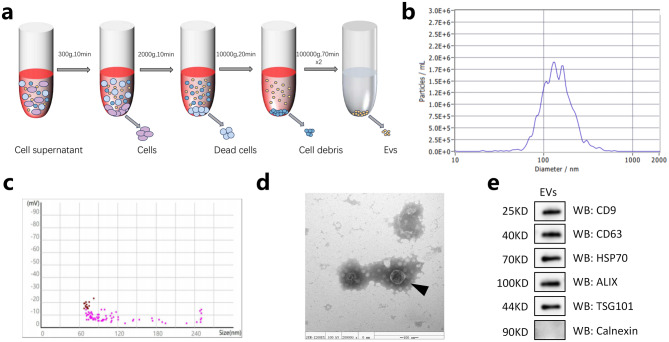
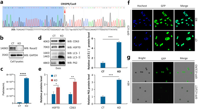
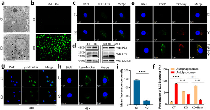
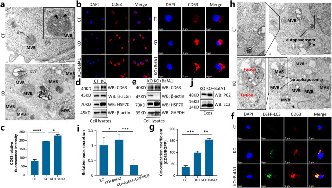
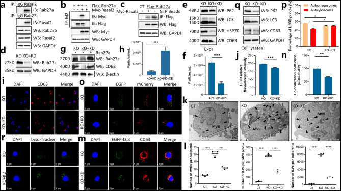
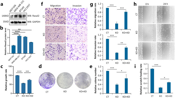
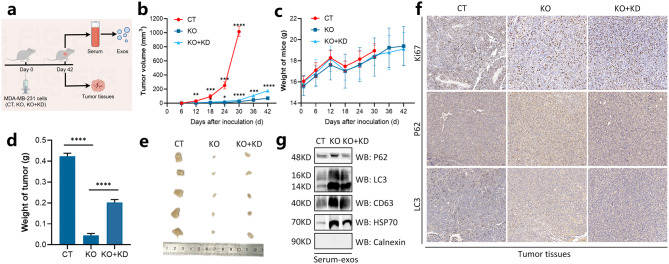
Methods: Rasal2 knock-out (KO) MDA-MB-231 cells were conducted by crispr-cas9 technique and Rab27a knock-down (KD) in Rasal2 KO MDA-MB-231 cells (KO + KD) were further established by siRNA-mate plus transfection Reagent. Control (CT)/KO MDA-MB-231 cells stably overexpressing GFP-LC3 were generated by using GFP-LC3 plamisd. Transmission electron microscope (TEM), nanoparticle tracking analysis (NTA) and western blot analysis (WB) were used to identify exos derived from TNBC. Confocal microscopy was used to observe the autophagic flux and the colocalization of autophagosomes and multivesicular bodies (MVBs). Co-immunoprecipitation analysis was performed to determine the interaction between Rasal2 and Rab27a. Immunohistochemical analysis were used to detect the expression levels of autophagy-related proteins in tumor tissues of xenograft mice inoculated with CT/KO/KO + KD MDA-MB-231 cells.

Results: In this paper, we found that Rasal2 KO disrupts autophagic flux and induces secretory autophagy to promote autophagic-exos secretion in TNBC. Moreover, Rasal2 inhibits the activity of Rab27a which regulates vesicles transport and fusion, and Rab27a mediates Rasal2 KO-induced autophagic-exos secretion. Additionally, Rab27a KD inhibits Rasal2 KO-induced secretory autophagy, thereby promoting TNBC progression both in vivo and in vitro.

Conclusions: Collectively, these findings delineated the role of Rab27a in TNBC progression modulated by Rasal2 through autophagy-exos pathway and suggested that it is of great significance for the early diagnosis, targeted therapy and prognosis judgement of TNBC from the perspective of tumor microenvironment.

Keywords: Autophagic-exos; Rab27a; Rasal2; Secretory autophagy; TNBC progression.


Conflict of interest statement

Declarations. Ethics approval and consent to participate: Animal experiments were approved by the Animal Ethical Committee of Qingdao University. Consent for publication: All authors agree to publish. Competing interests: The authors declare no competing interests.

Figures

Fig. 1
Fig. 1
Isolation and identification of exos. a, Isolation and purification of EVs from the conditioned media by ultracentrifugation. b, Size distribution of EVs analyzed by NTA. c, The ζ-potential of EVs. d, The morphology of EVs characterized by TEM, Scale bar = 100 nm. e, Proteins expression of exosomal positive and negative biomarkers detected by western blot analysis
Fig. 2
Fig. 2
Loss of Rasal2 promotes autophagic-exos secretion in TNBC. a, Construction of Rasal2 KO MDA-MB-231 cell lines by CRISPR/Cas9 technique. b, The protein expression of Rasal2 detected by western blot analysis. c, The effect of Rasal2 KO on exosomal secretion analyzed by NTA. d-e, Effects of Rasal2 KO on the expression levels of autophagic and exosomal marker proteins in exos. f, Establishment of CT and KO MDA-MB-231 cell lines with GFP-LC3 stable overexpression. g, Fluorescence intensity of magnetic beads-bound exos derived from CT and KO MDA-MB-231 cells stably overexpressing GFP-LC3
Fig. 3
Fig. 3
Rasal2 KO disturbs the autophagic pathway. a, Observation of autophagosomes produced in CT/KO MDA-MB-231 cells by TEM. b-c, Analysis of fluorescent aggregation plot in CT/KO MDA-MB-231 cells with transient transfection of EGFP-LC3 plasmid. d, Detection of autophagy-related marker proteins expression in CT/KO MDA-MB-231 cells with or without BafA1 treatment by western blotting. e-f, Observation of autophagic flux in CT/KO MDA-MB-231 cells transfected with mCherry-EGFP-LC3 by confocal fluorescence microscopy and quantitative analysis of red and yellow fluorescence aggregation plots. g-i, The acidic probe Lyso-Tracker Green was used to detect lysosomal acidity in CT/KO MDA-MB-231 cells by confocal fluorescence microscope
Fig. 4
Fig. 4
Rasal2 KO induces secretory autophagy. a, The morphology, size and number of MVBs and ILVs in CT/KO MDA-MB-231 cells observed by TEM. b-c, The intensity of fluorescent aggregation plots in BafA1-treated CT/KO MDA-MB-231 cells stained with CD63 antibody observed by confocal microscopy. d-e, The expression levels of exosomal marker proteins in CT/KO MDA-MB-231 cells with BafA1 treatment. f-g, The colocalization of autophagosomes and MVBs in CT/KO MDA-MB-231 cells with BafA1 treatment observed by confocal microscopy. h, The fusion of autophagosomes with MVBs in CT/KO MDA-MB-231 cells observed by TEM. i, The amount of exos secretion from KO MDA-MB-231 cells with BafA1/GW4869 treatment detected by NTA. j, The expression of autophagy marker proteins in exos derived from KO MDA-MB-231 cells with BafA1 treatment detected by western blotting
Fig. 5
Fig. 5
Rab27a mediates Rasal2 KO-induced autophagic-exos secretion. a, The interaction between endogenous Rab27a and Rasal2 determined by immunoprecipitation assay. b, The interaction between exogenous Rab27a and Rasal2 determined by immunoprecipitation assay. c, The effect of Rasal2 on the activity of Rab27a detected by using GTP-beads pull-down assay. d, Establishment of Rab27a KD in Rasal2 KO cell lines. e, The effect of Rab27a KD on the expression of autophagy marker proteins and exosomal marker proteins in exos-derived from Rasal2-KO cells. f, The effect of Rab27a KD on exosomal secretion from Rasal2 KO cells detected by NTA. g-h, The effect of Rab27a overexpression on exosomal secretion from (KO + KD) cells. i-j, The effect of Rab27a KD on the fluorescence intensity of CD63 in Rasal2 KO cells. k-l, The effect of Rab27a KD on the size and number of MVBs and ILVs in Rasal2 KO cells. m-n, The effect of Rab27a KD on the colocalization of autophagosomes and MVBs in Rasal2 KO cells. o-p, The effect of Rab27a KD on autophagic flux in Rasal2 KO cells. q, The effect of Rab27a KD on the expression of autophagic and exosomal marker proteins in Rasal2 KO cells. r, The effect of Rab27a KD on lysosomal acidity of Rasal2 KO cells
Fig. 6
Fig. 6
Inhibition of secretory autophagy promotes TNBC progression in vitro. a-b, The protein expression of Rasal2 in TNBC cells and normal breast cells. c-e, The effect of Rab27a on TNBC cell proliferation and clony formation regulated by Rasal2 evaluated by CCK-8 assay and clony formation assay. f-g, The effect of Rab27a on Rasal2-regulated TNBC cell migration and invasion measured by transwell assays. h-i, The effect of Rab27a on Rasal2-regulated TNBC cell migration analyzed by the wound healing assay
Fig. 7
Fig. 7
Inhibition of secretory autophagy promotes xenograft tumor growth in mice. a, xenograft animal models for MDA-MB-231 cells (CT, KO, KO + KD) were established in BALB/c nude mice. b, The growth curves of the tumor in mice after inoculating with MDA-MB-231 cells (CT/KO/KO + KD). c, The changes curve of mice weight after inoculating with MDA-MB-231 cells (CT/KO/KO + KD). d, Average tumor weight in CT/KO/KO + KD group. e, Photographs of tumors in CT/KO/KO + KD group. f, Immunohistochemistry analysis of proliferation-related and autophagy-related proteins. g, Expression of autophagic and exosomal marker proteins in exos derived from the serum of mice in CT/KO/KO + KD group

Similar articles

References

    1. Sung H, Ferlay J, Siegel RL, et al. Global Cancer statistics 2020: GLOBOCAN estimates of incidence and mortality worldwide for 36 cancers in 185 Countries[J]. CA Cancer J Clin. 2021;71(3):209–49. - PubMed
    1. Chiang YF, Huang KC, Chen HY et al. Hinokitiol inhibits breast Cancer cells in vitro Stemness-Progression and Self-Renewal with apoptosis and autophagy modulation via the CD44/Nanog/SOX2/Oct4 Pathway[J]. Int J Mol Sci. 2024;25(7). - PMC - PubMed
    1. Liedtke C, Mazouni C, Hess KR, et al. Response to neoadjuvant therapy and Long-Term survival in patients with Triple-Negative breast Cancer[J]. J Clin Oncol. 2023;41(10):1809–15. - PubMed
    1. Mehraj U, Mushtaq U, Mir MA, et al. Chemokines in triple-negative breast cancer heterogeneity: new challenges for clinical implications[J]. Semin Cancer Biol. 2022;86(Pt 2):769–83. - PubMed
    1. Hamdy NM, Basalious EB, El-Sisi MG, et al. Advancements in current one-size-fits-all therapies compared to future treatment innovations for better improved chemotherapeutic outcomes: a step-toward personalized medicine[J]. Curr Med Res Opin. 2024;40(11):1943–61. - PubMed

Substances

LinkOut - more resources