. 2025 Mar;15(3):1463-1479.
doi: 10.1016/j.apsb.2024.12.035. Epub 2025 Jan 3.

Novel carbazole attenuates vascular remodeling through STAT3/CIAPIN1 signaling in vascular smooth muscle cells

Affiliations

Novel carbazole attenuates vascular remodeling through STAT3/CIAPIN1 signaling in vascular smooth muscle cells

Joo-Hui Han et al. Acta Pharm Sin B. 2025 Mar.

Abstract

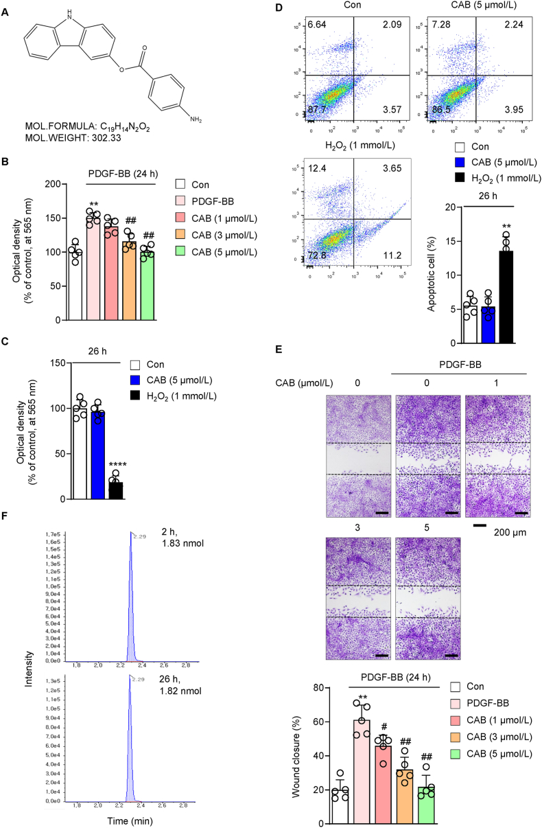
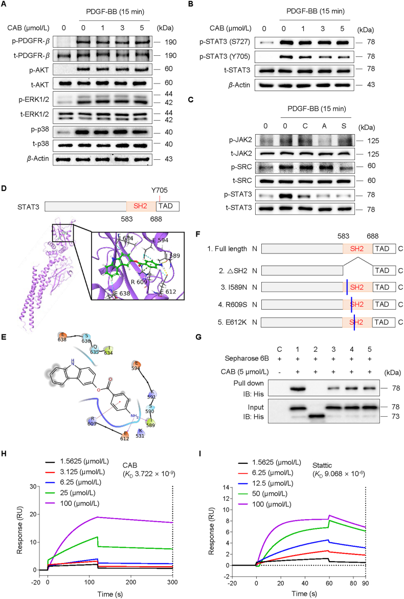
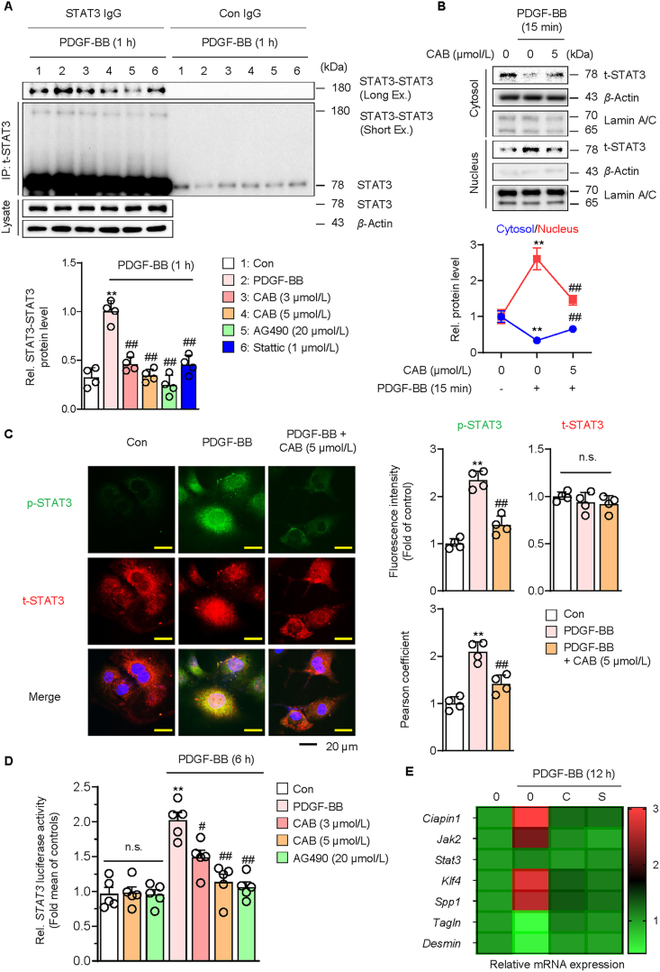
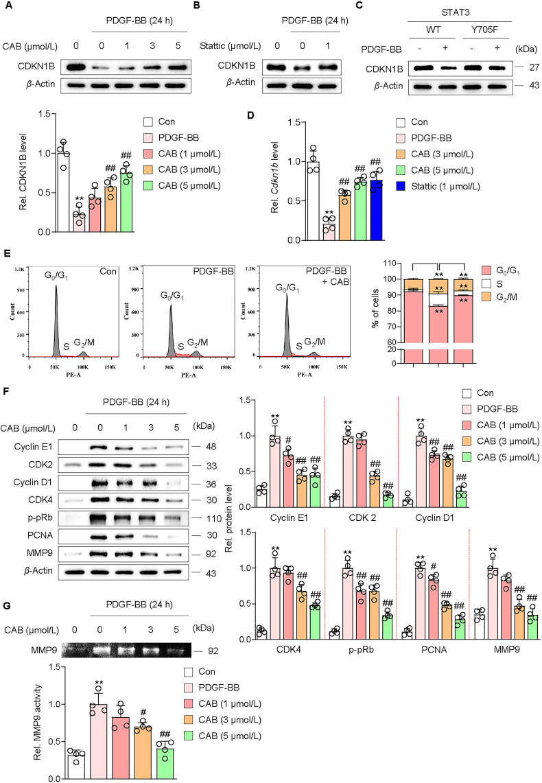
This study investigated the molecular mechanism of phenotypic switching of vascular smooth muscle cells (VSMCs), which play a crucial role in vascular remodeling using 9H-Carbazol-3-yl 4-aminobenzoate (CAB). CAB significantly attenuated platelet-derived growth factor (PDGF)-induced VSMC proliferation and migration. CAB suppressed PDGF-induced STAT3 activation by directly binding to the SH2 domain of STAT3. Downregulation of STAT3 phosphorylation by CAB attenuated CIAPIN1/JAK2/STAT3 axis through a decrease in CIAPIN1 transcription. Furthermore, abrogated CIAPIN1 decreased KLF4-mediated VSMC dedifferentiation and increased CDKN1B-induced cell cycle arrest and MMP9 suppression. CAB inhibited intimal hyperplasia in injury-induced neointima animal models by inhibition of the CIAPIN1/JAK2/STAT3 axis. However, CIAPIN1 overexpression attenuated CAB-mediated suppression of VSMC proliferation, migration, phenotypic switching, and intimal hyperplasia. Our study clarified the molecular mechanism underlying STAT3 inhibition of VSMC phenotypic switching and vascular remodeling and identified novel active CAB. These findings demonstrated that STAT3 can be a major regulator to control CIAPIN1/JAK2/STAT3 axis that may be a therapeutic target for treating vascular proliferative diseases.

Keywords: Atherosclerosis; Carbazole; Cytokine induced apoptosis inhibitor 1; Janus tyrosine kinase 2; Krüppel-like factor 4; Phenotyping switching; Signal transducer and activator of transcription 3; Vascular smooth muscle cell.


Conflict of interest statement

The authors declare that there is no conflict of interest.

Figures

Image 1
Graphical abstract
Figure 1
Figure 1
Inhibitory effect of CAB on PDGF-BB-induced VSMC proliferation and migration. (A) Chemical structure of 9H-Carbazol-3-yl 4-aminobenzoate (CAB). (B) MTT assay showing the effect of CAB (1–5 μmol/L) on PDGF-BB-induced VSMC proliferation (n = 5 per group). (C) MTT assay showing the cytotoxicity of CAB (5 μmol/L) and H2O2 (1 mmol/L, positive control) in VSMCs after 26 h (n = 5 per group). (D) Effect of CAB (5 μmol/L) on VSMCs apoptosis. Cells were treated with CAB (5 μmol/L) for 26 h (n = 5 per group). (E) Wound healing assay showing the effect of CAB (1–5 μmol/L) on PDGF-BB-induced VSMC migration (n = 5 per group). (F) Representative extracted ion chromatogram for quantification of the intracellular CAB concentration from VSMCs after 2 or 26 h. P-values were determined by the ANOVA followed by a post hoc Bonferroni's test. ∗∗P < 0.01 and ∗∗∗∗P < 0.0001 vs. Con, #P < 0.05 and ##P < 0.01 vs. PDGF-BB. The data are the mean ± SD.
Figure 2
Figure 2
Inhibitory effect of CAB on PDGF-BB-induced STAT3 activation in VSMCs. (A) Effect of CAB (1–5 μmol/L) on PDGFR-β, AKT, ERK1/2, and p38 phosphorylation in the PDGF-BB-induced VSMCs for 15 min. (B) Effect of CAB (1–5 μmol/L) on STAT3 phosphorylation in the PDGF-BB-induced VSMCs for 15 min Ser727: S727, Tyr705: Y705. (C) Effect of CAB (5 μmol/L), AG490 (20 μmol/L), and SU6656 (2 μmol/L) on JAK2, SRC, and STAT3 phosphorylation in the PDGF-BB-induced VSMCs for 15 min. (D) Docking results of the STAT3 SH2 domain (PDB accession number: 6TLC) in complex with CAB. STAT3 and CAB are shown in purple ribbon and green stick models, respectively. The surrounding residues associated with the CAB are shown in the gray stick model. The overall SH2 domain structure and magnified view of the binding pocket are shown. TAD: transactivation domain. (E) Ligand interaction diagrams of CAB docked to the STAT3 SH2 domain (PDB accession number: 6TLC). The 2D structure of CAB is drawn in a black line. The residues interacting with the compound are surrounded with pink arrows indicating three hydrogen bonds (I589, R609, and E612). (F) Schematic domain structures of STAT3 recombinant proteins. ΔSH2: SH2 deletion, I589N: Ile589 to Asn589, R609S: Arg609 to Ser609, E612K: Glu612 to Lys612. (G) His-tag pull-down assay with deletion and point mutation of predicted binding sites of STAT3 whether CAB binds to SH2 of STAT3. C: the group of full-length STAT3 protein mixed with CAB unconjugated Sepharose 6B. (H, I) Surface plasmon resonance (SPR) analysis of CAB or stattic binding to the STAT3 protein. The graph illustrates the sensorgram results for different concentrations of CAB (1.5625, 3.125, 6.25, 25, and 100 μmol/L) and stattic (1.5625, 6.25, 12.5, 50, and 100 μmol/L) binding to immobilized STAT3 protein (amino acids 127–688). The response (RU) is plotted against time (seconds), showing the association and dissociation phases for each concentration. The binding interaction was characterized by an equilibrium constant (KD) of CAB and stattic (3.722 × 10−9 mol/L and 9.068 × 10−6 mol/L, respectively), indicating the affinity between CAB and the STAT3 protein.
Figure 3
Figure 3
Inhibitory effect of CAB on PDGF-BB-induced STAT3 transcriptional activity in VSMCs. (A) Effect of CAB (3–5 μmol/L), AG490 (20 μmol/L) and stattic (1 μmol/L) on STAT3 dimerization in the PDGF-BB-induced VSMCs. Immunoblot of STAT3 monomers and dimers using STAT3 or control (Con) IgG (n = 4 per group). 1: Con, 2: PDGF-BB, 3: CAB (3 μmol/L), 4: CAB (5 μmol/L), 5: AG490 (20 μmol/L), and 6: Stattic (1 μmol/L) Short Ex.: short exposure, Long Ex.: long exposure (B) Effect of CAB on STAT3 nuclear translocation in the PDGF-BB-induced VSMCs for 15 min. The cells were fractionated into cytosolic and nuclear compartments as described in the Methods section (n = 4 per group). (C) Fluorescence images (left) and quantification data (right) of p-STAT3 and t-STAT3 in the PDGF-BB-induced VSMCs for 15 min. The fluorescence intensities of FITC (i.e., p-STAT3) and TRITC (i.e., t-STAT3) were quantified using ImageJ software. Colocalization of TRITC and DAPI (i.e., nuclei) was analyzed using the Pearson correlation coefficient (n = 4 per group). Scale bars: 20 μm. (D) Luciferase activities of STAT3 in the CAB-treated VSMCs after PDGF-BB stimulation for 6 h (n = 5 per group). The level of STAT3 binding promoter-reporter firefly luciferase activity is indicated relative to the activity of the Renilla luciferase control. (E) Heatmap of the mRNA expression of the indicated genes with or without CAB (5 μmol/L) or stattic (1 μmol/L) in the PDGF-BB-induced VSMCs for 24 h (n = 4 independent experiments). P-values were determined by the ANOVA followed by a post hoc Bonferroni's test. ∗∗P < 0.01 vs. Con, #P < 0.05 and ##P < 0.01 vs. PDGF-BB. n.s.: not significant. The data are the mean ± SD.
Figure 4
Figure 4
Inhibitory effect of CAB on PDGF-BB-induced CIAPIN1/JAK2/STAT3/KLF4 regulatory networks in VSMCs. (A) STAT3-associated protein networks generated using the STRING database. (B) Effect of PDGF-BB on STAT3 phosphorylation and CIAPIN1 expression until 24 h in VSMCs. Effect of (C) CAB (1–5 μmol/L), (D) stattic (1 μmol/L), (E) STAT3 Tyr705 mutant (Y705F) on CIAPIN1, p-JAK2, t-JAK2, p-STAT3, t-STAT3, KLF4 and SPP1 expression in the PDGF-BB-induced VSMCs for 24 h. WT: wild-type (F) Fluorescence images of p-STAT3 (Tyr705) and transgelin in the PDGF-BB-induced VSMCs for 24 h. The fluorescence intensities (Supporting Information Fig. S5) of FITC (i.e., p-STAT3) and TRITC (i.e., transgelin) were quantified using ImageJ software (n = 4 per group). Scale bars: 10 μm. (G) Representative images of the chromatin immunoprecipitation (ChIP)-PCR assay evaluated the binding of STAT3 to CIAPIN1 promoter in VSMCs treated with CAB for 2 h, followed by stimulation with PDGF-BB for 6 h. ChIP was performed with anti-STAT3 antibody and normal rabbit IgG. The PCR amplifications were performed using the franking primers for the STAT3-response elements (REs). The specificity of STAT3 binding was verified using primers targeting irrelevant regions in the promoter. The input represents the electrophoresis of chromatin fragments that did not undergo immunoprecipitation. (H) qPCR assays for STAT3 and STAT3–RE1 binding. (I) Effect of STAT3–RE1 mutation on Ciapin1 promoter reporter activity in PDGF-BB-induced VSMCs for 6 h (n = 4 per group). The data are presented as fold induction compared with the activity in control cells. P-values were determined by the ANOVA followed by a post hoc Bonferroni's test. ∗∗P < 0.01, ∗∗∗P < 0.001 and ∗∗∗∗P < 0.0001 vs. Con, ##P < 0.01, ###P < 0.001 and ####P < 0.0001 vs. PDGF-BB. n.s.: not significant. The data are the mean ± SD.
Figure 5
Figure 5
Inhibitory effect of CAB on PDGF-BB induced STAT3/CDKN1B regulation leading to cell cycle arrest and MMP9 repression. Effect of (A) CAB (1–5 μmol/L), (B) stattic (1 μmol/L), (C) STAT3 Tyr705 mutant (Y705F) on CDKN1B expression in the PDGF-BB-induced VSMCs for 24 h (n = 4 per group). WT: wild-type (D) Effect of CAB (1–5 μmol/L) on Cdkn1b mRNA expression in the PDGF-BB-induced VSMCs for 24 h (n = 4 per group). (E) Effect of CAB on cell cycle progression in the PDGF-BB-induced VSMCs for 24 h. Representative flow cytometry histograms of cell cycle progression and quantification data (n = 4 per group) are presented. Each shown value was derived by counting at least 10,000 events, and the number of cells in the G0/G1, S, and G2/M phases is expressed as percentages of total cells. (F) Effect of CAB (1–5 μmol/L) on cyclin E1, CDK2, cyclin D1, CDK4, p-Rb, PCNA, and MMP9 expression in the PDGF-BB-induced VSMCs for 24 h (n = 4 per group). (G) Effect of CAB (1–5 μmol/L) on MMP9 activity in the PDGF-BB-induced VSMCs for 24 h. Representative gelatin zymography and quantification data (n = 4 per group) for MMP9 activity are presented. P-values were determined by the ANOVA followed by a post hoc Bonferroni's test. ∗∗P < 0.01 vs. Con, #P < 0.05 and ##P < 0.01 vs. PDGF-BB. The data are the mean ± SD.
Figure 6
Figure 6
Effect of CIAPIN1 overexpression on CAB-mediated inhibition of VSMC dedifferentiation, proliferation, and migration. (A) Verification of human CIAPIN1 (hCIAPIN1 or hCIA1) transduction in VSMCs using Western blot assay. (B) Effect of CAB and stattic on CIAPIN1 nuclear translocation in the PDGF-BB-induced VSMCs for 24 h. The cells were fractionated into cytosolic and nuclear compartments as described in the Methods section (n = 4 per group). P-values were determined by the ANOVA followed by a post hoc Bonferroni's test. ∗∗P < 0.01 vs. Con, and ##P < 0.01 vs. PDGF-BB. (C) Fluorescence images (top) and quantification data (bottom) of CIAPIN1 and transgelin in the PDGF-BB-induced VSMCs for 24 h. The fluorescence intensities of FITC (i.e., CIAPIN1) and TRITC (i.e., transgelin) were quantified using ImageJ software. Colocalization of FITC and DAPI (i.e., nuclei) was analyzed using the Pearson correlation coefficient (n = 4 per group). Scale bars: 10 μm. P-values were determined by the ANOVA followed by a post hoc Bonferroni's test. ∗∗P < 0.01 vs. each group. (D) MTT assay showing the effect of CAB (5 μmol/L) on PDGF-BB-induced CIAPIN1-overexpressing VSMC proliferation at 24, 48 and 72 h (n = 5 per group). P-values were determined by the unpaired Student's t-test. ∗∗P < 0.01 vs. LV-Con, #P < 0.05 vs. LV-Con + CAB (5 μmol/L) for each time. (E) Wound healing assay showing the effect of CAB (5 μmol/L) on PDGF-BB-induced CIAPIN1-overexpressing VSMC migration after 24 h (n = 5 per group). P-values were determined by the ANOVA followed by a post hoc Bonferroni's test. ∗∗P < 0.01 and ∗∗∗P < 0.001 vs. each group. The data are the mean ± SD.
Figure 7
Figure 7
Effect of CAB and CIAPIN1 overexpression on neointima formation. (A) Representative images showing H&E staining in CAB-administered rat carotid arteries 14 days after injury. Sham: sham-operated, BI: balloon-injured, rCIA1: rat CIAPIN1 overexpression, I: intima, M: media. Scale bar: 100 μm. (B) Quantification of the intima and media area, intima/media ratio, and circumference of the external elastic lamina (EEL) in histological sections (n = 7 per group). N.D.: not detected. (C) Representative images showing the immunohistochemistry staining for PCNA-positive cells (dark brown, red arrow) and quantification of percentages of stained cells in the sham, BI, and BI with CAB-administered rat carotid arteries 14 days post-injury. Scale bars: 50 μm (n = 5 per group). (D) Representative images and quantification of immunofluorescence analysis in rat carotid arteries 14 days post-injury and after CIAPIN1 overexpression and CAB administration. Nuclei were stained with DAPI (blue), CIAPIN1 (FITC, green), and SM α-actin (TRITC, red). Scale bars: 100 μm (n = 5 per group). (E) Effect of CAB (5 μmol/L) on CIAPIN1, JAK2, STAT3, KLF4, SPP1 and CDKN1B activation in the PDGF-BB-induced CIAPIN1-overexpressing VSMCs for 24 h. P-values were determined by the ANOVA followed by a post hoc Bonferroni's test. ∗∗P < 0.01 vs. each group. n.s.: not significant. The data are the mean ± SD.

References

    1. Basatemur G.L., Jorgensen H.F., Clarke M.C.H., Bennett M.R., Mallat Z. Vascular smooth muscle cells in atherosclerosis. Nat Rev Cardiol. 2019;16:727–744. - PubMed
    1. Park H.S., Han J.H., Jung S.H., Lee D.H., Heo K.S., Myung C.S. Anti-apoptotic effects of autophagy via ROS regulation in microtubule-targeted and PDGF-stimulated vascular smooth muscle cells. Korean J Physiol Pharmacol. 2018;22:349–360. - PMC - PubMed
    1. Petsophonsakul P., Furmanik M., Forsythe R., Dweck M., Schurink G.W., Natour E., et al. Role of vascular smooth muscle cell phenotypic switching and calcification in aortic aneurysm formation. Arterioscler Thromb Vasc Biol. 2019;39:1351–1368. - PubMed
    1. Lacolley P., Regnault V., Nicoletti A., Li Z., Michel J.B. The vascular smooth muscle cell in arterial pathology: a cell that can take on multiple roles. Cardiovasc Res. 2012;95:194–204. - PubMed
    1. Bennett M.R., Sinha S., Owens G.K. Vascular smooth muscle cells in atherosclerosis. Circ Res. 2016;118:692–702. - PMC - PubMed

LinkOut - more resources