. 2025 Mar;15(3):1255-1267.
doi: 10.1016/j.apsb.2024.12.018. Epub 2024 Dec 21.

Disrupting calcium homeostasis and glycometabolism in engineered lipid-based pharmaceuticals propel cancer immunogenic death

Affiliations

Disrupting calcium homeostasis and glycometabolism in engineered lipid-based pharmaceuticals propel cancer immunogenic death

Qiuxia Peng et al. Acta Pharm Sin B. 2025 Mar.

Abstract

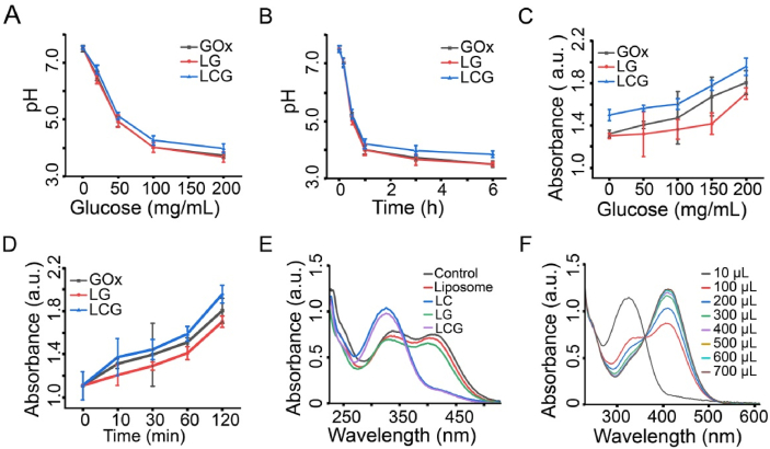
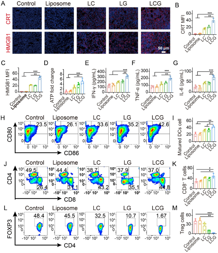
Homeostasis and energy and substance metabolism reprogramming shape various tumor microenvironment to sustain cancer stemness, self-plasticity and treatment resistance. Aiming at them, a lipid-based pharmaceutical loaded with CaO2 and glucose oxidase (GOx) (LipoCaO2/GOx, LCG) has been obtained to disrupt calcium homeostasis and interfere with glycometabolism. The loaded GOx can decompose glucose into H2O2 and gluconic acid, thus competing with anaerobic glycolysis to hamper lactic acid (LA) secretion. The obtained gluconic acid further deprives CaO2 to produce H2O2 and release Ca2+, disrupting Ca2+ homeostasis, which synergizes with GOx-mediated glycometabolism interference to deplete glutathione (GSH) and yield reactive oxygen species (ROS). Systematical experiments reveal that these sequential multifaceted events unlocked by Ca2+ homeostasis disruption and glycometabolism interference, ROS production and LA inhibition, successfully enhance cancer immunogenic deaths of breast cancer cells, hamper regulatory T cells (Tregs) infiltration and promote CD8+ T recruitment, which receives a considerably-inhibited outcome against breast cancer progression. Collectively, this calcium homeostasis disruption glycometabolism interference strategy effectively combines ion interference therapy with starvation therapy to eventually evoke an effective anti-tumor immune environment, which represents in the field of biomedical research.

Keywords: Calcium homeostasis disruption; Cancer plasticity; Engineered lipids; Glycometabolism interference; Immunogenic cell death; Lactic acid; Oxidative stress; Starvation therapy.


Conflict of interest statement

The authors declare that they have no competing interests.

Figures

Image 1
Graphical abstract
Scheme 1
Scheme 1
Schematic diagram illustrating LCG synthesis and how the combination of calcium homeostasis and glycometabolism in LCG pharmaceuticals unlock multifaceted events hamper Tregs infiltration and function, encourage CD8+ T recruitment and brought about cancer immunogenic death to repress breast cancer.
Figure 1
Figure 1
Construction and characterization of LCG. (A) Schematic diagram of LCG synthesis, scale bar = 50 nm; (B) TEM images of CaO2 nanoparticles, scale bar = 20 nm; (C) TEM images of LCG nanoparticles; (D, E) Particle size distribution (D) and zeta potential (E) of different nanoparticles. (F–H) XRD (F), XPS (G) and EDS (H) spectra of LCG; (I) UV absorption spectra of different nanoparticles; (J) Ca2+ release from LCG at different pH conditions. Data are expressed as mean ± SD, n = 3.
Figure 2
Figure 2
Multifaceted function tests in LCG including GOx enzymically-catalytic activity, H2O2 production and GSH depletion. (A) pH changes in GOX-, LG- or LCG-involved glucose solution with different glucose concentrations; (B) pH changes in GOX-, LG- or LCG-involved glucose solution at different periods. (C) Absorbance changes in H2O2 production by GOX, LG and LCG in glucose solution at different concentrations; and (D) Absorbance changes in H2O2 production by GOX, LG and LCG in glucose solution over time. (E) Absorbance changes in GSH content in solution after co-incubation of different materials with GSH (10 mmol/L); (F) Absorbance changes in GSH content in solution after co-incubation of different volumes of LCG with GSH (10 mmol/L). Data are expressed as mean ± SD (n = 3).
Figure 3
Figure 3
Cellular-level multifaceted events exploration and its role in promoting ROS production. (A) Fluorescence images of cells after different treatments (red fluorescence signal from LCG-DID, cells stained by DAPI and LysoTraker Green), scale bar = 5 μm; (B) FCM patterns of 4T1 cells after incubation with LCG-DID for different periods (0 and 4 h), both of which were used to assess the cellular phagocytosis of LCG-DID by 4T1 cells. (C) The concentration of Ca2+ in the culture medium following various cell treatments. (D–F) Intracellular contents of H2O2 (D), GSH (E) and LA (F) in 4T1 cells after co-incubation with different nanomaterials, scale bar = 20 μm; (G) CLSM images of 4T1 cells stained with DCFH-DA after co-incubation with different nanoparticles; and (H) FCM analysis of 4T1 cells stained with DCFH-DA after co-incubation with different nanoparticles, both of which could detect the intracellular ROS content after different treatments. Data are expressed as mean ± SD (n = 3); ∗P<0.05, ∗∗P<0.01, ∗∗∗P<0.001, ∗∗∗∗P<0.0001.
Figure 4
Figure 4
In vitro anti-tumor evaluations and cancer immunogenic death test. (A) Relative viability of 4T1 cells after incubations with different samples under varied concentrations for 24 h; (B) Relative viability of 4T1 cells after incubations with different samples under varied concentrations for different periods (n = 6); (C) CLSM images of PI-stained 4T1 cells in different experimental subgroups that experienced corresponding treatments for 4, 8 and 12 h, scale bar = 100 μm; (D) CLSM images of 4T1 cells co-stained with Annexin V-mCHerrry and SYTOX Green in different groups, scale bar = 20 μm; (E) FCM patterns of 4T1 cells stained with PI and Annexin V-FITC after co-incubation with different samples; (F) CLSM images of Fluo-4 AM-stained 4T1 cells after co-incubation with different nanoparticles for tracing intracellular Ca2+ levels, scale bar = 20 μm; and (G) corresponding MFI values. (H) Intracellular ATP content detection after co-incubation of 4T1 with different nanoparticles; (I–N) Immunofluorescence images (I–K) and corresponding MFI values (L–N) of CRT (I,L), HSP 70 (J, M) and HMGB1 (K, N), scale bar = 20 μm; (O) WB images of intracellular CRT and HMGB1 after different treatments. Data are expressed as mean ± SD (n = 3), ‘ns’ no significance, ∗∗P < 0.01, ∗∗∗P < 0.001 and ∗∗∗∗P < 0.0001.
Figure 5
Figure 5
Anti-tumor assay and mechanism exploration. (A) Schematic of imaging in small animals; (B) Fluorescence biodistribution imaging of LCG in mice under different time points post-injection of LCG-DID and free DID (Control); (C) Fluorescence images of vivo organ harvested from LCG-treated mice after 6 and 48 h post-injection of LCG-DID. (D) Schematic diagram of tumors treated with different nanomaterials; (E) Time-dependent changes in the average body weight of mice in different treatment groups; (F) Time-dependent changes in the average tumor volume of mice in different treatment groups; (G) Time-dependent changes in the average tumor body weight in different treatment groups; (H) Digital images of excised tumors from mice in different treatment groups at the end of experiment period; (I) Photos of 4T1 tumor-bearing mice before and after different treatments in different treatment groups; (J) Survival rate of mice treated with various formulations in different treatment groups; (K) Immunofluorescence images of tumor slices co-stained with FOXP3 and DAPI after different treatments for assaying Tregs infiltration, scale bar = 50 μm; (L) Immunofluorescence images of tumor slices co-stained with CD4, CD8 and DAPI after different treatments for assaying T-cell infiltration, scale bar = 50 μm; (M) ROS levels in tumor sections stained with dihydroethidium (DHE), scale bar = 50 μm; (N) Optical photos of tumor slices after different immunochemical staining in different treatment groups for realizing H&E, KI67 and TUNEL expression analysis, scale bar = 50 μm. Data are expressed as mean ± SD (n = 5); ∗P<0.05 and ∗∗P<0.01.
Figure 6
Figure 6
Induces ICD and activates immunity. (A–C) Fluorescence micrographs of 4T1 tumor tissue stained with CRT and HMGB1 (A), scale bar = 50 μm, along with the corresponding MFI (B,C); (D) Intratumoral ATP levels in tumor after different treatments (n = 4); (E–G) Secretion levels of IFN-γ (E), TNF-α (F), and IL-6 (G) in mouse serum based on Elisa kits (n = 4); (H,I) Representative FCM patterns (H) and quantitative analysis (I) of matured DCs (CD80+CD86+) in tumor tissues after various treatments; (J,K) Representative FCM patterns (J) and quantitative analysis (K) of CD4+ and CD8+ T cells in tumor tissues following various treatments (n = 3); (L, M) Representative FCM patterns (L) and quantitative analysis (M) of Tregs ((FOXP3+CD4+) in tumor tissues following various treatments (n = 3). Data are expressed as mean ± SD; ∗P<0.05, ∗∗P<0.01 and ∗∗∗P<0.001.

Similar articles

Cited by

References

    1. Siegel P.M., Massagué J. Cytostatic and apoptotic actions of TGF-β in homeostasis and cancer. Nat Rev Cancer. 2003;3:807–820. - PubMed
    1. DeBerardinis R.J., Chandel N.S. Fundamentals of cancer metabolism. Sci Adv. 2016;2 - PMC - PubMed
    1. Panieri E., Santoro M.M. ROS homeostasis and metabolism: a dangerous liaison in cancer cells. Cell Death Dis. 2016;7 - PMC - PubMed
    1. Liu Y.Y., Gu R.J., Gao M.R., Wei Y.W., Shi Y., Wang X., et al. Emerging role of substance and energy metabolism associated with neuroendocrine regulation in tumor cells. Front Endocrinol. 2023;14 - PMC - PubMed
    1. Zhang X.N., Zhao Q., Yang J.J., Wang T.X., Chen F.B., Zhang K. Tumor microenvironment-triggered intratumoral in-situ biosynthesis of inorganic nanomaterials for precise tumor diagnostics. Coord Chem Rev. 2023;484

LinkOut - more resources