. 2025 Mar;15(3):1383-1396.
doi: 10.1016/j.apsb.2025.01.017. Epub 2025 Jan 28.

Long-chain acylcarnitine deficiency promotes hepatocarcinogenesis

Affiliations

Long-chain acylcarnitine deficiency promotes hepatocarcinogenesis

Kaifeng Wang et al. Acta Pharm Sin B. 2025 Mar.

Abstract

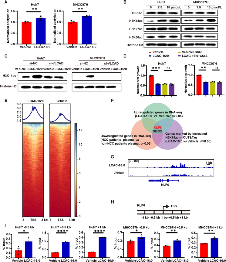
Despite therapy with potent antiviral agents, chronic hepatitis B (CHB) patients remain at high risk of hepatocellular carcinoma (HCC). While metabolites have been rediscovered as active drivers of biological processes including carcinogenesis, the specific metabolites modulating HCC risk in CHB patients are largely unknown. Here, we demonstrate that baseline plasma from CHB patients who later developed HCC during follow-up exhibits growth-promoting properties in a case-control design nested within a large-scale, prospective cohort. Metabolomics analysis reveals a reduction in long-chain acylcarnitines (LCACs) in the baseline plasma of patients with HCC development. LCACs preferentially inhibit the proliferation of HCC cells in vitro at a physiological concentration and prevent the occurrence of HCC in vivo without hepatorenal toxicity. Uptake and metabolism of circulating LCACs increase the intracellular level of acetyl coenzyme A, which upregulates histone H3 Lys14 acetylation at the promoter region of KLF6 gene and thereby activates KLF6/p21 pathway. Indeed, blocking LCAC metabolism attenuates the difference in KLF6/p21 expression induced by baseline plasma of HCC/non-HCC patients. The deficiency of circulating LCACs represents a driver of HCC in CHB patients with viral control. These insights provide a promising direction for developing therapeutic strategies to reduce HCC risk further in the antiviral era.

Keywords: Acetyl coenzyme A; CUT&Tag; Chemoprevention; H3K14; Hepatocellular carcinoma; KLF6; Long-chain acylcarnitine; Metabolomics.


Conflict of interest statement

The authors declare no conflicts of interest.

Figures

Image 1
Graphical abstract
Figure 1
Figure 1
Long-chain acylcarnitines (LCACs) are negatively correlated with hepatocellular carcinoma (HCC) risk. (A) Diagram showing experimental design (see Methods). (B) Relative cell viability of cells cultured using 10% baseline plasma of chronic hepatitis B (CHB) patients (non-HCC, n = 76; HCC, n = 76). (C) Colony-forming assays of cells incubated with 10% baseline plasma of CHB patients (n = 3). (D) The top 2 signaling pathways induced by baseline plasma from CHB patients who developed HCC in Huh7 cells. (E) Volcano plot of LC–MS-based metabolomics from the plasma of CHB patients (non-HCC, n = 76; HCC, n = 76). (F) Kaplan–Meier estimates and the risk of HCC ranked according to LCAC tertile levels (non-HCC, n = 76; HCC, n = 76). (G) Risk of HCC by 4.5 years according to LCAC tertile levels using a multivariable Cox proportional hazard model. (non-HCC, n = 76; HCC, n = 76). Unadjusted hazard ratio (black) and adjusted model (age, sex, total bilirubin, platelets, albumin, and elevated ALT; red). The line length indicates the 95% confidence interval. Data are presented as mean ± SEM. ∗P < 0.05, ∗∗P < 0.01. P values were calculated based on one-way ANOVA (B), log-rank test (F), or Wald test (G).
Figure 2
Figure 2
LCACs inhibit hepatocarcinogenesis. (A) Dose curve of a panel of cell lines treated LCAC-16:0 for 120 h (n = 3). All tested doses were compared to the vehicle group. (B) Colony-forming assays of cells treated with the indicated concentration of LCAC-16:0. (C) Mice bearing Hepa1-6 hepatoma were treated with LCAC-16:0. Representative images are shown (n = 5 for Vehicle, n = 5 for LCAC-16:0). (D) AKT/Ras mice were treated with LCAC-16:0. Representative images are shown (n = 7 for Vehicle, n = 7 for LCAC-16:0). (E) C57BL/6 mice were treated with diethylnitrosamine (DEN) plus tetrachloromethane (CCl4) to induce spontaneous hepatoma. Thereafter, the mice were treated with LCAC-16:0. Representative images are shown (n = 16 for Vehicle, n = 8 for LCAC-16:0). (F) Hepatic lobule structure of the DEN/CCl4 mice. (G) Hepatic expression of Ki67 in DEN/CCl4 mice. Data are presented as mean ± SEM. ∗P < 0.05, ∗∗P < 0.01, ∗∗∗P < 0.001, ∗∗∗∗P < 0.0001 vs. vehicle; ns, no significance. P values were calculated based on one-way ANOVA.
Figure 3
Figure 3
Uptake and metabolism of LCACs are required for their anti-tumor effect. (A) Intracellular LCAC-16:0 levels in Huh7 cells after incubation with 7.5 μmol/L LCAC-16:0 for 24 h (n = 3). (B) Intracellular acetyl-CoA levels in Huh7 cells after incubation with 7.5 μmol/L LCAC-16:0 for 24 h (n = 3). (C) Schematic of 13C-(1,2,3,4)-LCAC-16:0 facilitating incorporation of 13C into acetyl-CoA. (D) Isotopic tracing analysis of 13C-(1,2,3,4)-LCAC-16:0 in Huh7 cells (n = 3). (E) Inhibitory effect of LCAC-16:0 (15 μmol/L for 120 h) on HCC cells upon siRNA-mediated inhibition of very long-chain acyl-coenzyme A dehydrogenase (VLCAD) expression (n = 3). (F) Inhibitory effect of LCAC-16:0 (15 μmol/L for 120 h) on HCC cells upon CTPI-2 mediated inhibition of transport of acetyl-CoA out of mitochondria (n = 3). (G) Inhibitory effect of LCAC-16:0 (15 μmol/L for 120 h) on HCC cells upon SB-204990 mediated depletion of cytoplasmic acetyl-CoA (n = 3). Data are presented as mean ± SEM. ∗P < 0.05, ∗∗P < 0.01, ∗∗∗P < 0.001, ∗∗∗∗P < 0.0001; ns, no significance. P values were calculated based on one-way ANOVA.
Figure 4
Figure 4
LCACs act through modulating H3 histone acetylation. (A) ELISA showing acetylation levels of H3 in HCC cells treated with 7.5 μmol/L LCAC-16:0 for 24 h. (B) Western blot showing acetylation levels of H3K14, H3K27, and H3K56 of HCC cells treated with the indicated concentration of LCAC-16:0 for 24 h. (C) Western blot showing H3K14ac levels in HCC cells with si-NC or si-VLCAD in the presence or absence of LCAC-16:0 (15 μmol/L for 24 h) treatment. (D) Inhibitory effect of LCAC-16:0 (15 μmol/L for 120 h) on HCC cells upon C646 mediated inhibition of p300 (n = 3). (E) Heatmap showing the genomic occupancy of H3K14ac ± 3 kb flanking TSSs in Huh7 cells. The genes shown in rows are sorted in descending order by signal strength. (F) Bioinformatics analysis filtered KLF6 and SMOX as downstream targets of H3K14ac. (G) Normalized read densities for H3K14ac at the KLF6 gene. (H) Diagram depicting the locations in the KLF6 gene of amplicons used in chromatin immunoprecipitation (ChIP) and ChIP-qPCR assays. (I) ChIP-qPCR analysis showing KLF6 gene occupancy by H3K14ac in HCC cells treated with 7.5 μmol/L LCAC-16:0 for 24 h (n = 3). Data are presented as mean ± SEM. ∗P < 0.05, ∗∗P < 0.01, ∗∗∗P < 0.001, ∗∗∗∗P < 0.0001; ns, no significance. P values were calculated based on one-way ANOVA.
Figure 5
Figure 5
LCACs inhibit hepatocarcinogenesis via KLF6. (A) Western blot showing KLF6 expression levels of HCC cells treated with 7.5 μmol/L LCAC-16:0 for 96 h. (B) The correlation between relative protein expression of KLF6 in HCC tissues and plasma LCAC-16:0 levels of HCC patients (n = 12). (C) Inhibitory effect of LCAC-16:0 (15 μmol/L for 120 h) on HCC cells under conditions of siRNA-mediated KLF6 inhibition (n = 3). (D) Western blot showing KLF6 expression levels in HCC cells with si-NC or si-VLCAD in the presence or absence of LCAC-16:0 (15 μmol/L for 96 h) treatment. (E) Western blot showing KLF6 expression levels in HCC cells with or without CPTI-2 in the presence or absence of LCAC-16:0 (15 μmol/L for 96 h) treatment. (F) Western blot showing KLF6 expression levels in HCC cells with or without C646 in the presence or absence of LCAC-16:0 (15 μmol/L for 96 h) treatment. (G) qPCR showing KLF6 expression levels in HCC cells induced by baseline plasma of HCC/non-HCC patients (48 h) with si-NC or si-VLCAD (n = 3). Data are presented as mean ± SEM. ∗P < 0.05, ∗∗P < 0.01, ∗∗∗P < 0.001, ∗∗∗∗P < 0.0001; ns, no significance. P values were calculated based on one-way ANOVA.
Figure 6
Figure 6
LCACs inhibit hepatocarcinogenesis via p21. (A) Analysis of RNA-seq data using the TRRUST database. (B) Western blot showing p21 expression in HCC cells treated with 7.5 LCAC-16:0 for 96 h. (C) The correlation between relative protein expression of p21 in HCC tissues and plasma LCAC-16:0 levels of HCC patients (n = 12). (D) Inhibitory effect of LCAC-16:0 on HCC cells upon siRNA-mediated p21 inhibition (n = 3). (E) Inhibitory effect of LCAC-16:0 (25 mg/kg/day) on MHCC97H subcutaneous tumor growth in nude mice in response to shRNA-mediated inhibition of p21 expression (n = 6). (F) Western blot showing p21 expression levels in HCC cells with si-NC or si-KLF6 in the presence or absence of LCAC16:0 (15 μmol/L for 96 h) treatment. (G) qPCR showing p21 expression levels in HCC cells induced by baseline plasma of HCC/non-HCC patients (48 h) with si-NC or si-VLCAD (n = 3). Data are presented as mean ± SEM. ∗P < 0.05, ∗∗P < 0.01, ∗∗∗P < 0.001, ∗∗∗∗P < 0.0001; ns, no significance. P values were calculated based on one-way ANOVA.

References

    1. Forner A., Reig M., Bruix J. Hepatocellular carcinoma. Lancet. 2018;391:1301–1314. - PubMed
    1. Sung H., Ferlay J., Siegel R.L., Laversanne M., Soerjomataram I., Jemal A., et al. Global Cancer Statistics 2020: GLOBOCAN estimates of incidence and mortality worldwide for 36 cancers in 185 countries. CA Cancer J Clin. 2021;71:209–249. - PubMed
    1. Liu Z., Jiang Y., Yuan H., Fang Q., Cai N., Suo C., et al. The trends in incidence of primary liver cancer caused by specific etiologies: results from the Global Burden of Disease Study 2016 and implications for liver cancer prevention. J Hepatol. 2019;70:674–683. - PubMed
    1. Liaw Y.F., Sung J.J., Chow W.C., Farrell G., Lee C.Z., Yuen H., et al. Lamivudine for patients with chronic hepatitis B and advanced liver disease. N Engl J Med. 2004;351:1521–1531. - PubMed
    1. Singal A.G., El-Serag H.B. Hepatocellular carcinoma from epidemiology to prevention: translating knowledge into practice. Clin Gastroenterol Hepatol. 2015;13:2140–2151. - PMC - PubMed

LinkOut - more resources