

Feasibility and practicality of a novel teaching aid for microvascular anastomosis simulation training in neurosurgery generated by 3D printing
- PMID: 40370766
- PMCID: PMC12075562
- DOI: 10.3389/fsurg.2025.1546573
Feasibility and practicality of a novel teaching aid for microvascular anastomosis simulation training in neurosurgery generated by 3D printing
Abstract
Background: This study aimed to develop a novel teaching aid for microvascular anastomosis training in neurosurgery using 3D printing technology based on CT and MRI imaging data, and to evaluate its effectiveness and practicality.
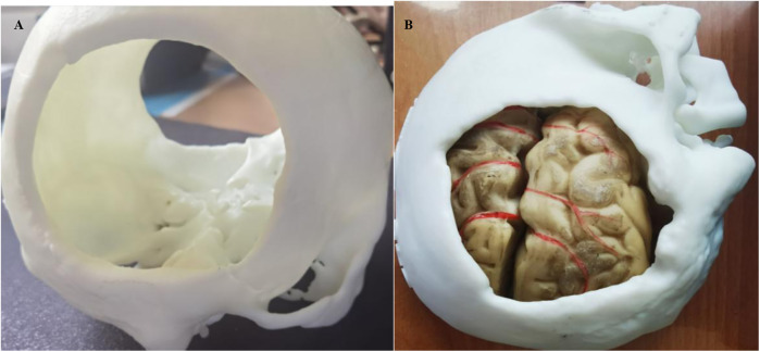
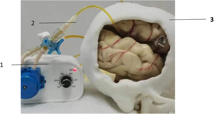
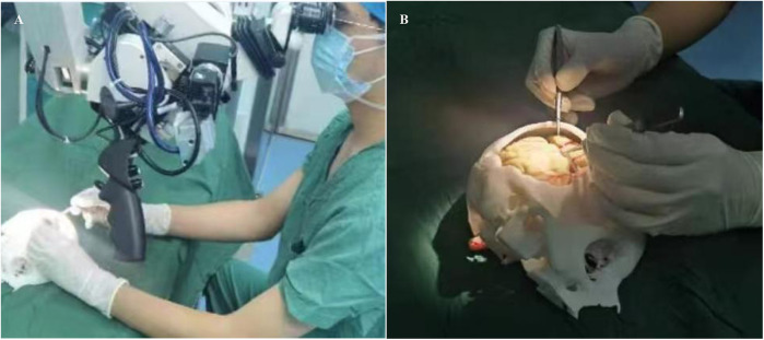
Methods: Based on CT or MRI imaging data, a 3D model integrating micro-vessels, skull, and brain tissue was fabricated and connected to a peristaltic pump and a pipeline system to create a teaching aid for microvascular anastomosis simulation training. Twenty senior medical students were recruited and divided into two groups: a control group, which trained using traditional soft rubber tubes, and an observation group, which trained using the 3D-printed teaching aid. Following the training, participants from both groups performed chicken wing artery anastomosis. The training outcomes, including the patency rate of vascular anastomosis, the time required to complete the anastomosis, and the trainees' surgical performance, were evaluated. Additionally, six experienced neurosurgeons were recruited to teach the course using both teaching aids for two hours each. They were then surveyed via a questionnaire to assess and rate the effectiveness of the teaching aids.
Results: The observation group demonstrated a significantly higher patency rate of vascular anastomosis, a shorter time to complete the anastomosis, and higher scores for surgical proficiency and procedural standardization compared to the control group (all P < 0.001). Additionally, the neurosurgeons provided positive evaluations of the novel 3D-printed teaching aid, awarding high scores for its practicality, scientific rigor, and overall effectiveness.
Conclusion: The novel 3D-printed teaching aid serves as an effective tool for microvascular anastomosis training in neurosurgery, offering significant advantages such as enhanced training effectiveness, high-fidelity simulation, cost efficiency, and customization capabilities.
Keywords: 3D printed teaching aid; 3D printing; microsurgery; neurosurgery; simulation training; vascular anastomosis.
© 2025 Shi, Ren, Zhao, Cui, Su, Yan and Bu.
Conflict of interest statement
The author declare that the research was conducted in the absence of any commercial or financial relationships that could be construed as a potential conflict of interest.
Figures
Similar articles
-
Validation of a dynamic 4D microsurgical bypass simulator for training and teaching microvascular anastomosis techniques with blood flow and fluorescence imaging.World Neurosurg X. 2024 Sep 21;24:100396. doi: 10.1016/j.wnsx.2024.100396. eCollection 2024 Oct. World Neurosurg X. 2024. PMID: 39399349 Free PMC article.
-
Creation of a novel simulator for minimally invasive neurosurgery: fusion of 3D printing and special effects.J Neurosurg Pediatr. 2017 Jul;20(1):1-9. doi: 10.3171/2017.1.PEDS16568. Epub 2017 Apr 25. J Neurosurg Pediatr. 2017. PMID: 28438070
-
3D-printed cranial models simulating operative field depth for microvascular training in neurosurgery.Surg Neurol Int. 2021 May 10;12:213. doi: 10.25259/SNI_849_2020. eCollection 2021. Surg Neurol Int. 2021. PMID: 34084640 Free PMC article.
-
Three-Dimensional Modeling in Training, Simulation, and Surgical Planning in Open Vascular and Endovascular Neurosurgery: A Systematic Review of the Literature.World Neurosurg. 2021 Oct;154:53-63. doi: 10.1016/j.wneu.2021.07.057. Epub 2021 Jul 19. World Neurosurg. 2021. PMID: 34293525
-
Review of 3-Dimensional Printing on Cranial Neurosurgery Simulation Training.World Neurosurg. 2016 Apr;88:188-198. doi: 10.1016/j.wneu.2015.12.031. Epub 2015 Dec 25. World Neurosurg. 2016. PMID: 26724615 Review.
References
-
- Cai JQ, Duan CB, Qi TF, Jiang CL. Application of 3D printing technology in clinical teaching of neurosurgery. Zhon Guo Wei Qin Xi Shen Jing WaI Ke Za Zhi. (2020) 25(05):238–40.
LinkOut - more resources
Full Text Sources