Case Reports
. 2025 Apr 30:12:1534966.
doi: 10.3389/fvets.2025.1534966. eCollection 2025.

Case Report: Use of veterinary cuttable plates to maintain reduction and reinforce polymethylmethacrylate (PMMA) fixation in two canine C2 vertebral fractures

Affiliations
Case Reports

Case Report: Use of veterinary cuttable plates to maintain reduction and reinforce polymethylmethacrylate (PMMA) fixation in two canine C2 vertebral fractures

Olivia M Snead et al. Front Vet Sci. .

Abstract

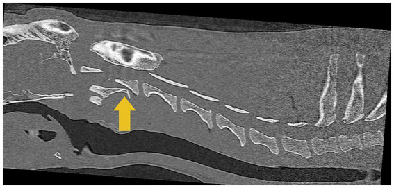
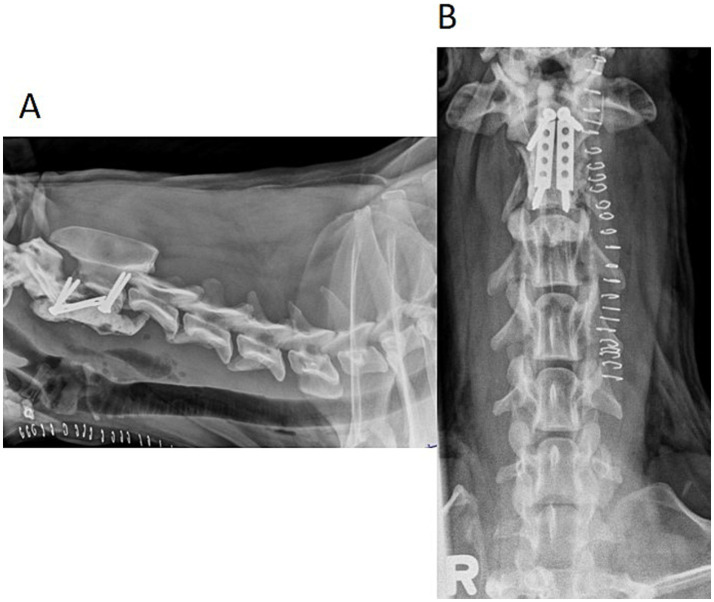
Veterinary cuttable plates (VCPs) were used to maintain reduction and provide reinforcement in screw and polymethylmethacrylate (PMMA) repairs of C2 vertebral fractures in two dogs. A 6-month-old male intact 3.8 kg Shih Tzu and a 4-year-old male intact 30.5 kg Golden Retriever presented after motor vehicle accidents. Computed tomography (CT) scans revealed fractures of the C2 vertebrae in both dogs. Fractures were reduced and stabilized with a combination of cortical screws, VCPs, and PMMA. The novel technique utilized veterinary cuttable plates positioned between screws cranial and caudal to the fracture for distraction and reduction of the fractures and can be incorporated into the acrylic. Post-operative imaging showed adequate fracture reduction and relief of spinal cord compression. Recheck examinations demonstrated a return to normal and near-normal neurologic function in both patients.

Keywords: PMMA; case report; cervical fracture; fixation; reduction; vertebral fracture.


Conflict of interest statement

The authors declare that the research was conducted in the absence of any commercial or financial relationships that could be construed as a potential conflict of interest.

Figures

Figure 1
Figure 1
Computed tomographic (CT) image of the cervical region shows a C2 vertebral fracture with the dorsal displacement of the caudal fragment causing spinal cord compression indicated by the yellow arrow.
Figure 2
Figure 2
Post-operative 3-D reconstruction of the patient’s cervical vertebral column with lateral (A) and ventrodorsal (B) views. Two cortical screws are located in the cranial fracture fragment of C2, and two cortical screws are located in the cranial aspect of C3. A VCP was placed diagonally between the cranial and caudal screws and was encased in PMMA. Post-operative transverse CT image (C) shows the screws placed the cranial fracture fragment of C2. Though all efforts were made to avoid breaching of the vertebral canal, the right pedicle screw did breach. The left screw penetrated the transverse foramen without adverse consequences in this size patient.
Figure 3
Figure 3
Computed tomographic (CT) image of the cervical region shows a C2 vertebral fracture with the dorsal displacement of the caudal fragment causing spinal cord compression, indicated by the yellow arrow.
Figure 4
Figure 4
Lateral (A) and ventrodorsal (B) post-operative radiographs of the patient’s cervical vertebral column. Two cortical screws are located in the cranial fracture fragment of C2, and two cortical screws are located in the caudal fracture fragment of C2. Two VCPs were placed parallel between the cranial and caudal screws and were encased in PMMA.

References

    1. Pike FS, Kumar MSA, Boudrieau RJ. Reduction and fixation of cranial cervical fracture/Luxations using screws and polymethylmethacrylate (PMMA) cement: a distraction technique applied to the base of the skull in thirteen dogs. Vet Surg. (2012) 41:235–47. doi: 10.1111/j.1532-950X.2011.00916.x, PMID: - DOI - PubMed
    1. Jeffery ND. Vertebral fracture and luxation in small animals. Vet Clin Small Anim. (2010) 40:809–28. doi: 10.1016/j.cvsm.2010.05.004, PMID: - DOI - PubMed
    1. Schmidli FE, Stein VM, Aikawa T, Boudrieau RJ, Jeandel A, Jeffery N, et al. . Fracture of the second cervical vertebra in 66 dogs and 3 cats: a retrospective study. Vet Comp Orthop Traumatol. (2019) 32:200–6. doi: 10.1055/s-0039-1678542 - DOI - PubMed
    1. Hawthorne JC, Blevins WE, Wallace LJ, Glickman N, Waters DJ. Cervical vertebral fractures in 56 dogs: a retrospective study. J Am Anim Hosp Assoc. (1999) 35:135–46. doi: 10.5326/15473317-35-2-135, PMID: - DOI - PubMed
    1. Shores A. Spinal trauma: pathophysiology and management of traumatic spinal injuries. Vet Clin N Am Small Anim Pract. (1992) 22:859–88. doi: 10.1016/S0195-5616(92)50080-8, PMID: - DOI - PubMed

Publication types

LinkOut - more resources