Case Reports
. 2025 May 7:2025:3531508.
doi: 10.1155/humu/3531508. eCollection 2025.

Deletion Testing of the DEGS1 Gene Should Be Part of the Diagnostic Pipeline for Hypomyelinating Leukodystrophy (HLD18)

Affiliations
Case Reports

Deletion Testing of the DEGS1 Gene Should Be Part of the Diagnostic Pipeline for Hypomyelinating Leukodystrophy (HLD18)

Mariateresa Zanobio et al. Hum Mutat. .

Abstract

Hypomyelinating leukodystrophies are a heterogeneous group of disorders characterized by abnormal myelin formation in the central nervous system. Thanks to the increased use of NGS, a growing number of pathogenic single nucleotide variants in DEGS1 have recently been reported to be responsible for hypomyelinating leukodystrophy 18 (HLD18), a rare and severe autosomal recessive form. DEGS1 is a small gene (4 exons and 17 kb) encoding Δ4-dihydroceramide desaturase, which catalyzes the final step in ceramide biosynthesis. Here, we present two patients from unrelated families affected by severe and progressive white matter disease with developmental delay with or without regression and severe intellectual disability. Trio exome sequencing (ES) revealed in both probands two homozygous missense variants in the DEGS1 gene, p.Asp16His and p.Asn255Ser, both inherited from their heterozygous healthy mothers and with a noncarrier father. This curious finding of inconsistent segregation data raises the need for further testing. There is no MLPA test available for this gene, as no deletions have been reported. However, we tried a customized high-resolution 1 M CGH array, which was surprisingly positive in both cases: a 63-kb heterozygous deletion encompassing the entire gene in one proband and a 7-kb heterozygous deletion of Exons 2-3 in the second case. Previously reported cases of HLD18 have all been found to carry single nucleotide pathogenic variants in DEGS1, and the two patients described here are the first to carry whole or partial microdeletions involving DEGS1 that unmask pathogenic missense variants on the other allele. These two cases report the first examples of microdeletions of DEGS1 that unmask recessive allele pathogenic variants, underscoring the importance of considering whole or partial gene deletions in the diagnostic pipeline.


Conflict of interest statement

The authors declare no conflicts of interest.

Figures

Figure 1
Figure 1
In the left panel, brain MRI scan of affected proband of Family 1; axial (A, B), coronal (C), and sagittal (D) T2-weighted sequences showing volumetric reduction and hyperintensity of periventricular and deep white matter (arrows in A). The thalami are atrophic and show contextual laminar hyperintensity (arrows in B). Hemispheric and vermian cerebellar atrophy is also present (arrows in C and D), as well as a markedly thinned corpus callosum (arrowheads in D). In the right panel, brain MRI scan of affected proband of Family 2; T1-weighted sagittal image (E, F), T2-weighted axial image (G), and FLAIR axial image (H) show mild dilatation of the lateral ventricles with diffuse hypomyelination, more pronounced in the frontal and occipital periventricular white matter (arrows in G and H), thinning of the corpus callosum (arrow in E), and mild cerebellar atrophy (arrows in E and F).
Figure 2
Figure 2
3D modeling of the wild-type (a) and mutant (b) DEGS1 protein using DynaMut2 online tool. Black arrows indicate the stabilizing bond between Arg314 and Lys317 at the C-terminus of the protein and Asp16. The zoomed-in figure within the rectangle of (b) demonstrated that this bond is disrupted due to the substitution of Asp16 with histidine.
Figure 3
Figure 3
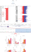
Family 1 pedigree with Sanger sequencing data of causative SNV in DEGS1 gene (a); results of custom CGH array from Cytogenomics software indicating the deletion of DEGS1 gene (b); Sanger sequencing data of breakpoints in DEGS1 gene (c). Ratio of unsaturated/saturated sphingolipids (Cer/DHCer and SM/DHSM) determined in the serum of Proband 1, his mother, two age-matched healthy controls (CTR C), and five healthy adult controls (CTR A), as determined by LC-MS/MS; statistical analysis was performed using one-way ANOVA coupled to Bonferroni post hoc test for multiple comparison (d).
Figure 4
Figure 4
3D modeling of the wild-type (a) and mutant (b) DEGS1 protein using the DynaMut2 online tool. Black arrows indicate the loss of physiological hydrophobic bonds between Asn255 and the aromatic ring of Tyr258 (green dotted line) as well as the newly formed polar bond between Ser255 and Leu273 (orange dotted line), due to amino acid substitution. These intramolecular rearrangements alter the proper folding of the enzyme and may negatively impact its function.
Figure 5
Figure 5
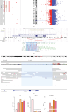
Family 2 pedigree with Sanger sequencing data of causative SNV in DEGS1 gene (a). Results of custom CGH array from Cytogenomics software indicating the deletion of DEGS1 gene (b). Sanger sequencing data of breakpoints in DEGS1 gene (c). IGV screen of LR-WGS by ONT, showing heterozygous deletion of DEGS1 (d). Ratio of unsaturated/saturated sphingolipids (Cer/DHCer and SM/DHSM) determined in RM1073 proband, her mother and father, and two age-matched healthy controls (CTR C) and five healthy adult controls (CTR A). Statistical analysis was performed using one-way ANOVA coupled with Bonferroni post hoc test for multiple comparison (e).
Figure 6
Figure 6
Graphical representation of variants in DEGS1 gene by ProteinPaint (https://proteinpaint.stjude.org/) [14, 15]. In red are reported the already published family variants; in light blue are patient variants. Evolutionary conservation sites are reported for the two missense variants identified in the two cases reported here.

References

    1. Torii T., Yamauchi J. Molecular Pathogenic Mechanisms of Hypomyelinating Leukodystrophies (HLDs) Neurology International . 2023;15(3):1155–1173. doi: 10.3390/neurolint15030072. - DOI - PMC - PubMed
    1. Yan H., Ji H., Kubisiak T., et al. Genetic Analysis of 20 Patients With Hypomyelinating Leukodystrophy by Trio-Based Whole-Exome Sequencing. Journal of Human Genetics . 2021;66(8):761–768. doi: 10.1038/s10038-020-00896-5. - DOI - PMC - PubMed
    1. Karsai G., Kraft F., Haag N., et al. DEGS1-Associated Aberrant Sphingolipid Metabolism Impairs Nervous System Function in Humans. Journal of Clinical Investigation . 2019;129(3):1229–1239. doi: 10.1172/JCI124159. - DOI - PMC - PubMed
    1. Pant D. C., Dorboz I., Schluter A., et al. Loss of the Sphingolipid Desaturase DEGS1 Causes Hypomyelinating Leukodystrophy. Journal of Clinical Investigation . 2019;129(3):1240–1256. doi: 10.1172/JCI123959. - DOI - PMC - PubMed
    1. Dolgin V., Straussberg R., Xu R., et al. DEGS1 Variant Causes Neurological Disorder. European Journal of Human Genetics . 2019;27(11):1668–1676. doi: 10.1038/s41431-019-0444-z. - DOI - PMC - PubMed

Publication types

LinkOut - more resources