Review
. 2025 Mar 31:6:e14.
doi: 10.1017/qrd.2025.3. eCollection 2025.

Unleashing virus structural biology: Probing protein and membrane intermediates in the dynamic process of membrane fusion

Affiliations
Review

Unleashing virus structural biology: Probing protein and membrane intermediates in the dynamic process of membrane fusion

Kelly K Lee. QRB Discov. .

Abstract

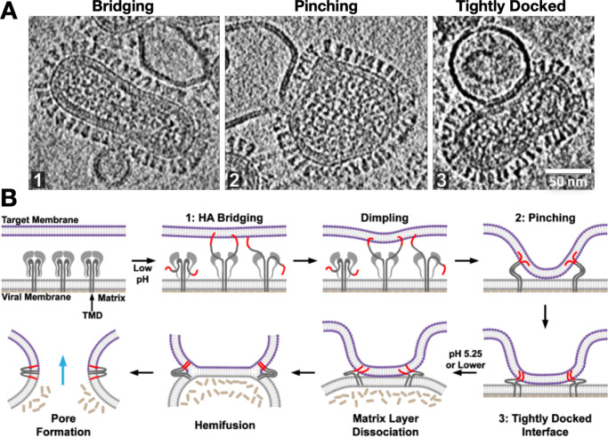
Viruses are highly dynamic macromolecular assemblies. They undergo large-scale changes in structure and organization at nearly every stage of their infectious cycles from virion assembly to maturation, receptor docking, cell entry, uncoating and genome delivery. Understanding structural transformations and dynamics across the virus infectious cycle is an expansive area for research that that can also provide insight into mechanisms for blocking infection, replication, and transmission. Additionally, the processes viruses carry out serve as excellent model systems for analogous cellular processes, but in more accessible form. Capturing and analyzing these dynamic events poses a major challenge for many structural biological approaches due to the size and complexity of the assemblies and the heterogeneity and transience of the functional states that are populated. Here we examine the process of protein-mediated membrane fusion, which is carried out by specialized machinery on enveloped virus surfaces leading to delivery of the viral genome. Application of two complementary methods, cryo-electron tomography and structural mass spectrometry enable dynamic intermediate states in intact fusion systems to be imaged and probed, providing a new understanding of the mechanisms and machinery that drive this fundamental biological process.

Keywords: CryoEM structural biology; dynamics and function; integrative structural biology; membranes; virology.


Conflict of interest statement

The author declares that he has no competing financial interests.

Figures

Figure 1.
Figure 1.
Viruses undergo dramatic conformation changes throughout their infectious cycles. (a) One example of this is seen in bacteriophage capsid maturation, where hundreds of protein subunits organized in an icosahedral shell reorganize while maintaining capsid integrity to produce an expanded mature capsid with thinner but more stable walls—due to greater inter-subunit contacts and fewer holes in the lattice—and greatly increased internal volume. A cross-section of HK97 bacteriophage is shown. Figure adapted from Wikoff et al. (2006)). This structural transformation is highly cooperative and is highly reminiscent of the expansion of (b) a mechanical Hoberman sphere, a “reversibly expandable, double-curved truss structure” Figure adapted from USA Patent US4942700A (Hoberman, 1988).
Figure 2.
Figure 2.
A common pathway has been proposed for protein-mediated membrane fusion leading to fusion pore formation via a “hemifused” intermediate.
Figure 3.
Figure 3.
Cryo-electron tomography reveals virus ultrastructure with resolution of individual fusion glycoproteins (blue arrows), membrane organization, and internal viral contents. (a) Central section for influenza A virus, (b) paraformaldehyde fixed SARS-CoV-2, and (c) HIV-1 virus particles. The differences in fusion protein abundance and distribution are clearly evident. (cryo-ET by Drs. Long Gui, Nancy Hom, and Vidya Mangala Prasad).
Figure 4.
Figure 4.
Advances in resolution of influenza virus-membrane interactions. (a) Some of the earliest glimpses of influenza’s membrane fusion event were captured for the virus inside of endosomes using heavy metal-stained, thin-section TEM of fixed cells (Matlin et al., 1981). The pathway of virus entry could be tracked, but direct resolution of protein and membranes and their structures was not possible with this approach. With the application of cryo-ET, a virus interacting with liposomes in vitro could be visualized under native buffer conditions in vitrified ice. Over the past several years, technical advances have increased resolution and data quality as is evident from the sharper images of proteins as well as membrane fine structure including resolution of leaflets in cryo-electron tomograms. For example (b) shows a central section through a tomogram of influenza virus interacting with a liposome that was collected using a cryo-EM configuration with a side-entry sample holder and charge-coupled device (CCD) (Lee, 2010), while (c) shows a tomogram collected on a contemporary microscope with a more sensitive, fast frame readout direct electron detector, an energy filter to filter out inelastically scattered electrons, and a more stable specimen stage (cryo-ET image provided by Dr. Vidya Mangala Prasad).
Figure 5.
Figure 5.
Snapshots and inferred pathway of influenza virus HA-mediated membrane fusion with liposomes visualized with cryo-ET. (a) Several intermediate membrane remodeling intermediates can be captured by initiating fusion reactions in vitro and flash-freezing the reaction (Figure adapted from (Benhaim and Lee, 2020), original data in (Gui et al., 2016)). (b) By carrying out a time course and monitoring the population kinetics of these intermediates, the sequence and pathway for HA-mediated fusion were inferred. These experiments highlighted the centrality of the tightly docked membranes following target membrane apposition to the virus surface through localized dimples but did not capture classical hemifused “stalks.” Cryo-ET also demonstrated that the internal matrix structural layer works in concert with HA to mediate fusion.
Figure 6.
Figure 6.
Fusion proteins in native contexts, such as on whole virions, function differently than isolated, soluble ectodomains that are typically the subject of structural studies. We used pulse deuteration HDX-MS to monitor activation and conformational change of influenza HA refolding pathway (a) on intact influenza virus (b) revealing the existence of a transient but highly populated dynamic intermediate state with exposed fusion peptides and dynamic B-loop motifs, in contrast to soluble HA ectodomain (c) that converted directly from pre- to post-fusion states (Benhaim et al., 2020). Pulse deuteration HDX-MS is thus a powerful means to take snapshots as the proteins traverse their pathways of conformational change and track the states of each peptide segment throughout the protein populates over time (insets on the right).

Similar articles

References

    1. Benhaim MA and Lee KK (2020) New biophysical approaches reveal the dynamics and mechanics of type i viral fusion machinery and their interplay with membranes. Viruses 12(4). 10.3390/v12040413. - DOI - PMC - PubMed
    1. Benhaim MA, Mangala Prasad V, Garcia NK, Guttman M and Lee KK (2020) Structural monitoring of a transient intermediate in the hemagglutinin fusion machinery on influenza virions. Science Advances 6(18), eaaz8822. 10.1126/sciadv.aaz8822. - DOI - PMC - PubMed
    1. Blijleven JS, Boonstra S, Onck PR, van der Giessen E and van Oijen AM (2016) Mechanisms of influenza viral membrane fusion. Seminars in Cell and Developmental Biology 60, 78–88. 10.1016/j.semcdb.2016.07.007. - DOI - PubMed
    1. Bond C, Santiago-Ruiz AN, Tang Q and Lakadamyali M (2022) Technological advances in super-resolution microscopy to study cellular processes. Molecular Cell 82(2), 315–332. 10.1016/j.molcel.2021.12.022. - DOI - PMC - PubMed
    1. Braet SM, Buckley TSC, Venkatakrishnan V, Dam KA, Bjorkman PJ and Anand GS (2023) Timeline of changes in spike conformational dynamics in emergent SARS-CoV-2 variants reveal progressive stabilization of trimer stalk with altered NTD dynamics. Elife 12, 1–21. 10.7554/eLife.82584. - DOI - PMC - PubMed

LinkOut - more resources