. 2025 Apr 30:16:1535352.
doi: 10.3389/fphar.2025.1535352. eCollection 2025.

Huangkui capsules regulate tryptophan metabolism to improve diabetic nephropathy through the Keap1/Nrf2/HO-1 pathway

Affiliations

Huangkui capsules regulate tryptophan metabolism to improve diabetic nephropathy through the Keap1/Nrf2/HO-1 pathway

Jiayu Su et al. Front Pharmacol. .

Abstract

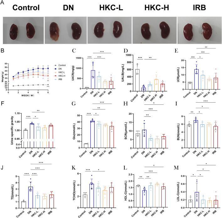
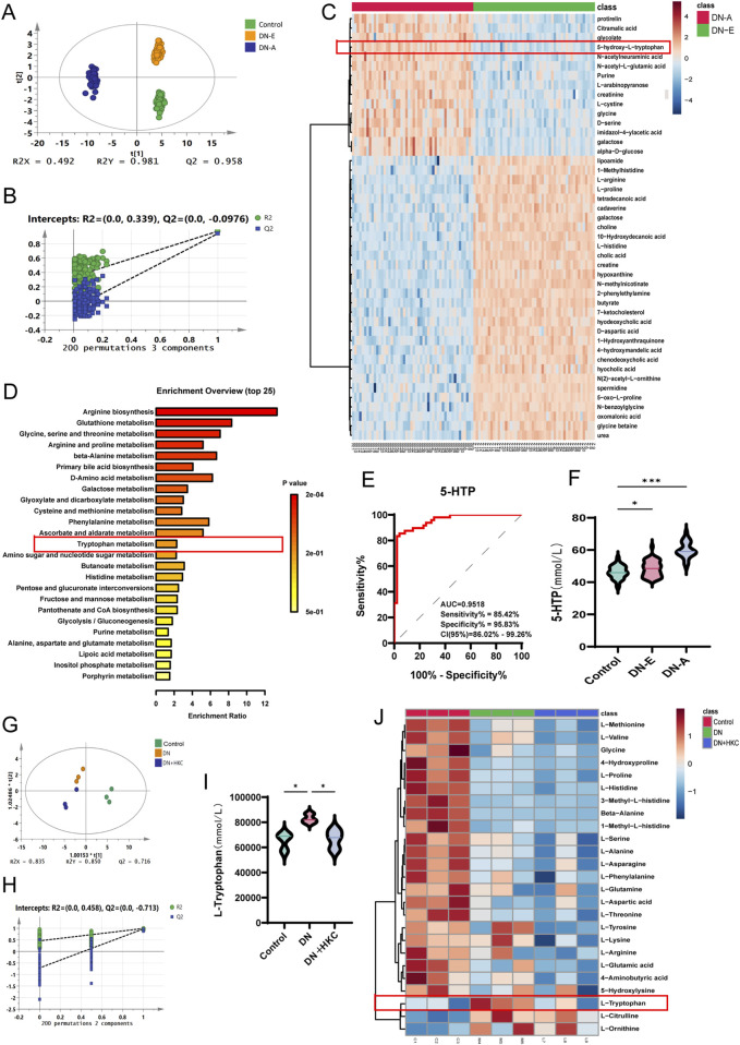
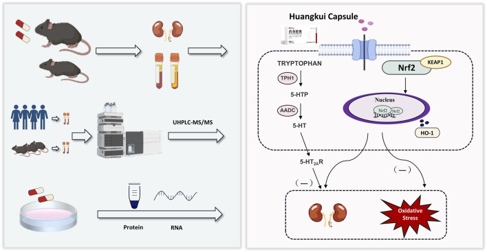
Background: Diabetic nephropathy (DN) is a serious complication of diabetes and one of the leading causes of end-stage renal disease. Huangkui capsule (HKC), a traditional Chinese patent medicine, is widely used in clinical practice for the treatment of chronic glomerulonephritis. However, the therapeutic effects and underlying mechanisms of HKC in DN remain poorly understood.

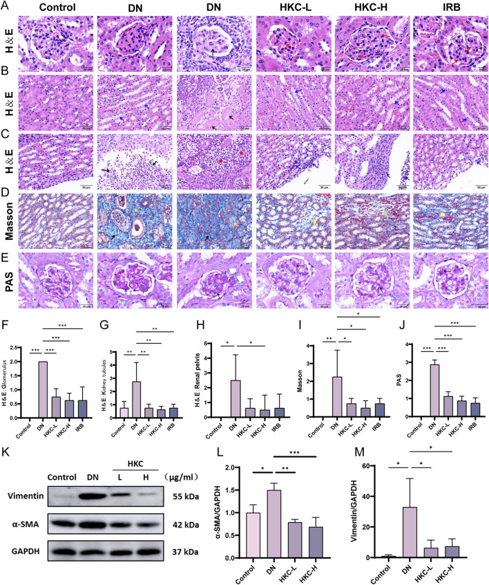
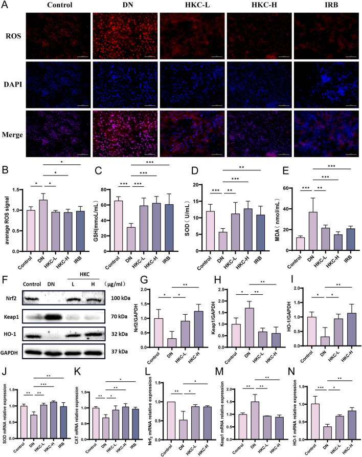
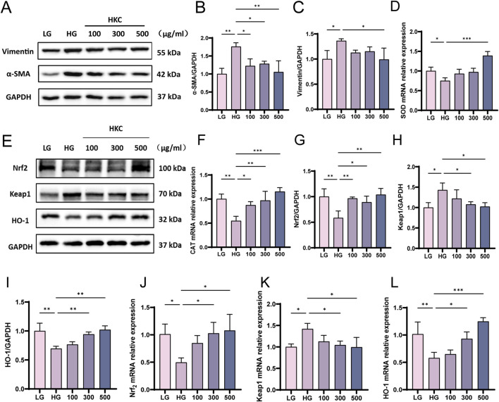
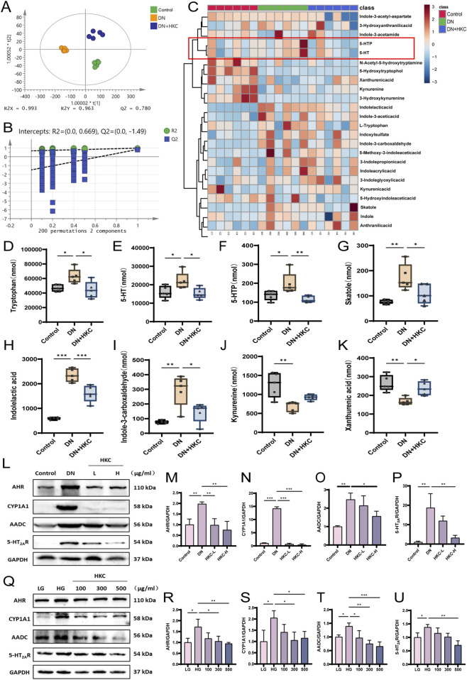
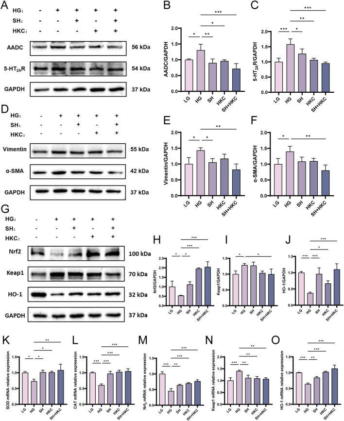
Methods: DN was induced in db/db mice, which were randomly divided into the DN, HKC-L, HKC-H and IRB groups, and db/m mice served as the Control group. Biochemical indices of blood and urine samples from the mice were measured, and HE staining, Masson staining and PAS staining were used to verify the anti-DN effect of HKC. The levels of ROS and the expression of Nrf2 pathway-related proteins and mRNAs were detected. Metabonomic analysis was used to investigate the role of tryptophan metabolism in the regulation of DN by HKC. HK-2 cells were used to establish a model of high-glucose (HG) injury in vitro, and HKC treatment was given for supplementary verification. Sarpogrelate hydrochloride (SH) combined with HKC, a 5-HT2AR inhibitor, was used to verify the effect of the 5-HT pathway in an in vitro model.

Results: Treatment with HKC significantly inhibited the increase in blood glucose and Urinary albumin/creatinine ratio (UACR), improved kidney injury signs in mice, reduced the level of ROS and improved oxidative stress injury through the Keap1/ Nrf2/HO-1 pathway. Metabonomic analysis revealed that tryptophan metabolism is involved in the process by which HKC improves DN, and HKC can regulate the 5-HT pathway to improve the renal injury by oxidative stress regulation. HKC treatment also significantly improved the renal and oxidative stress injuries in HG HK-2 cell model through the Nrf2 pathway in vitro. SH administration revealed that inhibiting 5-HT2AR could significantly inhibit the synthesis of 5-HT and improve the renal injury induced by HG.

Conclusion: Our study demonstrate that HKC can inhibit kidney injury and oxidative stress injury in db/db mice and HK-2 cells by regulating tryptophan metabolism and the Keap1/Nrf2/HO-1 pathway, which provides new insight for the clinical use of HKC for treatment of DN.

Keywords: Keap1/ Nrf2/HO-1 pathway; diabetic nephropathy; huangkui capsules; oxidative stress; tryptophan metabolism.


Conflict of interest statement

The authors declare that the research was conducted in the absence of any commercial or financial relationships that could be construed as a potential conflict of interest.

Figures

FIGURE 1
FIGURE 1
Effects of HKC on DN mice and analysis of blood and urine biochemical indexes. (A) Kidney morphology changes in mice; (B) Body weight changes in mice administered for 8 weeks; (C) Urinary albumin/creatinine ratio (ACR) level in urine; (D) Urinary albumin (UALB) level in urine; (E) Urinary creatinine (UCR) level in urine; (F) Specific gravity (SG) level in urine; (G) Blood glucose (Glu) level; (H) Serum creatinine (SCR) level; (I) Serum urea nitrogen (BUN) level; (J) Serum triglyceride (TG) level; (K) serum total cholesterol (TC) level; (L) serum high-density lipoprotein (HDL-C) level; (M) low-density lipoprotein (LDL-C) level. (*P < 0.05, **P < 0.01, ***P < 0.001, n = 6).
FIGURE 2
FIGURE 2
HKC ameliorates renal pathological injury in DN model mice. (A) HE staining to verify glomerular injury; (B) HE staining to verify renal tubular injury; (C) HE staining was used to verify the inflammatory injury in the renal pelvis; (D) Masson staining to verify glomerular injury; (E) PAS staining to verify glomerular basement membrane thickening; (F) Pathological score of glomerular injury (n = 4); (G) Pathological score of renal tubular injury (n = 4); (H) Pathological score of inflammatory injury in renal pelvis (n = 4); (I) Pathological score of glomerular injury (n = 4); (J) Pathological score of glomerular basement membrane thickening (n = 4); (K) Western blot analysis of α-SMA and Vimentin in mouse kidney; (L) Quantitative analysis of α-SMA (n = 3); (M) Quantitative analysis of Vimentin (n = 3). (*P < 0.05, **P < 0.01, ***P < 0.001).
FIGURE 3
FIGURE 3
HKC ameliorates DN by regulating oxidative stress through Keap1/Nrf2/HO-1 pathway in vivo. (A) Fluorescence probe to detect the level of ROS in mouse kidney; (B) Quantitative analysis of ROS level in mouse kidney (n = 4); (C) GSH content in serum of mice (n = 4); (D) SOD content in serum of mice (n = 4); (E) MDA content in serum of mice (n = 4); (F) Western blot analysis of Nrf2, Keap1 and HO-1 in mouse kidney; (G) Quantitative analysis of Nrf2; (H) Quantitative analysis of Keap1; (I) Quantitative analysis of HO-1; (J) SOD mRNA expression in mouse kidney; (K) CAT mRNA expression in mouse kidney; (L) Nrf2 mRNA expression in mouse kidney; (M) Keap1 mRNA expression in mouse kidney; (N) HO-1 mRNA expression in mouse kidney. (*P < 0.05, **P < 0.01, ***P < 0.001, n = 3).
FIGURE 4
FIGURE 4
HKC ameliorates DN by regulating oxidative stress through Keap1/ Nrf2/HO-1 pathway in vitro. (A) Western blot analysis of α-SMA and Vimentin in HK-2 cells; (B) Quantitative analysis of α-SMA; (C) Quantitative analysis of Vimentin; (D) SOD mRNA expression in HK-2 cells. (E) Western blot analysis of Nrf2, Keap1 and HO-1 in HK-2 cells; (F) CAT mRNA expression in HK-2 cells. (G) Quantitative analysis of Nrf2 protein expression; (H) Quantitative analysis of Keap1 protein expression; (I) Quantitative analysis of HO-1 protein expression; (J) Nrf2 mRNA expression in HK-2 cells; (K) Keap1 mRNA expression in HK-2 cells; (L) HO-1 mRNA expression in HK-2 cells. (*P < 0.05, **P < 0.01, ***P < 0.001, n = 3).
FIGURE 5
FIGURE 5
Targeted amino acid metabolomics analysis of clinical patient serum and non-targeted metabolomics analysis of mouse serum. (A) PLS-DA analysis of clinical non-targeted metabolomics (n = 50); (B) Permutation analysis of clinical non-targeted metabolomics; (C) heatmap analysis of clinical non-targeted metabolomics; (D) pathway enrichment analysis of clinical non-targeted metabolomics; (E) Receiver operating characteristic curve of 5-HTP in serum of clinical patients; (F) The level of 5-HTP in serum of clinical patients, (G) PLS-DA analysis of targeted metabolomics in mice (n = 3); (H) Permutation analysis of targeted metabolomics in mice; (I) L-tryptophan content in mouse serum; (J) Heatmap analysis of targeted metabolomics in mice.
FIGURE 6
FIGURE 6
Targeted tryptophan metabolomics analysis of mouse serum and Western blotting verification of tryptophan metabolite-related protein expression in vivo and in vitro. (A) PLS-DA analysis of targeted tryptophan metabolomics in mice (n = 5); (B) Permutation analysis of targeted tryptophan metabolomics in mice; (C) Heatmap analysis of targeted metabolomics in mice; (D) Tryptophan content level; (E) 5-HT content level; (F) 5-HTP content level; (G) Skatole content level; (H) Indolelactic acid content level; (I) Indlole-3-carboxaldehyde content level; (J) Kynureninee content level; (K) Xanthurenic content level; (L) Western blot analysis of AHR, CYP1A1, AADC and 5-HT2AR in mouse kidney. (M) Quantitative analysis of AHR in mice; (N) Quantitative analysis of CYP1A1 in mice; (O) Quantitative analysis of AADC in mice; (P) Quantitative analysis of 5-HT2AR in mice; (Q) Western blot analysis of AHR, CYP1A1, AADC and 5-HT2AR protein in cells; (R) Quantitative analysis of AHR in cells; (S) Quantitative analysis of CYP1A1 in cells; (T) Quantitative analysis of AADC in cells; (U) Quantitative analysis of 5-HT2AR in cells. (*P < 0.05, **P < 0.01, ***P < 0.001, n = 3).
FIGURE 7
FIGURE 7
Sarpogrelate hydrochloride inhibits the pharmacodynamic effect of 5-HT in high glucose HK-2 Cells in vitro. (A) Western blot analysis of AADC and 5-HT2AR in HK-2 cells; (B) Quantitative analysis of AADC in cells; (C) Quantitative analysis of 5-HT2AR in cells; (D) Western blot analysis of α-SMA and Vimentin in HK-2 cells; (E) Quantitative analysis of α-SMA in cells; (F) Quantitative analysis of Vimentin in cells; (G) Western blot analysis of Nrf2, Keap1 and HO-1 in HK-2 cells; (H) Nrf2 quantitative analysis; (I) Quantitative analysis of Keap1 in cells; (J) HO-1 quantitative analysis; (K) SOD mRNA expression; (L) CAT mRNA expression; (M) Nrf2 mRNA expression; (N) Keap1 mRNA expression; (O) HO-1 mRNA expression. (*P < 0.05, **P < 0.01, ***P < 0.001, n = 3).
FIGURE 8
FIGURE 8
Workflow of the present study. We validated the effect of HKC on DN in animal and cellular models, and in combination with metabolomics found that HKC could regulate tryptophan metabolism and Keap1/Nrf2/HO-1 pathway to ameliorate renal fibrosis injury.

Similar articles

References

    1. Albvr V. R., Tan S. H., Candasamy M., Bhattamisra S. K. (2019). Diabetic nephropathy: an update on pathogenesis and drug development. Diabetes Metab. Syndr. 13 (1), 754–762. 10.1016/j.dsx.2018.11.054 - DOI - PubMed
    1. An C., Jiao B., Du H., Tran M., Zhou D., Wang Y. (2022). Myeloid PTEN deficiency aggravates renal inflammation and fibrosis in angiotensin II-induced hypertension. J. Cell Physiol. 237 (1), 983–991. 10.1002/jcp.30574 - DOI - PMC - PubMed
    1. Anders H. J., Huber T. B., Isermann B., Schiffer M. (2018). CKD in diabetes: diabetic kidney disease versus nondiabetic kidney disease. Nat. Rev. Nephrol. 14 (6), 361–377. 10.1038/s41581-018-0001-y - DOI - PubMed
    1. Arellano-Buendía A. S., Tostado-González M., García-Arroyo F. E., Cristóbal-García M., Loredo-Mendoza M. L., Tapia E., et al. (2016). Anti-inflammatory therapy modulates nrf2-keap1 in kidney from rats with diabetes. Oxid. Med. Cell Longev. 2016, 4693801. 10.1155/2016/4693801 - DOI - PMC - PubMed
    1. Behl T., Kaur I., Sehgal A., Sharma E., Kumar A., Grover M., et al. (2021). Unfolding Nrf2 in diabetes mellitus. Mol. Biol. Rep. 48 (1), 927–939. 10.1007/s11033-020-06081-3 - DOI - PubMed

LinkOut - more resources