. 2025 Apr 30:16:1577942.
doi: 10.3389/fphar.2025.1577942. eCollection 2025.

Molecular pharmacokinetic mechanism of JBP485 against aristolochic acid I (AAI) -induced nephrotoxicity

Affiliations

Molecular pharmacokinetic mechanism of JBP485 against aristolochic acid I (AAI) -induced nephrotoxicity

Chong Wang et al. Front Pharmacol. .

Abstract

Introduction: In this study, we investigated the protective effect of JBP485 against aristolochic acid I (AAI)-induced nephrotoxicity and explored the pharmacokinetic mechanisms. The effects of JBP485 on AAI-induced cytotoxicity and nephrotoxicity were evaluated in vitro and in vivo, respectively.

Methods: To ascertain the protective effect of JBP485 against AAI-induced nephrotoxicity, we measured levels of urea nitrogen (BUN), creatinine (CRE), and indoxol sulfate in blood and urine; determined kidney weight-to-body weight ratio; and performed hematoxylin and eosin (H&E) staining. Cell viability and Western blotting assays, along with determination of malondialdehyde (MDA), superoxide dismutase (SOD), and intracellular reactive oxygen species (ROS) contents, were carried out to explore mechanisms underlying the protective effects of JBP485 against AAI-induced nephrotoxicity.

Results: JBP485 treatment attenuated AAI-induced injuries in rat kidney while decreasing the levels of indoxyl sulfate, CRE, and BUN in plasma and increasing those of indoxyl sulfate in urine compared to that in AAI alone-treated group. The co-administration of JBP485 with AAI significantly increased the concentration and AUC of AAI in plasma, while decreasing its cumulative urinary excretion and renal clearance. Moreover, JBP485 reduced the uptake of AAI in kidney slices and human organic anion transporter 1/3 (hOAT1/3)-transfected human embryonic kidney 293 (HEK293) cells, suggesting that JBP485 ameliorated AAI-induced nephrotoxicity by reducing renal exposure to AAI via OAT inhibition. Meanwhile, JBP485 modulated the abnormal expressions of Oat1, Oat3, organic cation transporter 2 (Oct2), P-glycoprotein (P-gp), multidrug resistance-associated protein 2 (Mrp2) and multidrug and toxin extrusion proteins 1 (Mate 1) in rat kidney, suggesting that JBP485 improved tubular secretion in AAI-treated rats. Moreover, JBP485 reversed the AAI-induced changes in the expression of heme oxygenase 1 (HO-1), NAD(P) H: quinone oxidoreductase-1 (NQO1), B-cell lymphoma-2 (Bcl-2) protein expressions and Bcl-2-like protein 4 (Bax) induced by AAI in rat kidney. JBP485 increased cell viability and reduced intracellular levels of ROS in NRK-52E cells treated with AAI.

Discussion: These results suggested that JBP485 protected against AAI-induced renal oxidative stress. All results indicated that JBP485 protected against AAI-induced nephrotoxicity by reducing renal exposure to AAI and alleviating oxidative stress. Our findings suggested that JBP485 has potential as a renoprotective agent for the prevention of AAI-induced nephrotoxicity.

Keywords: DDI; JBP485; OATs; aristolochic acid I; nephrotoxicity.


Conflict of interest statement

Author TK was employed by Japan Bioproducts Industry Co., Ltd. The remaining authors declare that the research was conducted in the absence of any commercial or financial relationships that could be construed as a potential conflict of interest.

Figures

FIGURE 1
FIGURE 1
JBP485 improved the morphology and pathology condition of the kidneys in rats. The changes in morphology (A), histopathology (B) and the kidney weight-to-body weight ratio (C) after co-administration of JBP485. Glomerular shrinkage (red arrows), tubule dilatation (green arrows), renal tubular epithelial cell deformation and vacuolation (blue arrows) and necrosis and shedding of cell (yellow arrows). Data are presented as mean ± S.D. **p < 0.01 compared with control group; ##p < 0.01 compared with AAI alone-treated group (n = 5).
FIGURE 2
FIGURE 2
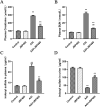
Protective effect of JBP485 on AAI nephrotoxicity in vivo. Blood and urine samples were collected for the determination of CRE (A), BUN (B), plasma (C) and urine indoxyl sulfate levels (D). Data are presented as mean ± S.D. **p < 0.01 compared with control group; ##p < 0.01 compared with AAI alone-treated group (n = 5).
FIGURE 3
FIGURE 3
JBP485 reduced AAI cytotoxicity on NRK-52E cells. (A) AAI toxicity in NRK-52E cells. (B) JBP485 toxicity in NRK-52E cells. (C) Protective effect of JBP485 on AAI cytotoxicity on NRK-52E cells. Data are presented as mean ± S.D. &&&p < 0.001 compared with control group; *p < 0.05, **p < 0.01 and ***p < 0.001 compared with AAI alone-treated group (n = 3).
FIGURE 4
FIGURE 4
Reduction of oxidative stress in kidney tissue by JBP485. Changes in renal MDA levels (A), renal SOD levels (B), HO-1 and NQO1 protein expression (C) and statistical analysis of them Western blot images (D, E). Data are presented as mean ± S.D. *p < 0.05, **p < 0.01 compared with control group; #p < 0.05, ##p < 0.01 compared with AAI alone-treated group (n = 5).
FIGURE 5
FIGURE 5
JBP485 reduced oxidative stress induced by AAI in NRK-52E cells. Control group (A), JBP485 alone group (B), AAI alone group (C), AAI+JBP485 group (D) (n = 3).
FIGURE 6
FIGURE 6
Inhibition of renal apoptosis by JBP485. Bcl-2 and Bax (A) protein expression and statistical analysis of them Western blot images (B, C). Data are presented as mean ± S.D. **p < 0.01 compared with control group; #p < 0.05, ##p < 0.01 compared with AAI alone-treated group (n = 5).
FIGURE 7
FIGURE 7
Effect of JBP485 on the pharmacokinetics of AAI in rats. Mean plasma concentration-time curves (A), cumulative urine excretion curves (B) after intravenous administration of AAI and JBP485 in rats. Data are expressed as the mean ± SD. ∗∗p < 0.01 compared with control (n = 5).
FIGURE 8
FIGURE 8
Effect of JBP485 on the uptake of AAI by rat kidney slices and hOAT1/3-HEK293 cells. Inhibition effect of JBP485 on the uptake of AAI in kidney slices (A). The time-dependent inhibitory effects of JBP485 on AAI uptake in hOAT1/3-HEK293 cells (B and E). The concentration-dependent inhibitory effects of JBP485 on AAI uptakein hOAT1/3-HEK293 cells (C and F). The inhibitory effect of JBP485 on the uptake of AAI in hOAT1/3-HEK293 cells (D and G). Data are expressed as the mean ± SD. p < 0.05 and ∗∗p < 0.01 compared with control (n = 3).
FIGURE 9
FIGURE 9
Effects of JBP485 on Mrp2, P-gp, Mate1, Oat1, Oat3 and Oct2 protein expression in the kidney. Changes on these transporters protein expression (A). Statistical analysis of Western blot images of the above transporter proteins in rats (B–G). Data are presented as mean ± S.D. *p < 0.05, **p < 0.01 compared with control group; #p < 0.05, ##p < 0.01 compared with AAI alone-treated group (n = 5).

Similar articles

References

    1. Aigbe F. R., Sofidiya O. M., James A. B., Sowemimo A. A., Akindere O. K., Aliu M. O., et al. (2019). Evaluation of the toxicity potential of acute and sub-acute exposure to the aqueous root extract of Aristolochia ringens Vahl. (Aristolochiaceae). J. Ethnopharmacol. 244, 112150. 10.1016/j.jep.2019.112150 - DOI - PubMed
    1. Anzai N., Kanai Y., Endou H. (2006). Organic anion transporter family: current knowledge. J. Pharmacol. Sci. 100 (5), 411–426. 10.1254/jphs.crj06006x - DOI - PubMed
    1. Bastek H., Zubel T., Stemmer K., Mangerich A., Beneke S., Dietrich D. R. (2019). Comparison of Aristolochic acid I derived DNA adduct levels in human renal toxicity models. Toxicolog 15, 29–38. 10.1016/j.tox.2019.03.013 - DOI - PubMed
    1. D'Autréaux B., Toledano M. B. (2007). ROS as signalling molecules: mechanisms that generate specificity in ROS homeostasis. Nat. Rev. Mol. Cell. Biol. 8 (10), 813–824. 10.1038/nrm2256 - DOI - PubMed
    1. Deguchi T., Ohtsuki S., Otagiri M., Takanaga H., Asaba H., Mori S., et al. (2002). Major role of organic anion transporter 3 in the transport of indoxyl sulfate in the kidney. Kidney Int. 61 (5), 1760–1768. 10.1046/j.1523-1755.2002.00318.x - DOI - PubMed

LinkOut - more resources