. 2025 Jun:111:19-100.
doi: 10.3114/sim.2025.111.02. Epub 2025 Feb 28.

Camarosporidiella, a challenge

Affiliations

Camarosporidiella, a challenge

W M Jaklitsch et al. Stud Mycol. 2025 Jun.

Abstract

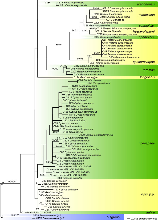
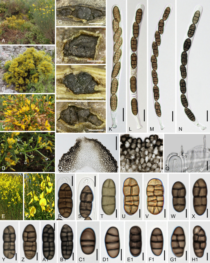
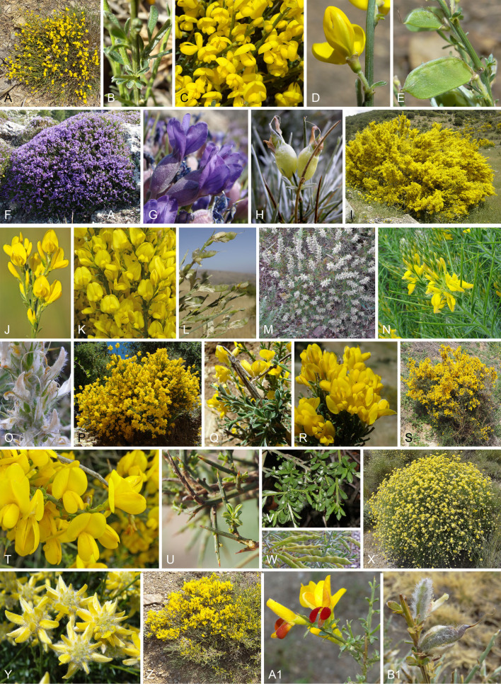
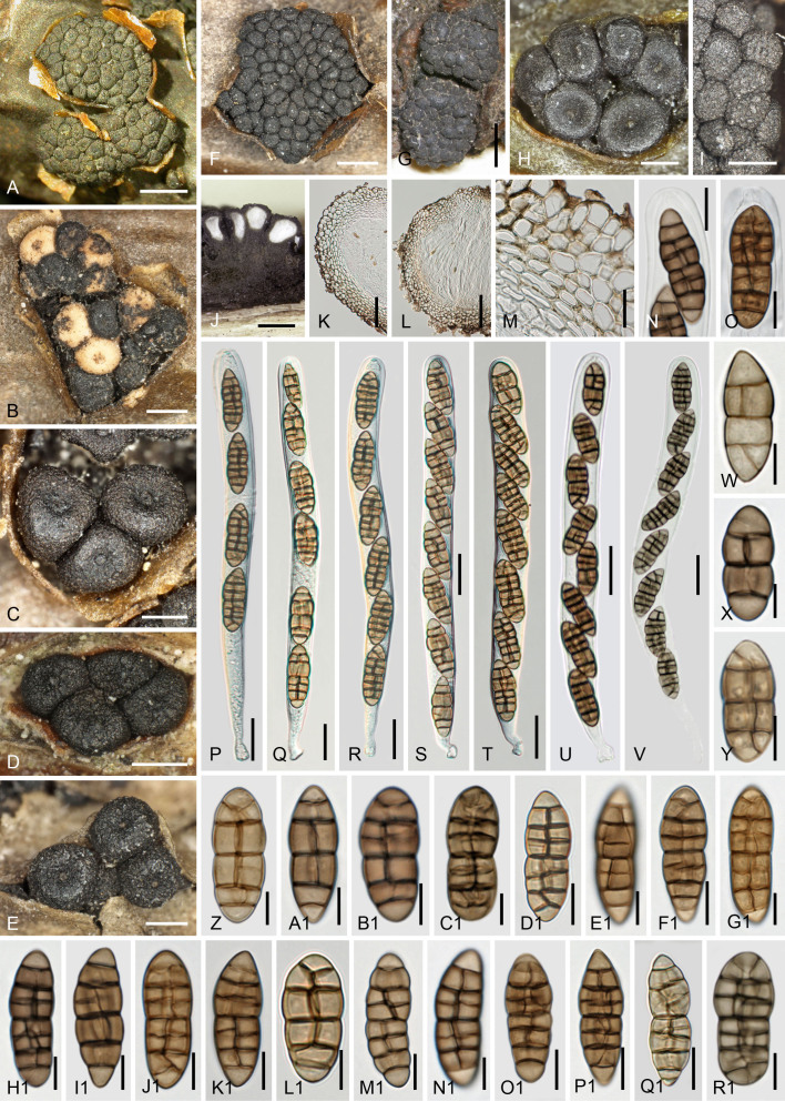
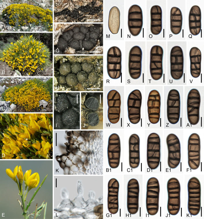
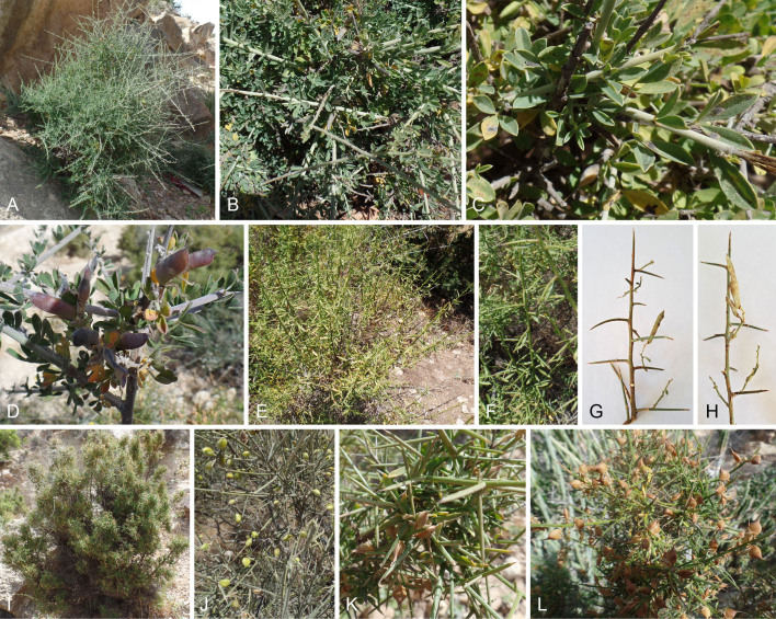
The genus Camarosporidiella is here assessed with respect to its phylogenetic structure and species composition. More than 160 pure cultures from ascospores and conidia of more than 150 fresh collections, mostly from Fabaceae, were prepared as DNA sources. Molecular phylogenetic analyses of a multigene matrix of partial nuSSU-, complete ITS, partial LSU rDNA, and tef1 exon sequences of our isolates and those of previous workers revealed that these markers are insufficient to provide a complete species resolution. From this reduced data matrix, however, we propose synonyms and accept taxa for previously described species, which could not be included in the final phylogenetic tree due to lack of rpb2, tef1 intron and tub2 sequences. The final phylogenetic tree, which was inferred from a combined nuSSU-ITS-LSU-rpb2-tef1-tub2 sequence matrix resolved our isolates into 27 statistically supported phylogenetic species, of which 15 are new. Altogether 34 species are here accepted in Camarosporidiella. Using type studies we stabilise old names, lectotypify Cucurbitaria asparagi, Cucurbitaria caraganae, Cucurbitaria coluteae, Cucurbitaria euonymi, Dichomera elaeagni Hendersonia mori, Sphaeria elongata, Sphaeria laburni Sphaeria spartii and epitypify them as well as Cucurbitaria cytisi, Cucurbitaria retamae and Cucurbitaria steineri to place them in their correct phylogenetic positions and fix their taxonomic concepts. Morphology alone is not suitable to identify these species, and therefore no determinative key to species can be given. However, if hosts are reliably identified, many species can be determined without molecular data. Host images are included with the figures of each fungal species. Taxonomic novelties: New species: Camarosporidiella aceris Jaklitsch & Voglmayr, Camarosporidiella aetnensis Jaklitsch & Voglmayr, Camarosporidiella aragonensis Jaklitsch & Voglmayr, Camarosporidiella asparagicola Jaklitsch & Voglmayr, Camarosporidiella astragalicola Jaklitsch & Voglmayr, Camarosporidiella cretica Jaklitsch & Voglmayr, Camarosporidiella echinosparti Jaklitsch & Voglmayr, Camarosporidiella hesperolaburni Jaklitsch & Voglmayr, Camarosporidiella longipedis Jaklitsch & Voglmayr, Camarosporidiella maroccana Jaklitsch & Voglmayr, Camarosporidiella ononidis Jaklitsch & Voglmayr, Camarosporidiella radiatae Jaklitsch & Voglmayr, Camarosporidiella spartioidis Jaklitsch & Voglmayr, Camarosporidiella sphaerocarpae Jaklitsch & Voglmayr, Camarosporidiella tridentatae Jaklitsch & Voglmayr. New combinations: Camarosporidiella asparagi (Maire) Jaklitsch & Voglmayr, Camarosporidiella caraganae (P. Karst.) Jaklitsch & Voglmayr, Camarosporidiella coluteae (Rabenh.) Jaklitsch & Voglmayr, Camarosporidiella cytisi (Mirza) Jaklitsch & Voglmayr, Camarosporidiella elaeagni (P. Karst.) Jaklitsch & Voglmayr, Camarosporidiella euonymi (Cooke) Jaklitsch & Voglmayr, Camarosporidiella retamae (Pat.) Jaklitsch & Voglmayr, Camarosporidiella steineri (Petr.) Jaklitsch & Voglmayr. New names: Camarosporidiella neomori Jaklitsch & Voglmayr, Camarosporidiella neospartii Jaklitsch & Voglmayr. Citation: Jaklitsch WM, Blanco MN, Rejos FJ, Tello S, Voglmayr H (2025). Camarosporidiella, a challenge. Studies in Mycology 111: 19-100. doi: 10.3114/sim.2025.111.02.

Keywords: Camarosporium; Cucurbitaria; Pleosporales; new taxa; taxonomy.


Figures

Fig. 1
Fig. 1
Phylogram of the best ML tree (-lnL = 7503.2187) revealed by RAxML from an analysis of the combined nSSU-ITS-LSU rDNA-tef1 matrix of Camarosporidiella, with two accessions of Staurosphaeria lycii as outgroup. Strain/culture numbers of sequences from GenBank are given following the taxon names; in the isolates of the current study the strain numbers are given first, followed by the hosts. ML and MP bootstrap support above 50 % are given at the first and second position, respectively, above or below the branches.
Fig. 1
Fig. 1
Phylogram of the best ML tree (-lnL = 7503.2187) revealed by RAxML from an analysis of the combined nSSU-ITS-LSU rDNA-tef1 matrix of Camarosporidiella, with two accessions of Staurosphaeria lycii as outgroup. Strain/culture numbers of sequences from GenBank are given following the taxon names; in the isolates of the current study the strain numbers are given first, followed by the hosts. ML and MP bootstrap support above 50 % are given at the first and second position, respectively, above or below the branches.
Fig. 2
Fig. 2
Phylogram of the best ML tree (-lnL = 24640.577) revealed by RAxML from an analysis of the combined nSSU-ITS-LSU rDNA-rpb2-tef1-tub2 matrix of Camarosporidiella, with two accessions of Staurosphaeria lycii as outgroup. Strain numbers are given first, followed by the hosts. ML and MP bootstrap support above 50 % are given at the first and second position, respectively, above or below the branches.
Fig. 2
Fig. 2
Phylogram of the best ML tree (-lnL = 24640.577) revealed by RAxML from an analysis of the combined nSSU-ITS-LSU rDNA-rpb2-tef1-tub2 matrix of Camarosporidiella, with two accessions of Staurosphaeria lycii as outgroup. Strain numbers are given first, followed by the hosts. ML and MP bootstrap support above 50 % are given at the first and second position, respectively, above or below the branches.
Fig. 3
Fig. 3
Camarosporidiella aceris (WU-MYC0037940). A, B. Host Acer campestre. C. Ascoma and two pycnidia in face view. D. Pycnidia in face view. E–G. Asci. H, I. Ascus apices (young in H). J–Q. Ascospores. R–V. Conidia. E–I, P, Q in 3 % KOH. Scale bars: C, D = 300 μm; E–G = 20 μm; H, L, P, Q = 10 μm; I–K, M–O, R–V = 7 μm.
Fig. 4
Fig. 4
Camarosporidiella aetnensis. A–E. Host Genista aetnensis (A. Tree; B. Tree with Spartium junceum in the foreground; C, D. Flower; E. Senescent open fruits). F, G. Ascomata in face view. H, I. Pycnidia (H. Face view; I. Cross section, white spots are ascoma initials). J. Ascoma in vertical section. K. Peridial cells in vertical section. L, M. Ascus apices. N–P. Asci. Q–G1. Ascospores (young in Q). H1–P1. Conidia (young in H1, I1). J–Q, T, V–X, F1, G1 in 3 % KOH. F, G, L, N, Q, T, V, W, G1, H1, K1. WU-MYC0037941; H–K, M, O, P, R, S, U, X–F1, I1, J1, L1–P1. WU-MYC0037942. Scale bars: F–I = 300 µm; J = 100 µm; K, N–P = 20 µm; L, M, Q–S, V–C1, E1, F1, G1 = 7 µm; T, U, H1–P1 = 5 µm; D1 = 10 µm.
Fig. 5
Fig. 5
Camarosporidiella aragonensis. A–D. Host Ononis aragonensis (A. Flowering shrub; B–D. Stems, flowers, leaves). E–G. Ascomata in face view (mixed with pycnidia in E and F). H. Pycnidia in face view. I. Peridial cells in vertical section. J–M. Asci (immature in J). N, O. Ascus apices. P–C1. Ascospores. D1–K1. Conidia. I–O, A1–C1 in 3 % KOH. E, H–O, Q–V, Y–B1, E1–K1. WU-MYC0037944; F, G, P, W, X, C1, D1. WU-MYC0037943. Scale bars: E–H = 500 µm; I–M = 20 µm; N, O, R–U, W, Z = 10 µm; P, Q, V, X, Y, A1–K1 = 7 µm.
Fig. 6
Fig. 6
Camarosporidiella asparagi. A–C. Host Asparagus albus. D–F. Ascomata in face view. G. Ascus apex. H–J. Asci. K, L. Vertical sections through ascomata. M. Lateral part of peridium in vertical section. N–V. Ascospores (young in N). G–J, W–F1 in 3 % KOH. D, E, G–K, M, N–T. WU-MYC0037945; F, L, U, V. WU-MYC0037946; W–F1. Lectotype MPU 886531. Scale bars: D = 1 mm; E, F = 300 µm; G, N–A1 = 7 µm; H–J = 20 µm; K, L = 100 µm; M = 50 µm; B1–F1 = 5 µm.
Fig. 7
Fig. 7
Camarosporidiella asparagicola. A, B. Host Asparagus acutifolius. C. Peridium in vertical section. D. Pycnidia. E, F. Ascomata in face view. G. Ascus apex. H. Paraphysis tip. I–K. Asci. L–W. Ascospores (young in L). X–B1. Conidia. C, G–K in 3 % KOH. C–H, J, L, N, P, W–B1. WU-MYC0037947; I, K, O, R–V. WU-MYC0037948; M, W. MPU 886530. Scale bars: C, I–K = 20 µm; D, F = 200 µm; E = 500 µm; G = 10 µm; H, M, O–Q, Z–B1 = 5 µm; L, N, R–Y = 7 µm.
Fig. 8
Fig. 8
Camarosporidiella astragalicola. A–C. Host Astragalus angustifolius. D. Ascomata mixed with pycnidia in face view. E. Cluster of partly sectioned pycnidia in face view. F. Ascus apex. G–T. Ascospores (young in G–I). U–D1. Conidia (young in U). G, H, P–T in 3 % KOH. D–T, V, W, Z–D1. WU-MYC0037949; U, X–Z. WU-MYC0037950. Scale bars: D = 300 µm; E = 1 mm; F, G, I–O, S–D1 = 7 µm; H, P–R = 5 µm.
Fig. 9
Fig. 9
Camarosporidiella caraganae. A, B, D. Host Caragana arborescens (A, B. Flowers and leaves; D. Fruits). C, E. Host Caragana halodendron (C. Flowers; E. Fruits). F, G. Vertical sections through ascomata (F. Oblique section). H–K. Ascomata in face view. L. Peridium in face view. M–P. Asci (young and fresh in M). Q. Ascus apex. R–B1. Ascospores (young and fresh in R). C1. Pycnidia. D1–I1. Conidia. N–Q, S, V, W, A1, B1, E1 in 3 % KOH. S. H 6006977; W, E1. Lectotype H 6006980; F, H–O, Q, R, T, V, A1–D1, G1–I1. WU-MYC0037952; G, P, Y, Z, F1. WU-MYC0037953; X, E1. C126; U. WU-MYC0037951. Scale bars: F, C1 = 150 µm; G = 50 µm; H, I = 1 mm; J, K = 350 µm; L, Q, U, Y–B1, D1 = 10 µm; M–P = 20 µm; R–T, V–X, E1 = 5 µm; F1–I1 = 7 µm.
Fig. 10
Fig. 10
Camarosporidiella coluteae. A–E. Hosts. A, C. Colutea arborescens (A, B. Flowers and leaves; C. Fruits). B, D, E. Colutea hispanica (flowers; in E also fruits). F–J. Ascomata in face view (immersed in F; with two central pycnidia in J; re-wetted in G, H). K. Pycnidia in face view. L, M. Upper peridium in vertical section. N–Q. Asci. R–D1. Ascospores. E1–L1. Conidia. L, M, P, Q, V, W, J1–L1 in 3 % KOH. F, J–M, T, G1. WU-MYC0037957; G, H. WU-MYC0037961; I, O, R, E1, H1. WU-MYC0037960; N, S, U–W, F1. WU-MYC0037956; P, Q, X–D1, K1, L1. WU-MYC0037958. I1. WU-MYC0052647. J1. WU-MYC0037959. Scale bars: F, G = 1 mm; H, J = 300 µm; I, K = 500 µm; L = 50 µm; M–Q = 25 µm; R, S, X, Y, E1–H1, J1–L1 = 7 µm; T–W, Z–D1, I1 = 10 µm.
Fig. 11
Fig. 11
Camarosporidiella coluteae. Type materials. A, B, E–V. Sphaeria coluteae (A, E–N. Lectotype HAL 3747; B, O–V. Isotype B 700100392). C, D, W–E1. Holotype of Hendersonia coluteae. A. Erumpent ascomata. B. Immersed pycnidia. C, D. Erumpent pycnidia. E–J. Ascospores. K–E1. Conidia. E–E1 in 3 % KOH. Scale bars: A, B = 500 µm; C = 300 µm; D = 150 µm; E–J, P, Q, T, V = 7 µm; K–O, R, S, U, W–E1 = 5 µm.
Fig. 12
Fig. 12
Camarosporidiella cretica. A–F. Hosts. A. Habitat with Genista acanthoclada and Spartium junceum. B–D. Genista acanthoclada (B. Shrub; C. Flowers, branching; D. Spines). E, F. Spartium junceum (E. Part of shrub; F. Flowers). G–I. Ascomata in face view (with pycnidia in G). J. Pycnidia in face view. K–N. Asci (immature in K). O. Upper peridium in vertical section. P. Peridial cells including upper surface cells. Q. Tips of paraphyses aside an immature ascus. R, S. Ascus apices. T–B1. Ascospores (immature in T; fresh in U, V). C1–H1. Conidia. N–Q, T, V, A1, B1 in 3 % KOH; C1–H1 in 1 % KOH. G, H, J, N, Q, T–V, Y, A1–H1. WU-MYC0037962; I, K, L, O, P, R, W, Z. WU-MYC0037963; M, S, X. WU-MYC0037964. Scale bars: G = 1 mm; H–J = 500 µm; K–N = 15 µm; O = 100 µm; P = 20 µm; Q–S = 7 µm; T–H1 = 5 µm.
Fig. 13
Fig. 13
Camarosporidiella cytisi, hosts. A–E. Cytisus balansae (A. Shrub; B. Branching and leaves; C, D. Flowers; E. Fruits). F–H. Erinacea anthyllis (F. Shrub; G. Flowers; H. Fruits). I–L. Genista cinerea (I. Shrub; J, K. Flowers; L. Fruits). M–O. Genista hirsuta (M. Mature plant; N. Flowers and spines; O. Fruits). P–R. Genista pseudopilosa (P. Shrub; Q. Leaves; R. Flowers). S–W. Genista scorpius (S. Shrub; T. Flowers; U. Spines and leaves; V. Leaves; W. Fruits). X, Y. Genista umbellata (X. Shrub; Y. Flowers). Z–B1. Genista versicolor (Z. Shrub; A1. Flowers; B1. Fruits).
Fig. 14
Fig. 14
Camarosporidiella cytisi. A–C. Ascomata in face view. D. Pycnidia in face view (left one vertically cut). E. Peridial cells in vertical section. F–J. Asci. K, L. Ascus apices. M–F1. Ascospores (immature in M). G1–P1. Conidia (immature in G1). E, W, F1, M1 in 3 % KOH. A, W, M1. Holotype ZT Myc 0067344; B, C, M, Q, R, E1. WU-MYC0037986; E, F, I, L, S, I1. WU-MYC0052599; D, G1, J1. WU-MYC0037987; G, H, V, K1. WU-MYC0037999; J, Y. WU-MYC0037993; K, P, U, Z. WU-MYC0037988; N, X. WU-MYC0037990; O. WU-MYC0052601; T, A1, C1. WU-MYC0037998; B1. WU-MYC0037995; D1, P1. WU-MYC0052600; F1. WU-MYC0037994; H1, N1. WU-MYC0037989; L1. WU-MYC0037992. O1. WU-MYC0037996. Scale bars: A, B, D = 500 µm; C = 300 µm; E–J = 15 µm; K–F1 = 7 µm; G1, J1–P1 = 5 µm, H1, I1 = 3 µm.
Fig. 15
Fig. 15
Camarosporidiella echinosparti. A–F. Host Echinospartum boissieri (A. Shrub; B. Branching and spines; C. Flower and opposite spines; D, E. Flowers; F. Fruits). G, H. Ascomata in face view. I, J. Ascomata and hypostromata (vertically sectioned in J). K. Hypostroma cells. L. Slightly excentric vertical section of peridium with basal elongation. M. Peridial cells. N–R. Asci (immature in P). S, T. Ascus apices (immature in S). U–R1. Ascospores. P–T, E1–R1 in 3 % KOH. G–I, K–P, S, T, V–X, Z–D1, H1, I1, N1, P1, R1. WU-MYC0037965; J, Q. R, U, Y, E1–G1, J1–M1, O1, Q1. WU-MYC0037966. Scale bars: G, H = 300 µm; I, J = 500 µm; K, M–R = 20 µm; L = 200 µm; S, T, V–K1, M1–R1 = 7 µm; U = 5 µm; L1 = 10 µm.
Fig. 16
Fig. 16
Camarosporidiella elaeagni. A–C. Host Elaeagnus angustifolia (A. Branch; B. Flowers and leaves; C. Fruits and leaves). D. Aspect of immersed ascomata in face view. E. Ascomata in face view. F–I. Asci. J. Ascomata in horizontal section. K. Peridium at the ostiole in section. L. Ascus apex. M–V. Ascospores. W–Y. Pycnidia in face view (single pycnidium with apical outgrowths in Y). Z–L1. Conidia (initial stage in Z). F–I, K, L, R–V, E1, H–J1, L in 3 % KOH. D–H, J, K, M, O, P, S, U, C1, F1. WU-MYC0037968; I, L, N, Q, R, T, V, Z, B1, G1. WU-MYC0037972; W. WU-MYC0037967; X, A1, H1. H 6061060; Y, E1, I1, L1. Lectotype H 6061062; D1, K1. WU-MYC0037969; J1, M1. WU-MYC0037970. Scale bars: D, E, J, W, Z = 300 µm; X, Y, A1 = 500 µm; F–I = 20 µm; K = 30 µm; L, T, V = 10 µm; M–S, U, C1, E1, G1, I1–L1 = 7 µm; B1–F1, H1 = 5 µm.
Fig. 17
Fig. 17
Camarosporidiella elongata. Sexual morph. A–F. Hosts. A–D. Robinia pseudoacacia (A. Tree with fruits; B. Flowers and leaves; C. Flowers; D. Fruits). E, F. Amorpha fruticosa (E. Shrub with leaves and flowers; F. Flowers). G. Aspect of superficial ascomata in face view. H–K. Ascomata in face view. L, Q. Peridium in vertical section (excluding ostiole). M–P. Asci. R. Ascus apex. S–H1. Ascospores (young in S). P, F1–H1 in 3 % KOH. G, P, G1, H1. WU-MYC0015369; H. WU-MYC0037981; I, J, N, W. WU-MYC0037977; K, R, S, V, X, Y, A1, B1, D1. WU-MYC0037979; L, M, O, Q, Z. WU-MYC0037976; T, U, F1. WU-MYC0037975; C1. WU-MYC0037983; E1. WU-MYC0037980. Scale bars: G = 1 mm; H, J, K = 500 µm; I = 200 µm; L–P = 20 µm; Q = 50 µm; R, T–V, X, Y, A1, C1, E1, H1 = 10 µm; S, W, Z, B1, D1, F1, G1 = 7 µm.
Fig. 18
Fig. 18
Camarosporidiella elongata. Asexual morph. A. Aspect of superficial pycnidia in face view. B–F. Pycnidia (B, C. Face view; F. Cross section). G–I. Phialides and conidiophores in 3 ٪ KOH. J–X. Conidia. A, F–I. WU-MYC0015369; B. WU-MYC0037980; C, R, V. WU-MYC0037981; D, S, X. WU-MYC0037973; E, K. WU-MYC0037976; J, L, P, Q, T. WU-MYC0037975; M. WU-MYC0037974; N, W. WU-MYC0037983; O. WU-MYC0037977; U. WU-MYC0037979. Scale bars: A = 750 µm; B–F = 300 µm; G–I, O–R, T, V–X = 7 µm; J–N, S, U = 5 µm.
Fig. 19
Fig. 19
Camarosporidiella euonymi. A–C. Host Euonymus europaeus (A, B. Leaves and flowers; C. Fruits). D. Bark with immersed ascomata in face view. E–G. Ascomata in face view in subiculum after bark removal, some (E) or all (G) cut horizontally. H. Ascoma in vertical section. I, J. Pycnidia (I. Face view; J. Cross section). K, L. Asci. M, N. Ascus apices (M. Immature, note sheaths around ascospores). O–D1. Ascospores (young in O). E1–I1. Conidia (immature in E1). K–P, U–I1 in 3 % KOH. D, E, O, P, U–Y, D1. Lectotype K(M) 181361; F–N, Q–T, Z–C1, E1–I1. Epitype WU-MYC0037985. Scale bars: D = 500 µm; E–G, I, J = 300 µm; H = 100 µm; K, L = 20 µm; M, N, R, X, Y, C1 = 10 µm; O–Q, S–W, Z–B1, D1, F1 = 7 µm; E1, G1–I1 = 5 µm.
Fig. 20
Fig. 20
Camarosporidiella hesperolaburni. A–C. Host Hesperolaburnum platycarpum (A. Shrub; B. Branch with fruits; C. Leaves and fruit). D–G. Ascomata in face view. H. Part of lateral peridium in vertical section. I. Peridial cells. J–M. Asci. N. Ascus apex. O–W. Ascospores (immature in O). X–G1. Conidia (young in X, Y). H, I, L, M, O, P, U–W, B1–G1 in 3 % KOH. D, F, H, J, R, V–X, A1, B1, D1–G1. WU-MYC0052605; E, I, L, M, O, P, T, U, Y, Z, C1. WU-MYC0052603; G, K, N, Q, S. WU-MYC0052604. Scale bars: D–G = 500 µm; H, I = 25 µm; J–M = 15 µm; N–W, Z–G1 = 7 µm; X, Y = 5 µm.
Fig. 21
Fig. 21
Camarosporidiella laburni, hosts. A–D. Laburnum anagyroides (A. Tree; B, C. Flowers; D. Fruits). E–H. Anthyllis cytisoides (E. Plant; F–H. Leaves and flowers). I, J. Chamaecytisus ratisbonensis (leaves and flowers). K–M. Coronilla glauca (K. Plant; L. Flowers and leaves; M. Fruits). N–Q. Hippocrepis emerus (N. Plant; O, P. Flowers and leaves; Q. Fruits). R–U. Hormathophylla spinosa (R, S. Plant; T. Flowers; U. Fruits). V–X. Linum suffruticosum (V. Plant; W, X. Flowers). Y. Sophora davidii (old flowers, young fruits). Z. Styphnolobium japonicum (flowers and leaves).
Fig. 22
Fig. 22
Camarosporidiella laburni. Sexual morph. A–I. Ascomata in face view (A. Fresh; B. With albinos; C. Rehydrated; H. Showing nearly smooth surface; I. Showing verrucose surface). J. Vertical section through immature ascomata and thick subiculum (note pycnidial cavity on the lower left side). K–M. Peridium (K, L. Vertical section; M. Peridial cells). N, O. Ascus apices. P–V. Asci. W–K1. Ascospores on Laburnum (young in W, Y). L1–R1. Ascospores on other hosts. N, P, U–X, B1, C1, H1, I1, R1 in 3 % KOH. A, O. WU-MYC0052624; B, C. WU-MYC0052625; D. WU-MYC0052606; E, N1. WU-MYC0052618; F, I–K, M, P, R, D1. WU-MYC0052610; G. Lectotype L 0117954; H, Q, S, T, W, C1. WU-MYC0052607; l, V, Q1, R1. WU-MYC0052615; N, B1, H1. WU-MYC0015308; U, X, I1. WU-MYC0016004; Y. WU-MYC0052623; Z, F1, G1. WU-MYC0015117; A1, E1, J1, K1. WU-MYC0032024; L1, M1. WU-MYC0052616; O1. WU-MYC0052617; P1. WU-MYC0052612. Scale bars: A, G = 1.5 mm; B, I, J = 500 µm; C–E, H = 300 µm; F = 1 mm; K, L = 100 µm; M, P–V = 20 µm; N, O, D1, F1, O1–Q1 = 10 µm; W–C1, E1, G1–N1, R1 = 7 µm.
Fig. 23
Fig. 23
Camarosporidiella laburni. Asexual morph. A–D. Pycnidia (C. Partially sectioned; C, D. Between and below ascomata). E–U. Conidia on Laburnum (young in F). V–L1. Conidia on other hosts (immature in U). L, N in 3 % KOH. A, I, T. WU-MYC0052610; B. WU-MYC0052613. C, E, L, M. WU-MYC0016004; J, K. WU-MYC0015117; D, F–H, D, P–S. WU-MYC0052607; U. WU-MYC0052621; V, W. WU-MYC0052609; X–C1. WU-MYC0052615; D1. WU-MYC0052614; E1, F1. WU-MYC0052617; G1, H1. WU-MYC0052618; I1–L1. WU-MYC0052619. Scale bars: A, D = 500 µm; B, C = 300 µm; E–G, I–M, O–Q, T, V–F1 = 5 µm; H, N, R, S, U, G1–L1 = 7 µm.
Fig. 24
Fig. 24
Camarosporidiella longipedis (WU-MYC0052686). A–E. Host Genista longipes. F–I. Pycnidia in face view (with yellow tint in H, showing warts in G and I). J. Two horizontally cut pycnidia. K. Pycnidial wall in section. L. Phialides. M–K1. Conidia (immature in M). Scale bars: F = 2 mm; G, H = 500 µm; I, J = 300 µm; K = 20 µm; L–K1 = 5 µm.
Fig. 25
Fig. 25
Camarosporidiella maroccana, hosts. A–D. Chamaecytisus mollis (=C. albidus; A. Shrub; B. Characteristic branching; C. Leaves; D. Fruits). E–H. Genista ifniensis (=G. ferox; E. Shrub; F. Spines, branching; G, H. Dried spines, fruits). I–L. Genista tricuspidata (I. Shrub; J. Young fruits; K, L. Spines, fruits).
Fig. 26
Fig. 26
Camarosporidiella maroccana. A–C. Ascomata in face view. D. Pycnidia in face view. E. Part of peridium in vertical section. F. Peridial cells. G, H. Ascus apices. I–L. Asci. M–C1. Ascospores (immature in M–O). D1–M1. Conidia (immature in D1). E, F, L, M, O, Q, R, X–C1, E1, I1 in 3 % KOH. A, I, J, L, Q, S–U, Z, D1, G1, J1. WU-MYC0052627; B, E, F, H, K, V, W, F1, H1, L1, M1. WU-MYC0052629; C, P, Y, K1. WU-MYC0052630; D, O, R, A1–C1, E1, 11. WU-MYC0052628; G, M, X. WU-MYC0052632; N. WU-MYC0052631. Scale bars: A–D = 500 µm; E, F = 20 µm; G, H, N, P, Q, S–C1 = 7 µm; I–L = 15 µm; M, O, R, D1–M1 = 5 µm.
Fig. 27
Fig. 27
Camarosporidiella neomori. A, B. Hosts. A. Morus alba (with flowers, young fruits and leaves). B. Morus nigra (fruits and leaves). C, D. Ascomata in face view. E. Peridium in vertical section. F–H. Asci. I. Ascus apex. J–T. Ascospores. U–W. Pycnidia (U, V. Face view; W. Vertical section). X–B1. Conidia (young in X). F–I, O–T, X–D1 in 3 % KOH. C–W, A1–D1. WU-MYC0052633; X–Z. Lectotype B 700100395. Scale bars: C, D, U = 500 µm; E–H = 15 µm; I–T = 7 µm; V = 250 µm; W = 150 µm; X–D1 = 5 µm.
Fig. 28
Fig. 28
Camarosporidiella neospartii, hosts. A–E. Cytisus scoparius (A. Shrub; B. Shrub with flowers; C, D. Flowers; E. Fruits). F–J. Adenocarpus hispanicus (F, G. Branches with flowers; H, I. Flowers; J. Fruit). K–N. Cytisus fontanesii (K. Shrub; L. Leaves; M. Flowers; N. Fruits). O–S. Cytisus grandiflorus (O. Shrub; P. Leaves, flowers; Q, R. Flowers; S. Fruit). T–W. Cytisus multiflorus (T. Shrub; U–W. Flowers and fruits). X–A1. Cytisus oromediterraneus (X. Shrub; Y, Z. Leaves, flowers; A1. Fruits). B1, C1. Lotus dorycnium (B1. Flowers; C1. Fruits). D1. Gleditsia triacanthos (leaves, flowers). E1–H1. Spartocytisus supranubius (E1. Habitat: Cañadas del Teide, Tenerife; F1–H1. Plant with flowers).
Fig. 29
Fig. 29
Camarosporidiella neospartii. Sexual morph. A–G. Ascomata in face view. H. Ascomata in vertical section. I. Slightly eccentric vertical section of ascoma. J. Peridial cells in vertical section. K–P. Asci. Q, R. Ascus apices (young and immature in Q). S–V1. Ascospores. J, P1 in 3 % KOH. A, H–J, O, B1, C1. WU-MYC0052654; C, P1. B (sheet 834); D, T, X. WU-MYC0052646; E, Q. WU-MYC0052642; F. WU-MYC0052657; G, D1, E1, G1. WU-MYC0052637; K, M, Z, A1. WU-MYC0052635; L, I1. WU-MYC0052643; N, F1, S1. WU-MYC0052658; P, T1. WU-MYC0052663; R. WU-MYC0052645; S. WU-MYC0052652; U. WU-MYC0052639; V, K1, Q1, X1. WU-MYC0052650; W. WU-MYC0052647; Y. WU-MYC0052661; H1. WU-MYC0052655; J1. Lectotype HAL 3115F; L1, O1, R1. WU-MYC0052651; M1, N1. WU-MYC0052660; V1. WU-MYC0052662. Scale bars: A = 1 mm; B–C = 500 µm; E–G = 300 µm; H = 200 µm; I = 100 µm; J–P = 20 µm; Q = 10 µm, R–V1 = 7 µm.
Fig. 30
Fig. 30
Camarosporidiella neospartii. Asexual morph. A–C. Pycnidia in face view (largely obscured by subicular hyphae in B; with flattened top in C). D. Pycnidium sectioned horizontally. E–F1. Conidia (immature in E, F). G1, H1. Phialides. I1–O1. Microconidia. A, E, F. WU-MYC0052638; B, N, S. WU-MYC0052647; C, D, M, Q. WU-MYC0052639; G, F1. WU-MYC0052648; H, K, L, P, R. WU-MYC0052646; I, U, V, C1, D1. WU-MYC0052653; J, Z–B1. WU-MYC0052641; O. WU-MYC0052636; X. WU-MYC0052649; G1–O1. WU-MYC0052635. Scale bars: A = 500 µm; B, C = 300 µm; D = 200 µm; E–F1 = 7 µm; G1, H1 = 5 µm; I1–O1 = 3 µm.
Fig. 31
Fig. 31
Camarosporidiella ononidis. A. Host Ononis fruticosa (flowers and leaves). B–E. Ascomata in face view. F, G. Pycnidia in face view. H, I. Peridium in vertical section. J–N. Asci (young in J). O, P. Ascus apices. Q. Free end of paraphysis. R. Immature ascospores with sheaths. S–H1. Ascospores (young in S). I1–V1. Conidia. H, I, K, M–O, V, W, Y, Z, F1–H1 in 3 % KOH. B, E, F, L, O–Q, T, W–Z, D1, H1, K1–O1. WU-MYC0052665; CAMO: C, D, G–K, M, N, R, S, U, V, A1–C1, E1–G1, I1, J1, P1–V1. WU-MYC0052667. Scale bars: B, D = 1 mm; C, E = 500 µm; F, G = 250 µm; H = 100 µm; I–N, R = 25 µm; O–Q, S–H1 = 10 µm; I1–V1 = 7 µm.
Fig. 32
Fig. 32
Camarosporidiella radiatae (WU-MYC0052668). A–D. Host Genista radiata (A. Well-developed plant in a botanical garden, B. Flowers, C. Plant showing wooden branch, D. Plant showing characteristic branching pattern). E–H. Ascomata in face view (note depressed ring around papilla in F and H). I, J. Ascus apices. K, L. Asci. M. Ascoma in vertical section. N. Detail of peridium. O–B1. Ascospores (young in S–U). C1. Pycnidia (in face view, one sectioned). D1–I1. Conidia. I–N, S–B1 in 3 % KOH. Scale bars: E, G, H = 500 µm; F = 1 mm; I, J, O, P, D1–I1 = 5 µm; K, L, N = 15 µm; M = 100 µm; Q–B1 = 7 µm; C1 = 300 µm.
Fig. 33
Fig. 33
Camarosporidiella retamae. A–C. Host Retama monosperma (A. Flowering shrub; B. Flowers; C. Fruits). D, E. Host Retama sphaerocarpa (D. Shrub; E. Flowering shrub). F, G. Ascomatal groups in face view. H. Pycnidial group in face view. I–M. Asci (young in I). N. Peridium in section. O. Ascus apex. P–E1. Ascospores (young in P). F1–Q1. Conidia. N, T, E1 in 3 % KOH. G, R, S, Z, N1–Q1. Holotype FH 00781432; F, H, J, K, N, T, E1, J1. WU-MYC0052669; I, M, O, V. WU-MYC0052672; L, U, W, X, G1–I1, K1. WU-MYC0052670; P, Q, Y, A1–D1, F1, L1, M1. WU-MYC0052671. Scale bars: F, G = 500 mm; H = 200 µm; I–N = 15 µm; O, B1, D1 = 7 µm; P–A1, C1, E1, L1, M1 = 5 µm; F1–K1, N1–Q1 = 3 µm.
Fig. 34
Fig. 34
Camarosporidiella spartioidis (WU-MYC0052673). A–F. Host Genista spartioides (A. Shrub; B. Flowering shrub; C–E. Flowers; F. Fruits). G–I. Ascomata in face view (habit in G). J. Pycnidia among much bigger ascomata in face view. K. Vertical section of ascomata showing internally yellow peridium. L. Peridial cells in vertical section. M–O. Asci (partially immature in J, K). P. Ascus apex. Q–C1. Ascospores (young in Q). D1–H1. Conidia. L, V–C1 in 3 % KOH. Scale bars: G = 500 µm; H, I, K = 300 µm; J = 100 µm; L–O = 20 µm; P, Q, T, X–Z, B1–D1, F1–H1 = 7 µm; U = 10 µm; R, S, V, W, A1, E1 = 5 µm.
Fig. 35
Fig. 35
Camarosporidiella sphaerocarpae. A–D. Host Retama sphaerocarpa (A. Flowering shrub; B. Shrub; C. Flowers; D. Young fruits). E, F. Pycnidia (E. Face view; F. Transsection). G–J. Ascomata in face view (habitus of aggregated ascomata in G). K. Ascoma in vertical section. L. Peridial cells. M–Q. Asci. R–F1. Ascospores (young in R). G1–P1. Conidia (young in G1). L, P, Q, T, E1, F1 in 3 % KOH. E–G, U, V, X, A1, N1. WU-MYC0052678; H, Y, C1, D1. WU-MYC0052676; I, G1, I1, O1, P1. WU-MYC0052681; J. WU-MYC0052682; K, J1. WU-MYC0052679; L, O–Q, T, E1, F1. WU-MYC0052680; M, N, R, S, W, Z, H1. WU-MYC0052675; B1, K1–M1. WU-MYC0052677. Scale bars: E, F, I, J = 200 µm; G, H = 500 µm; K = 100 µm; L–Q = 20 µm; R–V, X, Y, A1–L1 = 7 µm; W, Z, M1–P1 = 10 µm.
Fig. 36
Fig. 36
Camarosporidiella steineri. A–D. Host Astragalus siculus (A. Habitat on Mount Etna; B. Plant colony; C, D. Flowers, leaves and spines). E–G. Ascomata in face (E, F) and side (G) view. H. Part of peridium in vertical section. I. Pycnidia. J–M. Asci. N, O. Ascus apices. P–Z. Ascospores (young in P, Q, S). A1–N1. Conidia (young in A1). H, J–R, C1, J1, N1 in 3 % KOH. E–H, J–Z. Holotype W 0207540; I, A1–N1. WU-MYC0052685. Scale bars: E = 500 µm; F, G, I = 300 µm; H, J–M = 15 µm; N, O, Q–B1, E1–N1 = 7 µm; P, C1, D1 = 5 µm.
Fig. 37
Fig. 37
Camarosporidiella tridentatae. A–C. Host Ononis tridentata (A. Flowering shrub; B, C. Flowers, leaves). D–F. Ascomata in face view. G–K. Asci. L, M. Peridium in vertical section. N. Ascus apex. O–Z. Ascospores. A1. Pycnidia in face view. B1–P1. Conidia (immature in B1). H–K, O, V–Z, O1 in 3 % KOH. D, H–O, V–A1, C1, E1–M1, P1. WU-MYC0052666; E–G, P–U, B1, D1, N1, O1. WU-MYC0052664. Scale bars: D = 1 mm; E, F, A1 = 500 µm; G–K, M = 20 µm; L = 100 µm; N–Z, C1–H1, K1, L1, O1, P1 = 7 µm; B1, I1, J1, M1, N1 = 5 µm.

References

    1. Ainouche A, Bayer RJ, Cubas P, Misset MT. (2003). Phylogenetic relationships within tribe Genisteae (Papilionoideae) with special reference to genus Ulex. In: Advances in Legume Systematics part 10, Higher Level Systematics (Klitgaard BB, Bruneau A, eds) Royal Botanic Gardens, Kew: 239–252. https://www.anbg.gov.au/cpbr/publications/bayer-publications/80.Ad.Leg.S...
    1. Barr ME. (1990). Some dictyosporous genera and species of Pleosporales in North America. Memoirs of the New York Botanical Garden 62: 1–92.
    1. Braun U. (2018). Annotated list of taxonomic novelties published in “Herbarium Vivum Mycologicum” issued by J. F. Klotzsch and G. L. Rabenhorst between 1832 and 1855. Schlechtendalia 34: 3–90. 10.25673/90163 - DOI
    1. Carbone I, Kohn LM. (1999). A method for designing primer sets for speciation studies in filamentous ascomycetes. Mycologia 91: 553–556. 10.1080/00275514.1999.12061051 - DOI
    1. De Hoog GS, Gerrits van den Ende AHG. (1998). Molecular diagnostics of clinical strains of filamentous basidiomycetes. Mycoses 41: 183–189. 10.1111/j.1439-0507.1998.tb00321.x - DOI - PubMed

LinkOut - more resources