Clinical Trial
. 2025 May 15;135(10):e183941.
doi: 10.1172/JCI183941.

Clemastine fumarate accelerates accumulation of disability in progressive multiple sclerosis by enhancing pyroptosis

Affiliations
Clinical Trial

Clemastine fumarate accelerates accumulation of disability in progressive multiple sclerosis by enhancing pyroptosis

Joanna Kocot et al. J Clin Invest. .

Abstract

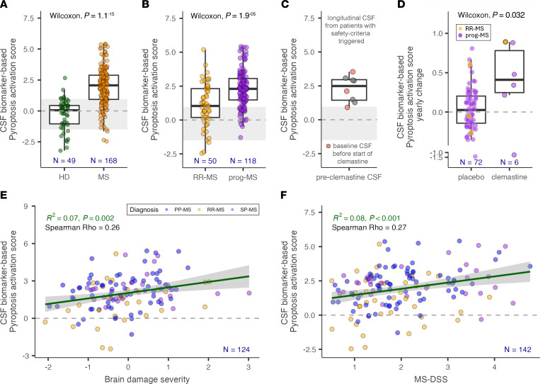
Multiple sclerosis (MS) is an immune-mediated demyelinating disease of the CNS. Clemastine fumarate, the over-the-counter antihistamine and muscarinic receptor blocker, has remyelinating potential in MS. A clemastine arm was added to an ongoing platform clinical trial, targeting residual activity by precision, biomarker-guided combination therapies of multiple sclerosis (TRAP-MS) (ClinicalTrials.gov NCT03109288), to identify a cerebrospinal fluid (CSF) remyelination signature and to collect safety data on clemastine in patients progressing independently of relapse activity (PIRA). The clemastine arm was stopped per protocol-defined criteria when 3 of 9 patients triggered individual safety stopping criteria. Clemastine-treated patients had significantly higher treatment-induced disability progression slopes compared with the remaining TRAP-MS participants. Quantification of approximately 7,000 proteins in CSF samples collected before and after clemastine treatment showed significant increases in purinergic signaling and pyroptosis. Mechanistic studies showed that clemastine with sublytic doses of extracellular adenosine triphosphate (ATP) activates inflammasome and induces pyroptotic cell death in macrophages. Clemastine with ATP also caused pyroptosis of induced pluripotent stem cell-derived human oligodendrocytes. Antagonist of the purinergic channel P2RX7, which is strongly expressed in oligodendrocytes and myeloid cells, blocked these toxic effects of clemastine. Finally, reanalysis of published single-nucleus RNA-Seq (snRNA-Seq) studies revealed increased P2RX7 expression and pyroptosis transcriptional signature in microglia and oligodendrocytes in the MS brain, especially in chronic active lesions. The CSF proteomic pyroptosis score was increased in untreated MS patients, was higher in patients with progressive than relapsing-remitting disease, and correlated significantly with the rates of MS progression. Collectively, this identifies pyroptosis as a likely mechanism of CNS injury underlying PIRA even outside of clemastine toxicity.

Keywords: Cellular immune response; Immunology; Multiple sclerosis; Neurodegeneration; Neuroscience.


Conflict of interest statement

Conflict of interest: The authors have declared that no conflict of interest exists.

Figures

Figure 1
Figure 1. CLM-induced changes in disability, metabolism, and inflammatory markers.
(A) Clinical safety was monitored by a continuous, machine-learning derived CombiWISE scale (range 0–100) that correlates strongly with EDSS (range 0–10). R-squared in Linear Regression was used. n = 935. (B) A minimum of 4 visits on stable therapy spanning at least 18 months is required to measure baseline (blue) CombiWISE slope. On-therapy CombiWISE slope (orange) is calculated based on 6-months follow-up data collected after therapy initiation. (C) Three out of 9 patients on CLM therapy triggered safety criteria, in contrast to none of the 63 patient-specific treatments of 6 other TRAP-MS therapies; P value of probability of this occurrence was based on χ2 test. (D) Progression CombiWISE slopes at baseline (B) and therapy (T) were compared between CLM arm (n = 9) and all other 6 TRAP-MS therapies (n = 63). Black horizontal line represents median value of the group. Red color indicates 3 patients that triggered safety-stopping criteria treatment slope exceeding 5× baseline slope. Displayed P value was generated from 2-sided unpaired Wilcoxon’s rank test comparing therapy-induced change in CombiWISE slope between the CLM arm and all other therapies. CLM induced increase of weight (E), LDL cholesterol (F), and total cholesterol (G) levels between baseline and treatment and these changes showed strong association as seen on example of weight change versus LDL cholesterol change (H). R-squared in linear regression was used. Furthermore, CLM induced increase of inflammatory biomarker CRP (I). Lipid panel was an optional laboratory test, and the results are missing for 1 patient. Blue line in H represents linear regression line; gray dashed line in DG and I represents 0 change. The lower and upper hinges of the boxplots correspond to the first and third quartiles (the 25th and 75th percentiles). The upper and lower whiskers extend from the hinge to the largest and smallest value, respectively.
Figure 2
Figure 2. CLM potentiation effect on ATP-induced inflammasome activation and GSDMD-driven pyroptotic cell death in THP-1 cells.
The THP-1 cells were primed with 200 ng/mL LPS overnight and then treated either with medium + DMSO (negative control, Ctrl), 10 μg/mL CLM, 2 mM ATP ± 10 μg/mL CLM, or 2.5 μM nigericin (positive control) for 3 hours, 6 hours, and 18 hours. Release of the proinflammatory cytokine IL-1β into the culture medium of THP-1 cells (A) and THP-1 GSDMD–/– cells (B), determined by ELISA assay. Activity of caspase-1 in the culture medium of THP-1 cells (C) and THP-1 GSDMD–/– cells (D), determined using bioluminescent assay. Lytic cell death of THP-1 cells (E) and THP-1 GSDMD–/– cells (F), determined by LDH activity released into the culture medium. Cell viability of THP-1 cells (G) and THP-1 GSDMD–/– cells (H) evaluated by the MTS assay. (AH) Data are presented as mean ± SD of 3 independent experiments, each performed in duplicate (n = 6). One-way ANOVA followed by Dunnett’s multiple-comparisons test was used to compare the testing groups with control group (Ctrl). One-way ANOVA test followed by Holm-Šidák’s multiple comparison was used to compare ATP and ATP+CLM group (to assess the potentiation effect of CLM). *P ≤ 0.05; **P ≤ 0.01; ***P ≤ 0.001; ****P ≤ 0.0001. Panel A, ATP+CLM at 3 hours (n = 1), nigericin at 3 hours (n = 3), 6 hours (n = 5), and 18 hours (n = 5) — above detection limit of the assay. Panel B, Ctrl at 3 hours (n = 5) and 6 hours (n = 3) — below detection limit of the assay. Nigericin at 18 hours (n = 6) — above detection limit of the assay. (I) Flow cytometry analysis of cell uptake of SYTOX Green in THP-1 cells primed with 200 ng/mL LPS overnight and then treated either with medium + DMSO (control), 10 μg/mL CLM, or 2 mM ATP ± 10 μg/mL CLM for 90 minutes. Representative plot of 2 independent experiments.
Figure 3
Figure 3. The potentiation effect of CLM on ATP-induced lytic cell death in primary monocyte-derived macrophages.
The primary monocyte-derived macrophages (AD) were primed with 200 ng/mL LPS overnight, then pretreated (JNJ and ATP+CLM+JNJ groups) with 30 nM JNJ-54175446 (a selective purine P2X7 receptor antagonist) for 1 hour, followed by treatment either with medium + DMSO (negative control, Ctrl), 10 μg/mL CLM, 2 mM ATP ± 10 μg/mL CLM, or 10 μM nigericin (positive control) for 3 hours, 6 hours, and 18 hours. Levels of the proinflammatory cytokine IL-1β (A) and caspase-1 (B) activity in the culture medium determined by ELISA and bioluminescence assay, respectively. Lytic cell death (C) determined by LDH activity released into the medium and cell viability (D) evaluated by the MTS assay. (AD) Data are represented as mean ± SEM of 3 independent experiments, each performed in duplicate (n = 6). One-way ANOVA followed by Dunnett’s multiple comparisons test was used to compare the testing groups with control group (Ctrl). One-way ANOVA test followed by Holm-Šidák’s multiple-comparison was used to compare ATP and ATP+CLM group (to assess the potentiation effect of CLM) and ATP+CLM versus ATP+CLM+JNJ group. Panel A, nigericin at 3 hours (n = 2), 6 hours (n = 1), and 18 hours (n = 4) — above detection limit of the assay. *P ≤ 0.05; **P ≤ 0.01; ***P ≤ 0.001; ****P ≤ 0.0001. (E) Representative fluorescence microscopy images depicting SYTOX Green-stained MDMs treated with drugs for 6 hours. Original magnification, ×10.
Figure 4
Figure 4. The potentiation effect of CLM on ATP-induced lytic cell death in iPSC-derived OLGs.
iPSC-derived OLG-enriched cells (A) were primed with 200 ng/mL LPS overnight followed by treatment either with medium + DMSO (negative control, Ctrl), 10 μg/mL CLM, 2mM ATP ± 10 μg/mL CLM, or 10 μM nigericin (positive control). LDH release after the treatments for 18 hours. Data are represented as mean ± SEM; 1 donor with 4 technical replicates (n = 4). One-way ANOVA followed by Dunnett’s multiple-comparisons test was used to compare the testing groups with control group (Ctrl). One-way ANOVA test followed by Holm-Šidák’s multiple-comparison test was used to compare ATP and ATP+CLM group (to assess the potentiation effect of CLM). *P ≤ 0.05; ****P ≤ 0.0001. iPSC-derived OLG cells (B) were primed with TNF-α (50 ng/mL) overnight, pretreated with 60 nM JNJ-54175446 for 1 hour, followed by treatment either with medium + DMSO (negative control, Ctrl), 10 μg/mL CLM, 2 mM ATP ± 10 μg/mL CLM, or 2.5 μM nigericin. LDH release after the treatments for 18 hours. Data are represented as mean ± SEM of 2 independent experiments, each performed in triplicate (n = 6) for control or in 4 replicates for the other groups. One-way ANOVA followed by Dunnett’s multiple-comparisons test was used to compare the testing groups with control group (Ctrl). One-way ANOVA test followed by Holm-Šidák’s multiple-comparison test was used to compare ATP and ATP+CLM group (to assess the potentiation effect of CLM) and ATP+CLM versus ATP+CLM+JNJ group. *P ≤ 0.05. ****P ≤ 0.0001.
Figure 5
Figure 5. Gene expression associated with pyroptosis signaling pathway in CNS tissue.
Published data of snRNA-seq from MS and control (GSE180759, GSE118257, and PRJNA544731) was reanalyzed, consisting of 21 MS subjects (2 PPMS and 19 SPMS) and 17 controls. Panels A and C illustrate the profiles of P2RX7, GSDM family, CASP1, and IL-1β across the lesion types. Meanwhile, panels B and D show gene expressions based on cell types.
Figure 6
Figure 6. MS severity–associated pyroptosis signature in CSF increases with CLM treatment.
CSF biomarker–derived pyroptosis activation score is significantly elevated in MS patients compared with HD (A) and in progressive MS (prog-MS) patients compared with RRMS patients (B). The gray area represents HD mean ± SD. (C) Longitudinal pre-CLM CSF samples of patients that triggered safety criteria show elevated pyroptosis activation score. CSF samples collected just prior to starting CLM are highlighted in red. (D) CLM-induced yearly change in pyroptosis activation score is significantly elevated compared with untreated MS patients. The lower and upper hinges of the boxplots correspond to the first and third quartiles (the 25th and 75th percentiles). The upper and lower whiskers extend from the hinge to the largest and smallest value, respectively. Pyroptosis activation scores are strongly associated with the rate of accumulation of cognitive and physical disability, represented by brain damage severity (E) and MS-DSS (F), respectively. The green line depicts linear regression line, and the gray shaded area shows 95% CI.

Update of

Similar articles

Cited by

References

    1. Weideman AM, et al. Meta-analysis of the age-dependent efficacy of multiple sclerosis treatments. Front Neurol. 2017;8:577. doi: 10.3389/fneur.2017.00577. - DOI - PMC - PubMed
    1. Wallin MT, et al. The prevalence of MS in the United States: a population-based estimate using health claims data. Neurology. 2019;92(10):e1029–e1040. doi: 10.1212/WNL.0000000000007035. - DOI - PMC - PubMed
    1. Wallin MT, et al. Global, regional, and national burden of multiple sclerosis 1990–2016: a systematic analysis for the Global Burden of Disease Study 2016. Lancet Neurol. 2019;18(3):269–285. doi: 10.1016/S1474-4422(18)30443-5. - DOI - PMC - PubMed
    1. Bruck W, et al. Remyelination in multiple sclerosis. J Neurol Sci. 2003;206(2):181–185. doi: 10.1016/S0022-510X(02)00191-0. - DOI - PubMed
    1. Mei F, et al. Micropillar arrays as a high-throughput screening platform for therapeutics in multiple sclerosis. Nat Med. 2014;20(8):954–960. doi: 10.1038/nm.3618. - DOI - PMC - PubMed

Publication types

MeSH terms

Associated data