

Oligodendrogenesis in Evolution, Development and Adulthood
- PMID: 40371693
- PMCID: PMC12313009
- DOI: 10.1002/glia.70033
Oligodendrogenesis in Evolution, Development and Adulthood
Abstract
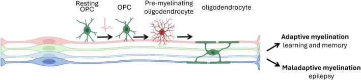
Oligodendrogenesis and myelin formation are important processes in the central nervous system (CNS) of jawed vertebrates, underpinning the highly efficient neural computation within the compact CNS architecture. Myelin, the dense lipid sheath wrapped around axons, enables rapid signal transmission and modulation of neural circuits. Oligodendrocytes are generated from oligodendrocyte precursor cells (OPCs), which are widely distributed in the adult CNS and continue to produce new oligodendrocytes throughout life. Adult oligodendrogenesis is integral to adaptive myelination, which fine-tunes neural circuits in response to neuronal activity, contributing to neuroplasticity, learning, and memory. Emerging evidence also highlights the role of oligodendrogenesis in specialized brain regions, linking oligodendrocytes to metabolic and homeostatic functions. In the aging and diseased brain, dysregulated oligodendrogenesis exacerbates myelin loss and may contribute to pathogenesis. In addition, maladaptive myelination driven by aberrant neuronal activity could sustain a dysfunction in conditions such as epilepsy. This review summarizes the current understanding of oligodendrogenesis, with insights into its evolution, regulation, and impact on aging and disease.
Keywords: adaptive myelination; development; evolution; maladaptive myelination; myelin; oligodendrocyte; oligodendrogenesis.
© 2025 The Author(s). GLIA published by Wiley Periodicals LLC.
Conflict of interest statement
The authors declare no conflicts of interest.
Figures
Similar articles
-
Role of growth factors and their interplay during oligodendroglial differentiation and maturation.Cytokine Growth Factor Rev. 2025 Aug;84:47-58. doi: 10.1016/j.cytogfr.2025.07.002. Epub 2025 Jul 10. Cytokine Growth Factor Rev. 2025. PMID: 40707311 Review.
-
Autophagy in Oligodendrocyte Lineage Cells Controls Oligodendrocyte Numbers and Myelin Integrity in an Age-dependent Manner.Neurosci Bull. 2025 Mar;41(3):374-390. doi: 10.1007/s12264-024-01292-1. Epub 2024 Sep 16. Neurosci Bull. 2025. PMID: 39283565
-
Uncommon Non-MS Demyelinating Disorders of the Central Nervous System.Curr Neurol Neurosci Rep. 2025 Jul 1;25(1):45. doi: 10.1007/s11910-025-01432-8. Curr Neurol Neurosci Rep. 2025. PMID: 40591029 Review.
-
C1ql1 expression in oligodendrocyte progenitor cells promotes oligodendrocyte differentiation.FEBS J. 2025 Jan;292(1):52-74. doi: 10.1111/febs.17256. Epub 2024 Sep 11. FEBS J. 2025. PMID: 39257292
-
Neurorehabilitation and white matter repair in traumatic spinal cord injury: a dialogue between clinical and preclinical studies.Front Neurol. 2025 Jun 18;16:1532056. doi: 10.3389/fneur.2025.1532056. eCollection 2025. Front Neurol. 2025. PMID: 40606124 Free PMC article. Review.
Cited by
-
Glial Diversity and Evolution: Insights from Teleost Fish.Brain Sci. 2025 Jul 11;15(7):743. doi: 10.3390/brainsci15070743. Brain Sci. 2025. PMID: 40722334 Free PMC article. Review.
References
-
- Atkinson‐Clement, C. , Sofia F., Fernandez‐Egea E., et al. 2020. “Structural and Functional Abnormalities Within Sensori‐Motor and Limbic Networks Underpin Intermittent Explosive Symptoms in Tourette Disorder.” Journal of Psychiatric Research 125: 1–6. 10.1016/j.jpsychires.2020.02.033. - DOI - PubMed
Publication types
MeSH terms
Grants and funding
- BB/L003236/1/BB_/Biotechnology and Biological Sciences Research Council/United Kingdom
- BB/S000844/1/BB_/Biotechnology and Biological Sciences Research Council/United Kingdom
- BB/Z516363/1/BB_/Biotechnology and Biological Sciences Research Council/United Kingdom
- UKRI537/MRC_/Medical Research Council/United Kingdom
LinkOut - more resources
Full Text Sources
