

Effect of Dual-Task Training on the Number of EEG Bands in Stroke Patients
- PMID: 40371695
- PMCID: PMC12079625
- DOI: 10.1002/pri.70065
Effect of Dual-Task Training on the Number of EEG Bands in Stroke Patients
Abstract
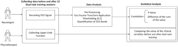
Background/objective: Dual-task training (DTT) positively impacts stroke recovery, but its effects on electroencephalography (EEG) using Fourier series analysis are under-researched. This study aimed to evaluate the effects of DTT on EEG in stroke patients by analyzing different EEG bands with fast Fourier transform (FFT).
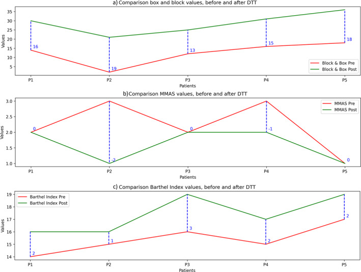
Methods: Five participants with unilateral ischemic stroke completed 12 sessions of 15-min DTT, three times a week for 4 weeks. EEG data were recorded before and after the intervention, and FFT analysis was conducted. Assessments of upper limb function, elbow flexor muscle tone, and daily living activities were also performed.
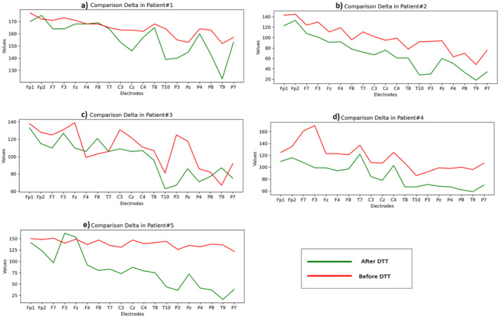
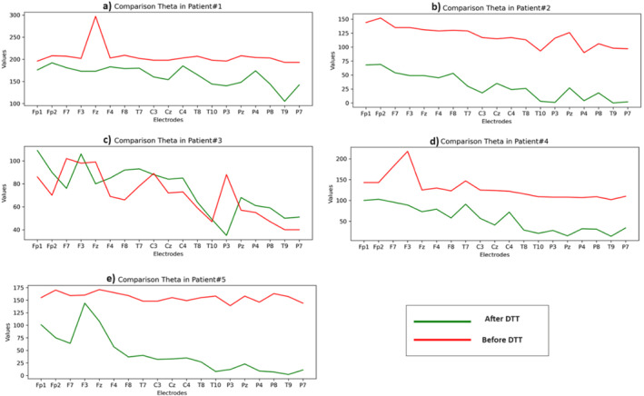
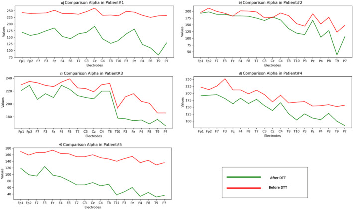
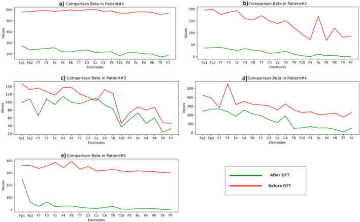
Results: FFT analysis showed a reduction in delta, theta, alpha, and beta bands post-DTT, while their correlation between measurement times remained consistent. These changes were somewhat reflected in the participants' improved clinical outcomes.
Conclusion: The results suggest that DTT positively affects EEG band frequencies, with a consistent correlation between pre- and post-intervention measurements. This indicates that FFT analysis could be a useful tool for assessing DTT's impact on stroke recovery.
Keywords: dual‐task training; electroencephalography; fast Fourier transform; signal analysis; stroke.
© 2025 The Author(s). Physiotherapy Research International published by John Wiley & Sons Ltd.
Conflict of interest statement
The authors declare no conflicts of interest.
Figures
Similar articles
-
Restoring of Interhemispheric Symmetry in Patients With Stroke Following Bilateral or Unilateral Robot-Assisted Upper-Limb Rehabilitation: A Pilot Randomized Controlled Trial.IEEE Trans Neural Syst Rehabil Eng. 2024;32:3590-3602. doi: 10.1109/TNSRE.2024.3460485. Epub 2024 Sep 27. IEEE Trans Neural Syst Rehabil Eng. 2024. PMID: 39269794 Clinical Trial.
-
Quantitative EEG data and comprehensive ADL (Activities of Daily Living) evaluation of stroke survivors residing in the community.J Physiol Anthropol Appl Human Sci. 2001 Jan;20(1):37-41. doi: 10.2114/jpa.20.37. J Physiol Anthropol Appl Human Sci. 2001. PMID: 11320778
-
Neurophysiological underpinnings of an intensive protocol for upper limb motor recovery in subacute and chronic stroke patients.Eur J Phys Rehabil Med. 2024 Feb;60(1):13-26. doi: 10.23736/S1973-9087.23.07922-4. Epub 2023 Nov 21. Eur J Phys Rehabil Med. 2024. PMID: 37987741 Free PMC article.
-
Literature review of stroke assessment for upper-extremity physical function via EEG, EMG, kinematic, and kinetic measurements and their reliability.J Neuroeng Rehabil. 2023 Feb 15;20(1):21. doi: 10.1186/s12984-023-01142-7. J Neuroeng Rehabil. 2023. PMID: 36793077 Free PMC article. Review.
-
The effects of error-augmentation versus error-reduction paradigms in robotic therapy to enhance upper extremity performance and recovery post-stroke: a systematic review.J Neuroeng Rehabil. 2018 Jul 4;15(1):65. doi: 10.1186/s12984-018-0408-5. J Neuroeng Rehabil. 2018. PMID: 29973250 Free PMC article.
References
-
- An, H.‐S. , and Kim D.‐J.. 2021. “Effects of Activities of Daily Living‐Based Dual‐Task Training on Upper Extremity Function, Cognitive Function, and Quality of Life in Stroke Patients.” Osong Public Health and Research Perspectives 12, no. 5: 304–313. 10.24171/j.phrp.2021.0177. - DOI - PMC - PubMed
-
- Arjmandi‐Rad, S. , Vestergaard Nieland J. D., Goozee K. G., and Vaseghi S.. 2024. “The Effects of Different Acetylcholinesterase Inhibitors on EEG Patterns in Patients With Alzheimer’s Disease: A Systematic Review.” Neurological Sciences 45, no. 2: 417–430. 10.1007/s10072-023-07114-y. - DOI - PubMed
MeSH terms
LinkOut - more resources
Full Text Sources
Medical
