. 2025 May 15:13:RP99670.
doi: 10.7554/eLife.99670.

AARS2 ameliorates myocardial ischemia via fine-tuning PKM2-mediated metabolism

Affiliations

AARS2 ameliorates myocardial ischemia via fine-tuning PKM2-mediated metabolism

Zongwang Zhang et al. Elife. .

Abstract

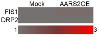
AARS2, an alanyl-tRNA synthase, is essential for protein translation, but its function in mouse hearts is not fully addressed. Here, we found that cardiomyocyte-specific deletion of mouse AARS2 exhibited evident cardiomyopathy with impaired cardiac function, notable cardiac fibrosis, and cardiomyocyte apoptosis. Cardiomyocyte-specific AARS2 overexpression in mice improved cardiac function and reduced cardiac fibrosis after myocardial infarction (MI), without affecting cardiomyocyte proliferation and coronary angiogenesis. Mechanistically, AARS2 overexpression suppressed cardiomyocyte apoptosis and mitochondrial reactive oxide species production, and changed cellular metabolism from oxidative phosphorylation toward glycolysis in cardiomyocytes, thus leading to cardiomyocyte survival from ischemia and hypoxia stress. Ribo-Seq revealed that Aars2 overexpression increased pyruvate kinase M2 (PKM2) protein translation and the ratio of PKM2 dimers to tetramers that promote glycolysis. Additionally, PKM2 activator TEPP-46 reversed cardiomyocyte apoptosis and cardiac fibrosis caused by AARS2 deficiency. Thus, this study demonstrates that AARS2 plays an essential role in protecting cardiomyocytes from ischemic pressure via fine-tuning PKM2-mediated energy metabolism, and presents a novel cardiac protective AARS2-PKM2 signaling during the pathogenesis of MI.

Keywords: AARS2; PKM2; cardiac remodeling; cardiomyocytes; cell biology; glycolysis; medicine; mouse.


Conflict of interest statement

ZZ, LZ, YC, YC, JH, CX, XZ, SZ, JX No competing interests declared

Figures

Figure 1.
Figure 1.. Cardiomyocyte-specific knockout of alanyl-tRNA synthetase (AARS2) leads to cardiac dysfunction and fibrosis in mice.
(A, B) Western blot and quantitative real-time PCR (qRT-PCR) analysis showing reduced expression of AARS2 proteins (A) or mRNA (B) of 3 d myocardial infarction (MI) hearts compared with sham hearts (n=3). (C) Construction diagram of α-MHC-MerCreMer (upper) and Aars2fl/fl mice (lower). (D) Western blots showing reduced AARS2 proteins in Aars2 cKO hearts compared with Aars2fl/fl hearts (n=3). (E) Schematic timelines of tamoxifen treatment and echocardiography (ECHO). (F) Representative M-mode tracings of ECHO in control and conditioned knockout (cKO) hearts before and after tamoxifen treatment. (G) Ejection fraction (EF) and fractional shortening (FS) of Aars2fl/fl and Aars2 cKO hearts at different time points after tamoxifen induction (n=10–11). (H) Cardiac output of Aars2fl/fl and Aars2 cKO mice at 28 d after tamoxifen induction (n=8–10). (I) Left ventricular end-diastolic diameter (LVEDD) and left ventricular end-systolic diameter (LVESD) in Aars2fl/fl and Aars2 cKO hearts at 28 d after tamoxifen induction (n=8–10). (J) Survival percentage of Aars2fl/fl and Aars2 cKO mice at 28 d after tamoxifen treatment (n=8–10). (K) WGA immunofluorescence showing cardiomyocyte hypertrophy on heart slices of Aars2 cKO group compared with Aars2fl/fl group at 28 d after tamoxifen induction (scale bar, 100 μm; n=5). (L) Masson staining and quantitative analysis showing increased cardiac fibrosis in Aars2 cKO hearts compared with Aars2fl/fl control hearts at 28 d after tamoxifen induction (scale bar, 1 mm; n=8). Mean ± s.e.m.
Figure 1—figure supplement 1.
Figure 1—figure supplement 1.. Genotyping of cardiomyocyte-specific Aars2 knockout mice.
(A) Genotyping of Aars2fl/fl and Aars2 cKO mice (n=3; Mut, mutant). (B) Western Blot showing normal expression of AARS2 proteins in the liver, lung, and skeletal muscle of Aars2fl/fl and Aars2 cKO mice (n=3).
Figure 2.
Figure 2.. Cardiomyocyte-specific conditioned knockout (cKO) of alanyl-tRNA synthetase (AARS2) results in cardiomyocyte apoptosis and energy metabolism deficiency.
(A, B) Immunofluorescence staining showing increased numbers of cTnT+ TUNEL+ cardiomyocytes (A) and Western blot showing reduced anti-apoptotic protein Bcl-2 and increased pro-apoptotic protein BAX (B) in Aars2 cKO hearts compared with Aars2fl/fl control hearts at 28 d post-tamoxifen treatment (scale, 200 μm; n=6 for panel A; n=3 for panel B). (C) Seahorse analysis showing reduced oxygen consumption rate (OCR) of cardiac mitochondria in Aars2 cKO hearts compared with Aars2fl/fl control hearts at 28 d after tamoxifen induction (n=4). (D) Seahorse analysis showing reduced extracellular acidification rate (ECAR) of adult mouse cardiomyocytes in Aars2 cKO hearts compared with Aars2fl/fl control hearts at 28 d after tamoxifen induction (n=4). (E–F) Seahorse analysis showing reduced OCR (E) and ECAR (F) of NRCMs in AARS2 siRNA group compared with control group at 3 d after transfection (n=4). Mean ± s.e.m.
Figure 2—figure supplement 1.
Figure 2—figure supplement 1.. Evaluating Aars2 siRNA for knockout efficiency of alanyl-tRNA synthetase (AARS2) proteins in neonatal rat cardiomyocytes (NRCMs).
(A) Western blots showing knockdown efficiency of three different Aars2 siRNAs in NRCMs, respectively (n=3). (B) Western blots confirming knockdown efficiency of Aars2 siRNA#3 in NRCMs (n=3). Ctrl, control; si, small interfering; mean ± s.e.m.
Figure 3.
Figure 3.. Cardiomyocyte-specific alanyl-tRNA synthetase (AARS2) overexpression improves cardiac function and decreases cardiac fibrosis in mice post-MI.
(A) Schematic diagram of α-MHC-MerCreMer, and CAG-Aars2 mice that is driven by the CAG promoter. (B) Western blots showing transgenic overexpression of AARS2 proteins in the hearts of Aars2 Tg/+compared with Aars2 WT control mice (n=3). (C) Experimental protocols for Tamoxifen induction for 5 d, and then recovery for 7 d before echocardiography (ECHO) and myocardial infarction (MI). (D) Representative M-mode of ECHO in control and Aars2 Tg/+mouse hearts at 3 d or 28 d post-MI. (E) Ejection fraction (EF) and fractional shortening (FS) of the Aars2 WT and Aars2 Tg/+mouse hearts were measured at different time points before and after MI (n=10–11). (F) The cardiac output of Aars2 WT and Aars2 Tg/+mice was measured before MI and 28 d after MI (n=10–11). (G) Left ventricle end-diastolic diameter (LVEDD) and left ventricle end-systolic diameter (LVESD) of Aars2 WT and Aars2 Tg/+ mice before MI and 28 d after MI (n=10–11). (H) Masson’s staining showing decreased fibrotic area in the hearts of Aars2 Tg/+compared with Aars2 WT mice at 28 d after MI (scale bar, 1 mm, n=10–11). Mean ± s.e.m.
Figure 3—figure supplement 1.
Figure 3—figure supplement 1.. Cardiomyocyte-specific overexpression of alanyl-tRNA synthetase (AARS2) in the heart but not in the liver, lung, and skeletal muscle.
(A) Genotyping of wild-type (WT), α-MHC-MerCreMer; Aars2 WT (Aars2 WT) control, and α-MHC-MerCreMer; Aars2 Tg/+ (Aars2 Tg/+) mice (n=3). (B) Immunofluorescence staining showing ectopic AARS2 proteins in cardiomyocytes of Aars2 Tg/+transgenic mice compared with Aars2 WT control mice (scale bar, 50 μm). (C) Western blots showing comparable expression of AARS2 proteins in the liver, lung, and skeletal muscle of Aars2 WT control and Aars2 Tg/+ transgenic mice (n=3). Mean ± s.e.m.
Figure 3—figure supplement 2.
Figure 3—figure supplement 2.. Overexpression of alanyl-tRNA synthetase (AARS2) in cardiomyocytes has no apparent effect on cardiomyocyte proliferation, hypertrophy, and angiogenesis after myocardial infarction (MI).
(A–B) At 7 d after MI, immunofluorescence staining showing comparable cTnT+/Ki67+ (A) and cTnT+/pH3+ (B) cardiomyocytes in the heart sections of Aars2 WT and Aars2 Tg/+groups (scale, 200 μm; n=5). (C–D) Immunofluorescence staining showing comparable numbers of cTnT+/Ki67+ (C) and cTnT+/pH3+ (D) cardiomyocytes at 48 h after infection of neonatal rat cardiomyocytes (NRCMs) with Mock lentivirus or Aars2 lentivirus (scale, 50 μm; n=3). (E) At 7 d after MI, immunofluorescence staining showing comparable CD31+/α-SMA+ coronary vessels in the heart sections of Aars2 WT and Aars2 Tg/+groups (scale, 200 μm; n=5). (F) Wheat germ agglutinin (WGA) staining showing no evident cardiomyocyte hypertrophy in heart slices of Aars2 WT and Aars2 Tg/+groups at 28 d after MI (scale, 100 μm; n=6). Mean ± s.e.m.
Figure 4.
Figure 4.. Overexpression of alanyl-tRNA synthetase (AARS2) attenuates cardiomyocyte apoptosis.
(A) Immunofluorescence staining showing reduced cTnT+/TUNEL+ cardiomyocytes in Aars2 Tg/+hearts compared with Aars2 WT control hearts at 7 d after myocardial infarction (MI) (scale bar, 200 μm, n=6). (B) Western blots showing increased anti-apoptotic protein Bcl-2 and decreased pro-apoptotic protein BAX in Aars2 Tg/+compared with Aars2 WT control hearts at 7 d after MI (n=3). (C) The serum level of lactate dehydrogenase (LDH) decreased in Aars2 Tg/+ hearts compared with Aars2 WT control hearts at 28 d after MI (n=10). (D) Immunofluorescence staining and quantitative analysis showing reduced MitoSOX in Aars2 OE neonatal rat cardiomyocytes (NRCMs) after 12 hr of hypoxia followed by 1 hr of reoxygenation (H/R, scale bar, 20 μm; n=4). (E) Western blots showing increased Bcl-2 and decreased BAX in NRCMs overexpressing Aars2 (Aars2 OE) compared with control NRCMs after 12 hr of hypoxia followed by 1 hr of reoxygenation (n=3). (F) The level of LDH decreased in Aars2 OE NRCMs after 12 hr of hypoxia followed by 1 hr of reoxygenation (n=6). Mean ± s.e.m.
Figure 5.
Figure 5.. Cardiomyocyte overexpression of alanyl-tRNA synthetase (AARS2) regulates cardiac metabolism.
(A) Mass spectrometry showing increased lactate and pyruvate but reduced acetyl-CoA in Aars2 Tg/+ hearts compared with Aars2 WT hearts after 7 d of myocardial infarction (MI) (n=5). (B) Mass spectrometry showing increased lactate and pyruvate but decreased acetyl-CoA in neonatal rat cardiomyocytes (NRCMs) overexpressing Aars2 (Aars2 OE) for 3 d (n=9). (C) Seahorse analysis showing oxygen consumption rate (OCR) of cardiac mitochondria in Aars2 WT and Aars2 Tg/+ mice at 28 ds after tamoxifen induction (n=3). (D) Seahorse analysis showing extracellular acidification rate (ECAR) and quantitative analysis of adult mouse cardiomyocytes in Aars2 WT and Aars2 Tg/+ mice at 28 d after tamoxifen induction (n=4). (E–F) Seahorse analysis showing OCR (E) and ECAR (F) of NRCMs in Mock control and Aars2 OE groups at 3 d after transfection (n=4). Mean ± s.e.m.
Figure 6.
Figure 6.. Overexpression of alanyl-tRNA synthetase (AARS2) increases the protein level of glycolytic pyruvate kinase M2 (PKM2) via enhancing PKM2 translation.
(A) Ribosome RNA-seq showing elevated translation of signaling pathways of glycolysis in the Aars2 OE NRCMs compared to the Mock neonatal rat cardiomyocytes (NRCMs). (B) Western Blots showing the level of AARS2, PDK4, and LDHA proteins in the hearts of Aars2 WT control and Aars2 Tg/+ transgenic mice (n=3). (C) Western Blots showing the level of AARS2 and PKM1 proteins in the hearts of Aars2 WT control and Aars2 Tg/+ transgenic mice (n=3). (D–F) Western Blots showing the level of AARS2 and PKM2 proteins in the hearts of Aars2 WT control and Aars2 Tg/+ transgenic mice (n=3) (D), in Mock control and Aars2 OE NRCMs (E) (n=3), and in the hearts of Aars2fl/fl and Aars2 cKO mice (F) (n=3). (G–H) Western Blots by non-denatured gels (G) and statistics (H) showing the amounts of PKM2 monomers, dimers, and tetramers in the hearts of Aars2 WT control and Aars2 Tg/+transgenic mice, and in the hearts of Aars2fl/fl and Aars2 cKO mice (n=4). (I) Mass spectrometry analysis measuring the amounts of alanine (Ala) from homogenates of heart tissues (n=6) and NRCM lysates (n=6). (J) Ratio of quantitative results of PKM2 dimers and tetramers in the hearts of Aars2 WT control and Aars2 Tg/+ transgenic mice of panel H (n=4). (K) Co-immunoprecipitation reveals no evident interactions between PKM2 and AARS2 in NRCMs. (L–M) qRT-PCR showing the comparative level of Pkm2 mRNA in the hearts of control sibling and Aars2 Tg/+transgenic hearts; control sibling and Aars2 cKO hearts (L); and in control, Aars2 OE, and AARS2siRNA NRCMs (M) (n=3). FC, Fold changes; Mean ± s.e.m.
Figure 6—figure supplement 1.
Figure 6—figure supplement 1.. Overexpression of alanyl-tRNA synthetase (AARS2) increases the translation level of some cellular proteins.
(A-D) Ribosome RNA-Seq showing elevated translation of signaling pathways of genes encoded by mitochondria(A), lipoprotein metabolic process (B), cellular response to hypoxia (C), and sodium ion transport (D) in the Aars2 OE neonatal rat cardiomyocytes (NRCMs) compared to the Mock NRCMs. FC, fold changes.
Figure 7.
Figure 7.. Pyruvate kinase M2 (PKM2) activator TEPP-46 improves cardiomyopathy in Aars2 conditioned knockout (cKO) mice.
(A) Experimental scheme and time points for tamoxifen induction, echocardiography (ECHO), and TEPP-46 administration. (B) Ejection fraction (EF) and fractional shortening (FS) of Aars2 cKO mouse hearts at different time points after administration of control solvent and TEPP-46 (n=7–8). (C) Representative M-mode of ECHO in different groups of 4 wk mice. (D) Cardiac outputs of Aars2 cKO mouse hearts at different time points after administration of control solvent and TEPP-46 (n=7–8). (E) Left ventricular end-diastolic diameter (LVEDD) and left ventricular end-systolic diameter (LVESD) at 28 d of Aars2 cKO mice after administration of control solvent and TEPP-46 (n=7–8). (F) Masson staining showing cardiac fibrosis of Aars2 cKO mouse hearts at 28 d after administration of either control solvent or TEPP-46 (scale bar, 1 mm; n=7–8). (G) Measurements of lactate dehydrogenase (LDH) release in neonatal rat cardiomyocytes (NRCMs) from the control group and TEPP-46 group (20 μM) after Aars2 siRNA or control siRNA treatment for 72 hr (n=5). (H) Quantitative analysis of MitoSOX immunofluorescence in NRCMs from the control group and TEPP-46 group (20 μM) after Aars2 siRNA or control siRNA treatment for 72 hr (scale bar, 20 μm; n=5). Mean ± s.e.m.
Author response image 1.
Author response image 2.
Author response image 3.
Author response image 4.
Author response image 5.
Author response image 6.
Author response image 7.

Update of

  • doi: 10.1101/2024.06.04.597368
  • doi: 10.7554/eLife.99670.1
  • doi: 10.7554/eLife.99670.2

Similar articles

References

    1. Chen L, Shi Y, Liu S, Cao Y, Wang X, Tao Y. PKM2: the thread linking energy metabolism reprogramming with epigenetics in cancer. International Journal of Molecular Sciences. 2014;15:11435–11445. doi: 10.3390/ijms150711435. - DOI - PMC - PubMed
    1. Chuang H-M, Su H-L, Li C, Lin S-Z, Yen S-Y, Huang M-H, Ho L-I, Chiou T-W, Harn H-J. The role of butylidenephthalide in targeting the microenvironment which contributes to liver fibrosis amelioration. Frontiers in Pharmacology. 2016;7:112. doi: 10.3389/fphar.2016.00112. - DOI - PMC - PubMed
    1. Dallabona C, Diodato D, Kevelam SH, Haack TB, Wong L-J, Salomons GS, Baruffini E, Melchionda L, Mariotti C, Strom TM, Meitinger T, Prokisch H, Chapman K, Colley A, Rocha H, Ounap K, Schiffmann R, Salsano E, Savoiardo M, Hamilton EM, Abbink TEM, Wolf NI, Ferrero I, Lamperti C, Zeviani M, Vanderver A, Ghezzi D, van der Knaap MS. Novel (ovario) leukodystrophy related to AARS2 mutations. Neurology. 2014;82:2063–2071. doi: 10.1212/WNL.0000000000000497. - DOI - PMC - PubMed
    1. Dayton TL, Gocheva V, Miller KM, Israelsen WJ, Bhutkar A, Clish CB, Davidson SM, Luengo A, Bronson RT, Jacks T, Vander Heiden MG. Germline loss of PKM2 promotes metabolic distress and hepatocellular carcinoma. Genes & Development. 2016a;30:1020–1033. doi: 10.1101/gad.278549.116. - DOI - PMC - PubMed
    1. Dayton TL, Jacks T, Vander Heiden MG. PKM2, cancer metabolism, and the road ahead. EMBO Reports. 2016b;17:1721–1730. doi: 10.15252/embr.201643300. - DOI - PMC - PubMed

LinkOut - more resources