

Antibiofilm activity of manogepix, ibrexafungerp, amphotericin B, rezafungin, and caspofungin against Candida spp. biofilms of reference and clinical strains
- PMID: 40372013
- PMCID: PMC12135511
- DOI: 10.1128/aac.00137-25
Antibiofilm activity of manogepix, ibrexafungerp, amphotericin B, rezafungin, and caspofungin against Candida spp. biofilms of reference and clinical strains
Abstract
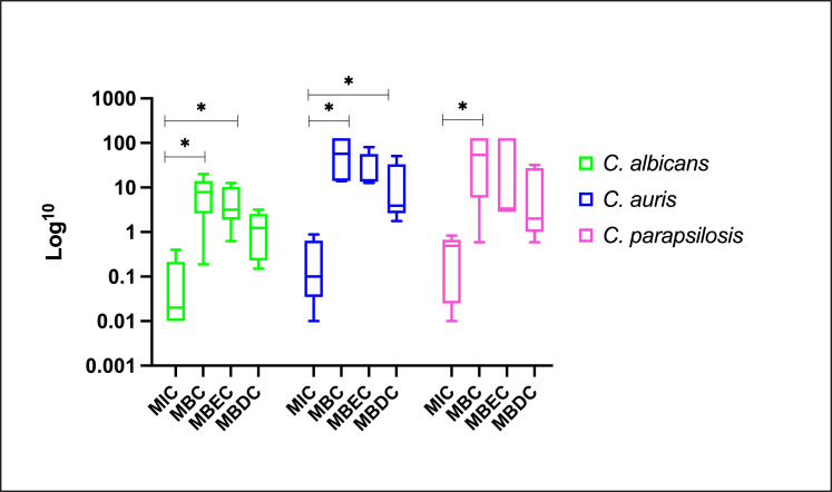
This study comprehensively assessed the activity of manogepix (MNGX), ibrexafungerp (IBF), amphotericin B (AMB), rezafungin (RZF), and caspofungin (CAS) against planktonic cells and mature biofilms of Candida spp.-reference and clinical strains using the Calgary biofilm device. Mature-phase biofilms of C. albicans, C. auris (clades I, II, III, IV), and C. parapsilosis were exposed to a range of drug concentrations (0.12-128 µg/mL). Minimum Inhibitory Concentration (MIC) values for planktonic cells were ≤2 µg/mL for all strains; however, biofilm-associated MICs, minimum biocidal concentration (MBC), minimum biofilm eradication (MBEC), and minimum biofilm damaging concentration (MBDC) were significantly higher (2-4,119 times). Geometric mean (GM) of MBEC values indicated that MNGX had the highest antifungal activity within Candida species, with a GM-MBEC of 5.9 µg/mL. Despite its overall potency, MNGX was less effective against C. auris biofilms from clade IV strains, where IBF showed superior activity. While not the most potent agent overall, AMB induced the smallest fold-change increases (2- to 32-fold) in biofilm-associated states data compared to planktonic MICs. Conversely, CAS exhibited the lowest activity against Candida spp. biofilms. The eradication of C. auris and C. parapsilosis biofilms required substantially higher concentrations than C. albicans, with some agents, such as RZF and CAS, necessitating up to 42-fold increases in dosage. In conclusion, our in vitro model highlights the antibiofilm activity of novel antifungals against major Candida species, revealing significant differences in efficacy among species. MNGX demonstrated the highest activity, underscoring its potential as a promising candidate for the treatment of biofilm-related infections.
Keywords: Calgary biofilm device; Candida biofilms; antibiofilm activity; novel antifungals.
Conflict of interest statement
The authors declare no conflict of interest.
Figures

Similar articles
-
Candida auris: a comparison between planktonic and biofilm susceptibility to antifungal drugs.J Med Microbiol. 2019 Sep;68(9):1353-1358. doi: 10.1099/jmm.0.001036. Epub 2019 Jul 4. J Med Microbiol. 2019. PMID: 31271350
-
In vitro activity of micafungin against biofilms of Candida albicans, Candida glabrata, and Candida parapsilosis at different stages of maturation.Folia Microbiol (Praha). 2018 Mar;63(2):209-216. doi: 10.1007/s12223-017-0555-2. Epub 2017 Oct 5. Folia Microbiol (Praha). 2018. PMID: 28983822
-
Geraniol inhibits both planktonic cells and biofilms of the Candida parapsilosis species complex: Highlight for the improved efficacy of amphotericin B, caspofungin and fluconazole plus Geraniol.Med Mycol. 2024 Nov 12;62(11):myae105. doi: 10.1093/mmy/myae105. Med Mycol. 2024. PMID: 39474890
-
Lack of efficacy of echinocandins against high metabolic activity biofilms of Candida parapsilosis clinical isolates.Braz J Microbiol. 2020 Sep;51(3):1129-1133. doi: 10.1007/s42770-019-00219-7. Epub 2020 Jan 2. Braz J Microbiol. 2020. PMID: 31898245 Free PMC article.
-
Candida and candidaemia. Susceptibility and epidemiology.Dan Med J. 2013 Nov;60(11):B4698. Dan Med J. 2013. PMID: 24192246 Review.
References
MeSH terms
Substances
Grants and funding
LinkOut - more resources
Full Text Sources
Miscellaneous
