. 2025 May 15;49(4):198.
doi: 10.1007/s11259-025-10764-2.

Three-dimensional reconstruction of gustatory papillae and its taste buds in short-hair cats (Felis Catus domestica, felidae, Carnivora)

Affiliations

Three-dimensional reconstruction of gustatory papillae and its taste buds in short-hair cats (Felis Catus domestica, felidae, Carnivora)

Plewa Barbara et al. Vet Res Commun. .

Abstract

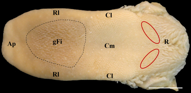
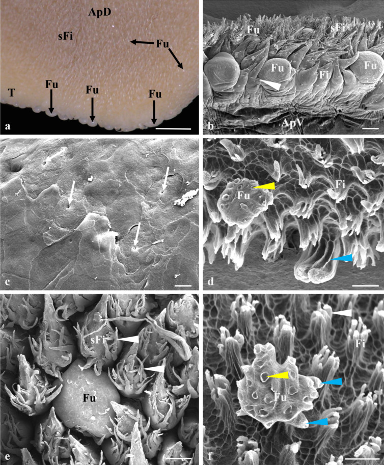
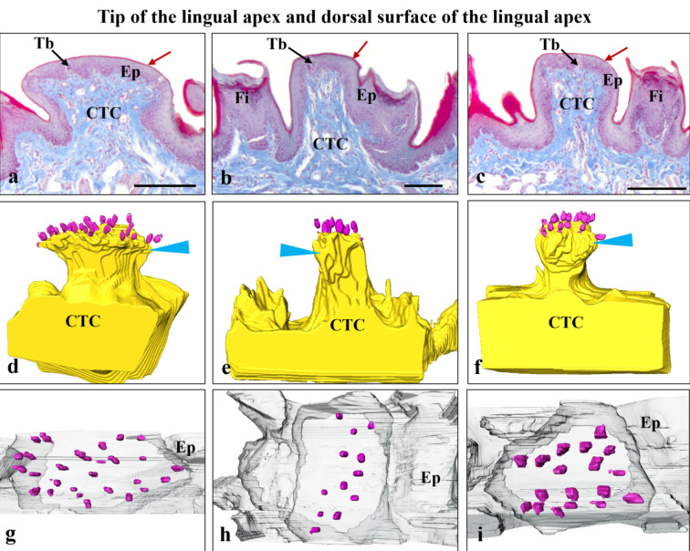
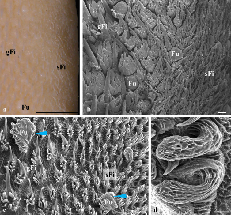
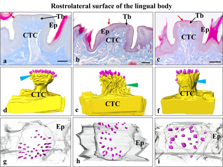
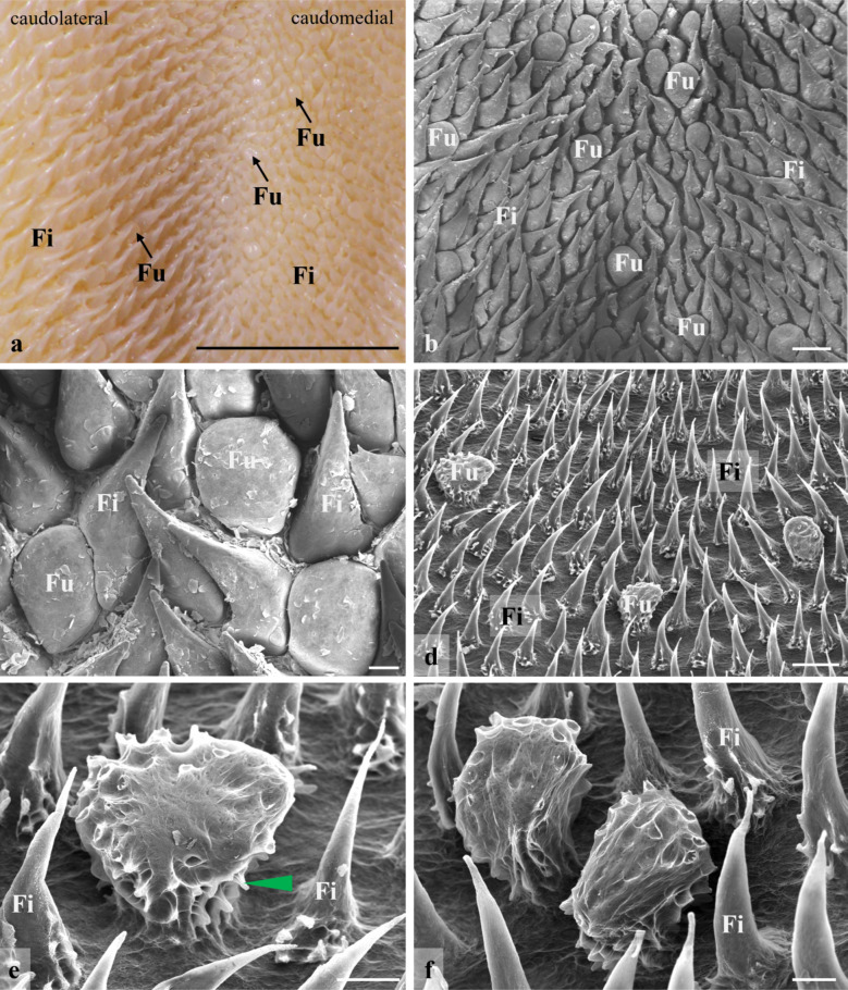
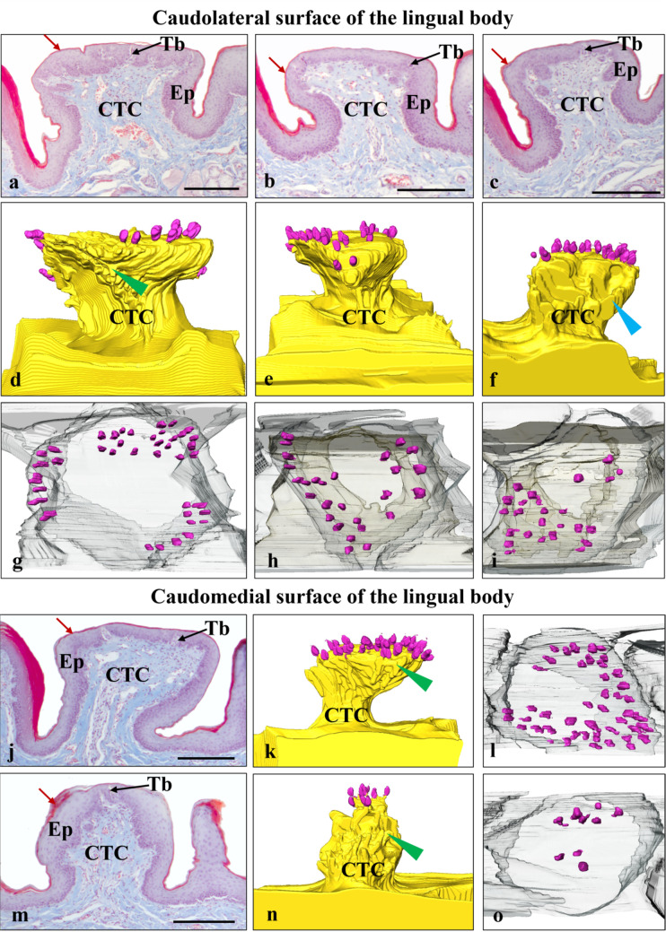
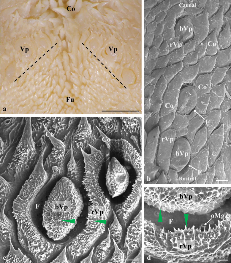
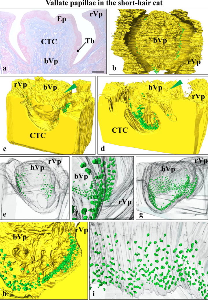
Previous studies on the cat's tongue focused on the anatomy of the tongue and on the lingual papillae micromorphology. According to new challenges in studies on gustatory papillae, we adopted a modern computer-aided method of 3D reconstruction of gustatory papillae from serial 2D cross-sections in the description of the taste bud system. The study in short-hair cats aimed for the first time in carnivores to analyze fungiform (Fu papillae) and vallate papillae (Vp papillae) with spatial visualization of connective tissue cores (CTCs) and the arrangement and exact number of taste buds (Tbs). Results indicate the diversity in size and internal connective tissue microstructure of Fu and Vp papillae in short-hair cats. Four types of CTCs were distinguished in Fu papillae: mushroom-like, club-like, columnar-like, and bud-like, whereas CTC in Vp papillae was mushroom-like. The Tbs of Fu and on Vp papillae were either evenly distributed or grouped. Tbs of Fu papillae analyzed in particular parts of the tongue and on Vp papillae revealed differences in its number. The estimated total number of Tbs on the tongue was up circa 8265. Our 2D and 3D Tbs analyses highlight the functional importance of different tongue regions: the lingual apex for food preselection, the lateral lingual surfaces for analyzing chewed food, and the caudal part of the tongue for final food tasting before swallowing. Comparing our results in cats with previous studies on herbivores, we recommend using proposed 3D analyses as effective tools for further comparative studies of mammalian gustatory structures.

Keywords: 3D reconstruction and modelling; Domestic Cat; Gustatory papillae; Taste buds.


Conflict of interest statement

Declarations. Ethics approval: According to Polish law and the EU directive (no. 2010/63/EU), the studies conducted within the study do not require approval of the Local Ethical Committee for Experiments on Animals in Poznan. Consent to participate: Not applicable. Consent to publish: Not applicable. Competing interests: The authors declare no competing interests.

Figures

Fig. 1
Fig. 1
Dorsal view of a short-hair cat tongue. Ap – lingual apex, Rl – rostrolateral surface of the lingual body, Cl – caudolateral surface of the lingual body, Cm – caudomedial surface of the lingual body, R – lingual root, gFi – giant filiform papillae, red circles – vallate papillae. Scale bar = 1 cm
Fig. 2
Fig. 2
The tongue tip and dorsal surface of the lingual apex in the short-hair cat. (a) – The macroscopic view. T – the tongue tip, ApD – the dorsal surface of the apex, Fu – fungiform papillae, sFi – small filiform papillae. Scale bar = 200 μm; (b) Fu – fungiform papillae, Fi – filiform papillae, sFi – small filiform papillae, ApV – ventral surface of the tongue, white arrowheads – keratinized small processes of Fi papillae. SEM, Scale bar = 200 μm. (c) – The magnification of the apical surface of the fungiform papillae on the tongue tip. White arrows – taste pores. Scale bar = 100 μm (d) The CTC of macerated Fu papillae of the tongue tip. Fu – fungiform papillae, Fi – filiform papillae, blue arrowhead – lateral laminae on Fu papillae CTCs, yellow arrowhead – depressions on the Fu papillae CTC surface, SEM, Scale bar = 100 μm. (e) – Fu – fungiform papillae on the lingual apex, sFi – small filiform papillae, white arrowheads – keratinized small processes of sFi papillae. SEM, Scale bar = 100 μm. (f) – The CTC of macerated Fu papillae of the lingual apex. Fu – fungiform papillae, Fi – filiform papillae, white arrowheads – keratinized small processes of Fi papillae, blue arrowhead – lateral laminae on Fu papillae CTCs, yellow arrowhead – depressions on the Fu papillae CTC surface SEM. Scale bar = 100 μm
Fig. 3
Fig. 3
The tongue tip and dorsal surface of the lingual apex in the short-hair cat (a, b) – cross-sections of Fu papillae on the tongue tip. Red arrow – keratinized layer, CTC – connective tissue core, Ep – multilayered epithelium, Tb – taste buds. LM, Scale bar = 100 μm (b), 200 μm (a). (c) – the cross-section of Fu papilla on the lingual apex. Red arrow – keratinized layer, CTC – connective tissue core, Ep – multilayered epithelium, Tb – taste buds, Fi – filiform papillae. LM. Scale bar = 100 μm (d, e) – the lateral view of 3D models of Fu papillae CTC on the tongue tip. CTC – connective tissue core, pink dots – taste buds, blue arrowhead – lateral laminae on Fu papillae CTCs. (f) – the lateral view of 3D models of Fu papillae CTC on the lingual apex. CTC – connective tissue core, pink dots – taste buds, blue arrowhead – lateral laminae on Fu papillae CTCs. (g, h) – top view of Fu papillae of the tongue tip. Ep – transparent multilayered epithelium, pink dots – taste buds. (i) top view of Fu papillae of the lingual apex. Ep – transparent multilayered epithelium, pink dots – taste buds
Fig. 4
Fig. 4
The rostrolateral surface of the lingual body in the short-hair cat. (a) gFi – giant filiform papillae, sFi – small filiform papillae, Fu – fungiform papillae. Scale bar = 0.5 cm. (b) gFi – giant filiform papillae, sFi – small filiform papillae, Fu – fungiform papillae. SEM. Scale bar = 200 μm. (c) the NaOH macerated CTC of lingual papillae. gFi – giant filiform papillae, sFi – small filiform papillae, Fu – fungiform papillae. SEM. Scale bar = 300 μm. (d) Fu papillae on the lateral margins of the tongue. SEM. Scale bar = 100 μm
Fig. 5
Fig. 5
The rostrolateral surface of the lingual body in the short-hair cat. (a-c) Cross-sections on Fu papillae. CTC – connective tissue core, Ep – multilayered epithelium, Tb – taste buds, red arrows – keratinized layer. LM. Scale bar = 100 μm. (d-f) The lateral view of 3D models of Fu papillae CTC. CTC – connective tissue core, pink dots – taste buds, blue arrowhead – lateral laminae on Fu papillae CTCs (d, f), green arrowheads – short and sharpened protrusions (e). (g-i) Top view of Fu papillae. Pink dots – taste buds
Fig. 6
Fig. 6
The caudal surface of the lingual body in the short-hair cat. (a) Fu – fungiform papillae, Fi – filiform papillae. Scale bar = 0.5 cm. (b) Caudomedial area of the lingual body. Fu – fungiform papillae, Fi – filiform papillae. SEM. Scale bar = 400 μm. (c) The caudomedial area of the lingual body. Fu – fungiform papillae, Fi – filiform papillae. SEM. Scale bar = 100 μm. (d) NaOH macerated CTC of Fu papillae (Fu) and filiform papillae (Fi) on the caudomedial area of the lingual body. SEM. Scale bar = 300 μm. (e, f) The CTC of the Fu papilla on the caudomedial area of the lingual body. Fi – filiform papillae, green arrowhead – collagenous protrusions. SEM. Scale bar = 100 μm
Fig. 7
Fig. 7
The caudal surface of the lingual body in the short-hair cat. (a-c) Caudolateral area of the lingual body. CTC – connective tissue cores, Ep – multilayered epithelium, Tb – taste buds, red arrows – keratinized layer. LM. Scale bar = 200 μm. (d-f) Lateral view of 3D models of CTC of Fu papillae. CTC – connective tissue core, pink dots – taste buds, green arrowhead – short collagenous protrusions (d), blue arrowhead – lateral laminae (f). (g-i) The top view of Fu papillae. Ep – transparent multilayered epithelium, pink dots – taste buds. (j, m) - Cross-sections of Fu papillae on the caudomedial area of the lingual body. CTC – connective tissue cores, Ep – multilayered epithelium, Tb – taste buds, red arrows – keratinized layer. LM. Scale bar = 200 μm. (k, n) - lateral view of 3D models of CTC of Fu papillae. CTC – connective tissue core, pink dots – taste buds, green arrowhead – collagenous protrusions. (l, o) - The top view of Fu papillae. Ep – transparent multilayered epithelium, pink dots – taste buds
Fig. 8
Fig. 8
Vallate papillae in the short-hair cat. (a) Macroscopic view. Vp – vallate papillae, Co – conical papillae, Fu – fungiform papillae. Scale bar = 0.5 cm. (b) Co – conical papillae, F – papillary furrow, bVp – body of the vallate papillae, rVP – ridge of the vallate papillae. SEM. Scale bar = 400 μm. (c) The CTC of Vp papillae. Co – conical papillae, F – papillary furrow, bVp – body of the vallate papillae, rVP – ridge of the vallate papillae, green arrowheads – short collagenous protrusions. SEM. Scale bar = 300 μm. (d) The magnification of the papillary furrow. F – papillary furrow, bVp – body of the vallate papillae, rVP – ridge of the vallate papillae, oMg – openings of mucous glands, green arrowheads – short collagenous protrusions. SEM. Scale bar = 100 μm
Fig. 9
Fig. 9
Vallate papillae in the short-hair cat. (a) bVP – the body of the vallate papillae, rVp – the ridge of vallate papillae, CTC – connective tissue core, Ep – multilayered epithelium, Tb – taste buds. LM. Scale bar = 200 μm. (b) The top view of 3D model of vallate papillae. bVP – the body of the vallate papillae, rVp – the ridge of vallate papillae, green dots – taste buds. (c, d) Lateral view of 3D models of vallate papillae. bVP – the body of the vallate papillae, rVp – the ridge of vallate papillae, CTC – connective tissue core, green dots – taste buds, green arrowheads – short collagenous protrusions. (e, g) The transparent 3D models of vallate papillae with the arrangement of taste buds (green dots). bVP – the body of the vallate papillae, rVp – the ridge of vallate papillae. (f, h) The magnification of 3D model of vallate papillae furrow with the arrangement of taste buds (green dots). bVP – the body of the vallate papillae, rVp – the ridge of vallate papillae. (i) The lateral view of the Vp body with the distribution of taste buds (green dots)

Similar articles

References

    1. Chamorro CA, Sandoval J, Fernandez JG, Fernandez M, de Paz P (1987) Compared studies of the lingual papillae of the cat (Felis catus) and the rabbit (Oryctolagus cuniculus) by scanning electron microscopy. Anat Histol Embryol 16:37–47. 10.1111/j.1439-0264.1987.tb00722.x - PubMed
    1. Crouch JE (1969) Text-Atlas of Cat anatomy. LEA & FEBIGER, Philadelphia, pp 131–138
    1. Dyce KM, Sack WO, Wensing CJG (2010) Textbook of veterinary anatomy, 4th edn. SAUNDERS ELSEVIER, St. Louis, Missouri, pp 399–405
    1. Eizirik E, Murphy WJ (2009) Carnivores (Carnivora). In: Hedges SB, Kumar S (eds) Timetree of Life, Oxford University Press, pp 504–507
    1. Emura S (2018a) Morphology of the lingual papillae in the Asian golden Cat. Okajimas Folia Anat Jpn 95:19–22. 10.2535/ofaj.95.19 - PubMed

LinkOut - more resources