. 2025 May 15;74(7):205.
doi: 10.1007/s00262-025-04054-8.

Mast cells-intestinal cancer cells crosstalk is mediated by TNF-alpha and sustained by the IL-33/ST2 axis

Affiliations

Mast cells-intestinal cancer cells crosstalk is mediated by TNF-alpha and sustained by the IL-33/ST2 axis

Chiara Dal Secco et al. Cancer Immunol Immunother. .

Abstract

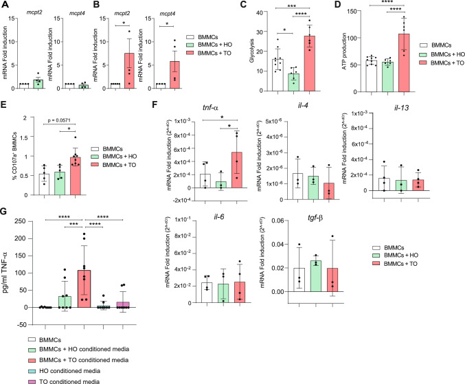
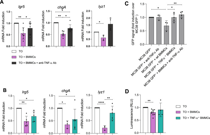
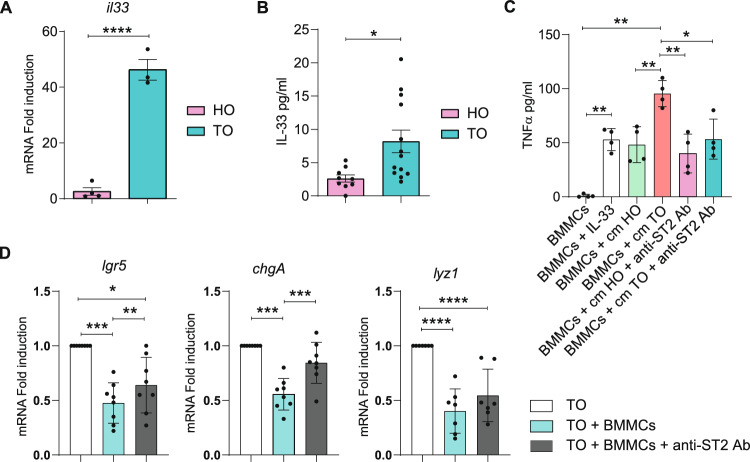
It is common knowledge that mast cells (MCs) exert different roles in the gastrointestinal tract, from the maintenance of homeostasis to the onset and propagation of different gut diseases such as food allergies, infections, inflammation, and cancer. However, the mechanisms through which MCs dialog and influence the intestinal tissue are not completely known. To get insight into the bidirectional crosstalk between MCs and the intestinal microenvironment, both in homeostatic and pathological settings, colon organoids from intestinal epithelium of healthy mice and adenomas from AOM/DSS-treated mice have been exploited and co-cultured with MCs. The influence of MCs on organoid architecture and the effect of healthy and tumoral organoids on the phenotype and responsiveness of MCs have been addressed. We observed that MCs interact with intestinal organoids and contribute to the differentiation of healthy organoids by upregulating the expression of mucin-2, chromogranin A, cadherin-1, and claudin 4. On the contrary, in co-culture with tumoral organoids a decrease in cell proliferation, chromogranin A, and lysozyme expression was observed. Tumoral organoids have been shown to activate MCs via the IL-33/ST2 axis leading to increased release of TNF-α which in turn was responsible for the observed effects on tumoral organoids. Our results indicate that MCs are important mediators of intestinal tissue homeostasis and that a different environment can shape and direct MCs toward the dampening or propagation of the inflammatory response. Ultimately, our MC-organoid co-cultures represent a valid in vitro tool to investigate the role of MCs in the gut.

Keywords: Co-cultures; IL-33; Intestinal differentiation and architecture; Mast cells; Organoids; TNF-α.


Conflict of interest statement

Declarations. Conflict of interest: The authors declare no competing interests.

Figures

Fig. 1
Fig. 1
Healthy and tumoral organoids differently affect the behavior of MCs. (A-B) qPCR analysis of mcpt2 and mcpt4 on BMMCs alone and co-cultured with HO (A) or TO (B) for 48 h. (C) Glycolysis and (D) ATP production evaluation in BMMCs alone and co-cultured with HO or TO. (E) Percentage of CD107a+ expressing BMMCs alone and co-cultured for 24 h with HO or TO. (F) qPCR analysis of tnf-α, il-4, il-13, il-6, and tgf-β on BMMCs alone and co-cultured with HO or TO organoids for 4 h. (G) TNF-α quantified by ELISA in supernatant from BMMCs cultured in normal medium or cultured in organoid-derived conditioned media and from organoids conditioned media. Data are expressed as mean + SD from at least n = 3–5 experiments. Statistical analysis was performed with paired Student t-test or with one-way ANOVA with Dunnet correction (* = p < 0.05 ** = p < 0.01; *** = p < 0.001)
Fig. 2
Fig. 2
Effect of MCs on the polarization and organization of HO. (A) qPCR analysis of lgr5, muc2, chgA, cdh1, and cldn4 on HO after 72 h co-culture with resting or IgE/antigen-activated BMMCs (Act BMMCs). (B) Immunofluorescence staining of ZO-1, Ezrin, and claudin 4 (all green) in HO co-cultured with resting and activated MCs. Nuclei in blue. (C) Western blot analysis of Cldn4 expression in healthy colon organoids cultured alone, and co-culture with resting or activated BMMCs. Right panel shows densitometry analysis calculated over actin expression and normalized versus organoid alone. Data are expressed as mean + SD from n = 3–6 experiments; statistical analysis were performed with one-way ANOVA with Dunnet correction (* = p < 0.05 ** = p < 0.01; *** = p < 0.001)
Fig. 3
Fig. 3
Effect of MCs on the polarization and organization of TO. (A) qPCR analysis of lgr5, muc2, chgA, lyz1, and cdh1 on tumoral colon organoids after 72 h of co-culture with resting or IgE/antigen-activated BMMCs. (B) Percentage of Ki67hi and Ki67 gMFI of tumoral organoid cells after 48 h of co-culture with resting BMMCs. (C) Proliferation of TO through luminescent assay or of (D) MC38-GFP cells, through evaluation of GFP fluorescence, cultured alone or in the presence of BMMCs. In the case of GFP-MC38 cells, data were normalized over GFP-MC38 cultured alone. Data are expressed as mean + SD from n = 3 experiments. Statistical analysis was performed with paired Student t-test ore one-way ANOVA with Dunnet correction (* = p < 0.05 ** = p < 0.01; *** = p < 0.001)
Fig. 4
Fig. 4
Blocking MC-derived TNF-α attenuates the effects produced on TO. (A) qPCR analysis of lgr5, chgA, and lyz1 in TO after 72 h of co-culture with BMMCs in the presence or absence of TNF-α blocking antibody. (B) qPCR analysis of lgr5, chgA, and lyz1 on TO after 72 h co-culture with wild type (wt) or TNF-α−/− BMMCs. (C) Proliferation rates of GFP-MC38 cultured alone or in presence of TNF-α blocking antibody, wt BMMCs, TNF-α−/− BMMCs, or wt BMMCs together with TNF-α blocking ab. (D) Proliferation rates of TO cultured alone or in presence TNF-α −/− BMMCs or wt BMMCs. Data are expressed as mean + SD from n = 3–8 experiments. Statistical analysis was performed with one-way ANOVA with Dunnet correction (* = p < 0.05 ** = p < 0.01; *** = p < 0.001)
Fig. 5
Fig. 5
The ST2/IL-33 axis plays a role in MCs-organoids interaction. (A) qPCR analysis of Il-33 expression in HO and TO. (B) Quantification of soluble IL-33 released from HO or TO in the culture medium was determined by ELISA. (C) Quantification by ELISA of TNF-α secreted by BMMCs alone, after incubation with 50 ng/ml of recombinant IL-33 and after cell culture in organoid-conditioned media (cm) in the presence/absence of ST2-blocking antibody. (D) qPCR analysis of lgr5, chgA, and lyz1 expression on TO organoids after 72 h of co-culture with BMMC in the presence or absence of ST2-blocking antibody. Data are expressed as mean + SD from n = 3–8 experiments. Statistical analysis was performed with Student t-test or one-way ANOVA with Dunnet correction (* = p < 0.05 ** = p < 0.01; *** = p < 0.001)

References

    1. Dwyer DF, Barrett NA, Austen KF et al (2016) Expression profiling of constitutive mast cells reveals a unique identity within the immune system. Nat Immunol 17:878–887. 10.1038/NI.3445 - PMC - PubMed
    1. Bischoff SC, Krämer S (2007) Human mast cells, bacteria, and intestinal immunity. Immunol Rev 217:329–337. 10.1111/J.1600-065X.2007.00523.X - PubMed
    1. Bischoff SC (2009) Physiological and pathophysiological functions of intestinal mast cells. Semin Immunopathol 31:185–205. 10.1007/s00281-009-0165-4 - PubMed
    1. Merluzzi S, Frossi B, Gri G et al (2010) Mast cells enhance proliferation of B lymphocytes and drive their differentiation toward IgA-secreting plasma cells. Blood 115:2810–2817. 10.1182/blood-2009-10-250126 - PubMed
    1. Groschwitz KR, Ahrens R, Osterfeld H et al (2009) Mast cells regulate homeostatic intestinal epithelial migration and barrier function by a chymase/Mcpt4-dependent mechanism. Proc Natl Acad Sci 106:22381–22386. 10.1073/pnas.0906372106 - PMC - PubMed

Substances

LinkOut - more resources