. 2025 May 15;10(12):e186646.
doi: 10.1172/jci.insight.186646. eCollection 2025 Jun 23.

Rheumatoid arthritis synovial fluid induces JAK-dependent intracellular activation of human sensory neurons

Affiliations

Rheumatoid arthritis synovial fluid induces JAK-dependent intracellular activation of human sensory neurons

Yuening Li et al. JCI Insight. .

Abstract

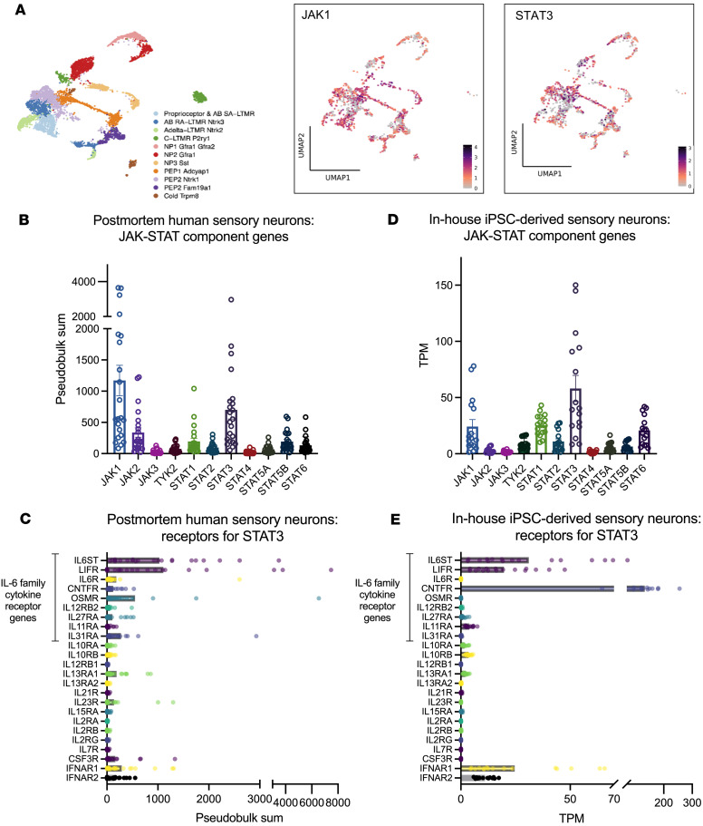
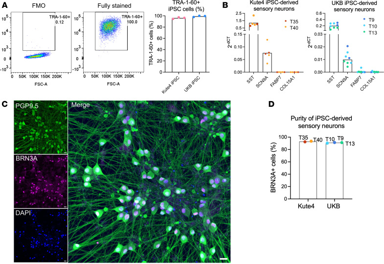
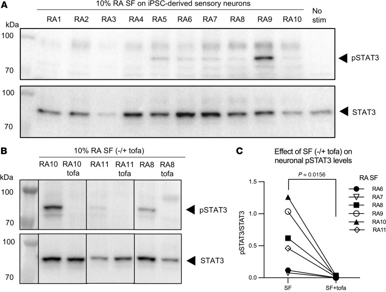
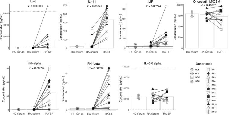
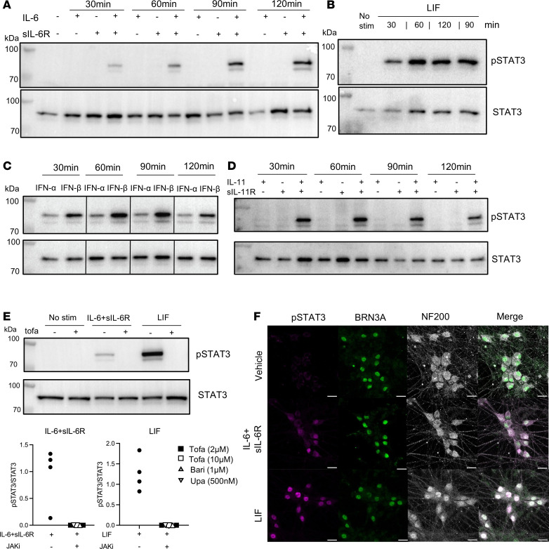
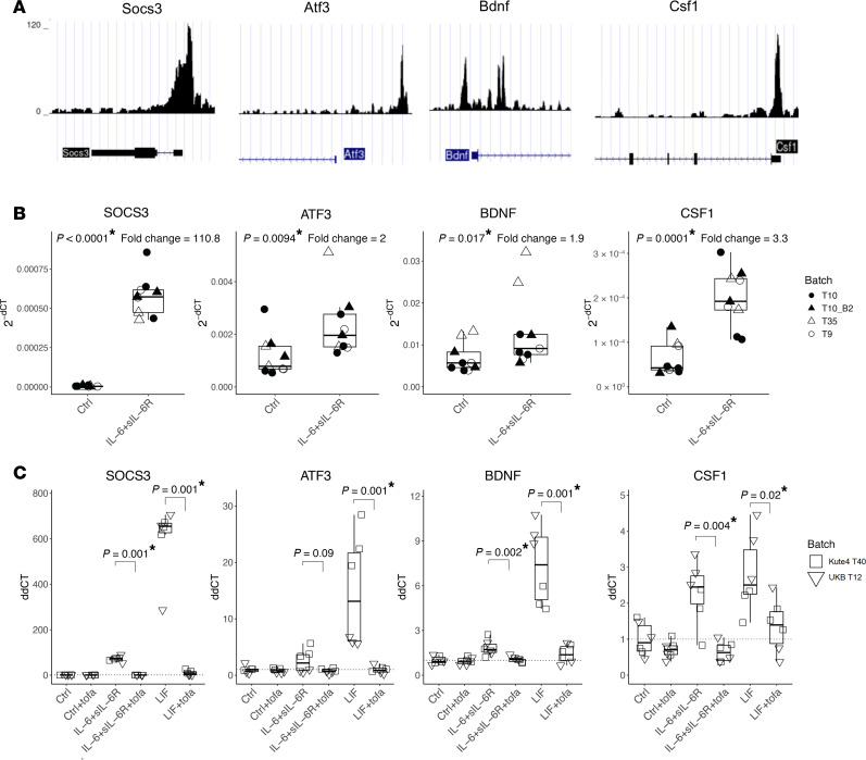
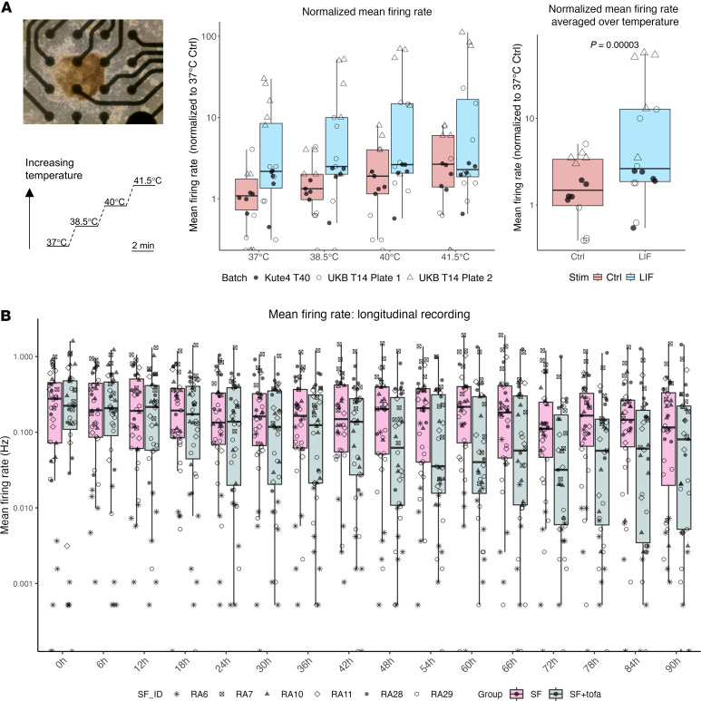
JAK inhibitors (JAKi) are widely used antiinflammatory drugs. Recent data suggest that JAKi have superior effects on pain reduction in rheumatoid arthritis (RA). However, the underlying mechanisms for this observation are not fully understood. We investigated whether JAKi can act directly on human sensory neurons. We analyzed RNA-seq datasets of sensory neurons and found that they expressed JAK1 and STAT3. Addition of cell-free RA synovial fluid to human induced pluripotent stem cell-derived (iPSC-derived) sensory neurons led to phosphorylation of STAT3 (pSTAT3), which was completely blocked by the JAKi tofacitinib. Compared with paired serum, RA synovial fluid was enriched for the STAT3 signalling cytokines IL-6, IL-11, LIF, IFN-α, and IFN-β, with their requisite receptors present in peripheral nerves postmortem. Accordingly, these recombinant cytokines induced pSTAT3 in iPSC-derived sensory neurons. Furthermore, IL-6 + sIL-6R and LIF upregulated expression of pain-relevant genes with STAT3-binding sites, an effect that was blocked by tofacitinib. LIF also induced neuronal sensitization, highlighting this molecule as a putative pain mediator. Finally, over time, tofacitinib reduced the firing rate of sensory neurons stimulated with RA synovial fluid. Together, these data indicate that JAKi can act directly on human sensory neurons, providing a potential mechanistic explanation for their suggested superior analgesic properties.

Keywords: Immunology; Inflammation; Neuroscience; Pain; Rheumatology; iPS cells.


Figures

Figure 1
Figure 1. Genes related to JAK/STAT signalling are expressed in human sensory neurons and iPSC-derived sensory neurons.
(A) Single-nuclei RNA-seq datasets (XSpecies DRG Atlas: http://research-pub.gene.com/XSpeciesDRGAtlas/) (13) were analyzed for the expression of JAK1 and STAT3 across different subtypes of human postmortem sensory neurons. Color scales indicate scaled average expression across samples. (BE) Pseudobulk analyses of single-nuclei RNA-seq of human postmortem (13, 14) (B) and in-house iPSC-derived sensory neurons for the indicated JAK and STAT molecules (17) (D). Expression patterns of the receptors for the indicated STAT3 signalling cytokines in human postmortem (C) and in-house iPSC-derived sensory neurons (E). TPM, transcripts per million. Error bars in B and D indicate SEM.
Figure 2
Figure 2. Characterization of iPSC-derived sensory neurons.
(A) IPSC-derived sensory neurons were differentiated from Kute4 and UKB lines and stained for the expression of TRA-1-60, a marker of undifferentiated cells. Representative flow cytometry plots and cumulative data (n = 3) showing the percentage of TRA-1-60+ cells in UKB iPSC at passage 14, as compared to the fluorescence minus one (FMO) control (left panel). (B) Bar plots showing gene expression of neuronal markers (SST, SCN9A) and nonneuronal genes (FABP7, COL15A1) by sensory neurons derived from Kute4 (left) and UKB (right) iPSC lines. Within each independent differentiation batch (indicated by trial (T) number and different colors) n = 2–3 biological repeats were used. (C and D) IPSC-derived sensory neurons were differentiated to over day 50 and stained for the sensory neuron marker BRN3A and panneuronal marker PGP9.5. Representative IHC of day 81 neuron staining shown in C (Scale bar: 20 μm) and cumulative data showing the purity of the neuronal culture, quantified as BRN3A+ cells within DAPI+ cells (D, n = 5 independent differentiation batches of Kute4 and UKB iPSC lines).
Figure 3
Figure 3. RA-derived synovial fluid can induce pSTAT3 in human sensory neuron cultures, which is blocked by tofacitinib.
(A) IPSC-derived sensory neurons (day 55) were incubated for 1 hour with 10% RA SF (n = 10) or media alone (No stim). Cells were lysed and probed for pSTAT3 (upper panel) and STAT3 (lower panel) using Western blot. (B and C) IPSC-derived sensory neurons were stimulated as in A in the absence or presence of tofacitinib (2 μM) for 1 hour and pSTAT3/STAT3 expression analyzed. Representative Western blot (B) and cumulative data (C) showing the quantification of the ratio of pSTAT3/STAT3 signal of neurons from two batches (day 55–65) stimulated with 10% RA SF from n = 6 different individuals with RA in the absence or presence of tofacitinib. Data were analyzed using a paired nonparametric 1-tailed t test.
Figure 4
Figure 4. Cytokines upstream of STAT3 signaling are enriched in RA SF.
Healthy control (HC) serum (n = 4) and paired RA serum and SF (n = 12) were analyzed for the presence of IL-6, IL-11, LIF, OSM, IFN-α, IFN-β, and IL-6R-α by Luminex assay. Statistical significance was assessed using paired nonparametric 2-tailed t tests (RA SF versus serum); P < 0.008 was considered significant after Bonferroni correction for the 6 cytokines. The dotted lines for IL-6 and IL-6R-α graphs indicate the highest standard value. The dashed lines indicate the lowest standard value. The legend symbols are representative of individual HC or RA samples; the bars represent the data means. Standard values, sensitivity, and all individual means (+SD) of the assay are listed in Supplemental Table 4.
Figure 5
Figure 5. RA-relevant recombinant cytokines induced neuronal pSTAT3 in peripheral sensory neurons.
(AD) IPSC-derived sensory neurons were incubated for 30, 60, 90, and 120 minutes with human recombinant (A) IL-6, sIL-6R, or both (100 ng/mL), (B) LIF (100 ng/mL), (C) IFN-α (300 U/mL), IFN-β (300 U/mL), or (D) IL-11, sIL-11R, or both (100 ng/mL). Cell lysates were probed for pSTAT3 and STAT3 by Western blot. (E) Neurons from 2 batches were incubated with IL-6 + sIL-6R or LIF without or with JAKi for 1 hour. Representative blot of neurons blocked using 10 μM tofacitinib and cumulative data using tofacitinib, baricitinib or upadacitinib (n = 4–5). (F) Neurons (day 77) were stained for pSTAT3 and sensory neuron markers BRN3A and NF200 by immunocytochemistry, shown in individual or merged channels. Scale bar: 20 μm.
Figure 6
Figure 6. IL-6 + sIL-6R and LIF increase the expression of pain-relevant genes with pSTAT3 binding sites, which is reversed by JAKi.
(A) ChIP sequencing dataset (23) showing pSTAT3 binding sites in Socs3, Atf3, Bdnf, and Csf1 in mouse DRG neurons. (B) IPSC-derived sensory neurons were treated without or with 100 ng/mL IL-6 + sIL-6R for 24 hours, followed by qPCR analysis. Boxplots showing gene expression of the indicated genes. Data derived from n = 9 culture wells using three independent differentiations (designated by the prefix “T”) from two iPSC lines (day 67–76). (C) IPSC-derived sensory neurons were treated without or with IL-6 + sIL-6R or LIF (all at 100 ng/mL) for 24 hours in the absence or presence of tofacitinib (2 μM), followed by qPCR analysis. Boxplots showing expression of the indicated genes. Data are derived from n = 6 neuronal wells derived from two independent iPSC lines (day 57–61). Statistical significance was determined by 1-tailed nonparametric t tests to compare between with or without IL-6 + sIL-6R (B) or with or without tofacitinib in the presence of IL-6 + sIL-6R or LIF (C). Neurons aged day 57–76 were used. P < 0.013 was deemed significant after Bonferroni correction and marked by an asterisk.
Figure 7
Figure 7. Modulation of neuronal firing by LIF and JAK inhibitors.
(A) Neurons plated on an MEA plate (scale bar: 20 μm) treated without or with 100 ng/mL LIF for 92 hours and subjected to increased temperature. Boxplots of Ctrl (red) and LIF (blue) showing normalized mean firing rates at each temperature (middle) or averaged across temperature (right). The P-value derives from a nonparametric t-test to assess the effect of LIF. Legend symbols represent wells from 3 plates and 2 iPSC lines (n = 14–16, neuronal age: day 64–75). (B) Neurons plated on an MEA plate treated with 10% RA SF without or with 2 μM tofacitinib (tofa) for 92 hours (92 h). Boxplots of RA SF without tofa (magenta) and with tofa (grey) showing mean firing rates of neurons every 6 hours (h) from the addition of RA SF (baseline, 0h) to 90 hours (90h). A repeated-measures ANOVA was used to assess the effect of time, group (SF vs SF+tofa) and any interaction. Legend symbols represent wells from 2 plates from the Kute4 iPSC line (T52, T55) with the respective RA SF stimulation (Plate 1 - RA6, 28, 29; Plate 2 – RA7, 10, 11. N = 5–7 per SF ± tofa, neuronal age D54-59).

Similar articles

Cited by

References

    1. McInnes IB, Schett G. The pathogenesis of rheumatoid arthritis. N Engl J Med. 2011;365(23):2205–2219. doi: 10.1056/NEJMra1004965. - DOI - PubMed
    1. Almutairi KB, et al. The prevalence of rheumatoid arthritis: a systematic review of population-based studies. J Rheumatol. 2021;48(5):669–676. doi: 10.3899/jrheum.200367. - DOI - PubMed
    1. Gullick NJ, et al. Real world long-term impact of intensive treatment on disease activity, disability and health-related quality of life in rheumatoid arthritis. BMC Rheumatol. 2019;3:6. doi: 10.1186/s41927-019-0054-y. - DOI - PMC - PubMed
    1. McInnes IB, et al. Cytokines in rheumatoid arthritis - shaping the immunological landscape. Nat Rev Rheumatol. 2016;12(1):63–68. doi: 10.1038/nrrheum.2015.171. - DOI - PubMed
    1. Tanaka Y, et al. Janus kinase-targeting therapies in rheumatology: a mechanisms-based approach. Nat Rev Rheumatol. 2022;18(3):133–145. doi: 10.1038/s41584-021-00726-8. - DOI - PMC - PubMed

MeSH terms