. 2025 May 15;20(5):e0323227.
doi: 10.1371/journal.pone.0323227. eCollection 2025.

Leishmania regulates host YY1: Comparative proteomic analysis identifies infection modulated YY1 dependent proteins

Affiliations

Leishmania regulates host YY1: Comparative proteomic analysis identifies infection modulated YY1 dependent proteins

Harsimran Kaur Brar et al. PLoS One. .

Abstract

The protein Yin-Yang 1 (YY1) is a ubiquitous multifunctional transcription factor. Interestingly, there are several cellular functions controlled by YY1 that could play a role in Leishmania pathogenesis. Leishmaniasis is a human disease caused by protozoan parasites of the genus Leishmania. This study examined the potential role of macrophage YY1 in promoting Leishmania intracellular survival. Deliberate knockdown of YY1 resulted in attenuated survival of Leishmania in infected macrophages, suggesting a role of YY1 in Leishmania persistence. Biochemical fractionation studies revealed Leishmania infection caused redistribution of YY1 to the cytoplasm from the nucleus where it is primarily located. Inhibition of nuclear transport by leptomycin B attenuates infection-mediated YY1 redistribution and reduces Leishmania survival. This suggests that Leishmania induces the translocation of YY1 from the nucleus to the cytoplasm of infected cells, where it may regulate host molecules to favour parasite survival. A label-free quantitative whole proteome approach showed that the expression of a large number of macrophage proteins was dependent on the YY1 level. Interestingly, several of these proteins were modulated in Leishmania-infected cells, revealing YY1-dependent host response and suggesting their potential role in Leishmania pathogenesis. Together, this study identifies YY1 as a novel virulence factor that promotes Leishmania survival inside host macrophages.


Conflict of interest statement

The authors declare that the research was conducted in the absence of any commercial or financial relationships that could be construed as a potential conflict of interest.

Figures

Fig 1
Fig 1. YY1 knockdown inhibits L. donovani survival in dTHP-1 cells.
(A) THP-1 cells were transfected with three unique YY1 siRNAs or a scrambled siRNA (scr siRNA) for 24 hours followed by differentiation with PMA, and subsequently infection with L. donovani at a 20:1 MOI for 24 hours. Cells were washed extensively and Western blotting was performed using the specified antibodies on whole-cell lysates of infected dTHP-1 cells. (B) Histogram representing densitometric analysis of YY1 knockdown normalized to Actin levels. Bars represent mean ± SD of three independent experiments. (C) Parasite rescue assay was performed in parallel as described in “Materials and methods”. Bars represent the mean ± SD of four independent experiments. Statistical significance was determined using two-sample two-tailed T-test (ns: not significant, *: p < 0.05, **: p < 0.01).
Fig 2
Fig 2. YY1 knockdown reduces L. donovani survival in hMDMs.
(A) Peripheral human blood monocytes were isolated and differentiated to macrophages as described in “Materials and methods”. hMDMs were transfected with two unique YY1 siRNAs or a scrambled siRNA (scr siRNA) for 24 h followed by infection with L. donovani at a 20:1 MOI for 24 h. The parasite rescue assay was then performed as described in “Materials and methods”. Bars represent mean ± SD of three independent experiments. Statistical significance was determined using two-sample two-tailed T-test (*: p < 0.05). (B) YY1 knockdown was verified by Western blot using the indicated antibodies on whole cell lysates of infected hMDMs.
Fig 3
Fig 3. L. donovani infection induces cytoplasmic translocation of YY1 in dTHP-1 cells.
dTHP-1 cells were infected with L. donovani at 20:1 MOI for 24 hours and cellular fractionation was performed as described in “Materials and methods”. (A) The cytoplasmic fractions of non-infected and infected cells were then Western blotted using the indicated antibodies. (B) Histograms representing densitometric analysis of Western blots of cytoplasmic fractions from dTHP-1 cells. (C) The nuclear fractions of non-infected and infected cells were Western blotted using the indicated antibodies. (D) Histograms representing densitometric analysis of western blots of nuclear fractions from dTHP-1 cells. The data shown are from three independent experiments.
Fig 4
Fig 4. Leptomycin B inhibits cytoplasmic translocation of YY1 in dTHP-1 cells.
(A) dTHP-1 cells were either untreated or treated with leptomycin B (Lepto) at a concentration of 10 nM or 20 nM for 3 hours prior to infection with L. donovani at a MOI of 20:1. After 24 hours, cells were fractionated as described in ”Materials and methods”. YY1 protein levels in cytoplasmic fractions were verified by Western blotting with the indicated antibodies. (B) Histogram representing densitometric analysis of YY1 expression in Western blot of cytoplasmic fractions from dTHP-1 cells. (C) Leptomycin B attenuates L. donovani survival in dTHP-1 cells. In parallel, parasite rescue assay was performed as described in “Materials and methods”. For this assay, dTHP-1 cells were either treated with vehicle alone, or treated with leptomycin B at a concentration of 10 nM or 20 nM for 3 hours, followed by infection with L. donovani at a MOI of 20:1 for 24 hours. Bars represent the mean ± SD of three independent experiments. Statistical significance was determined using two-sample two-tailed T-test (**: p < 0.01).
Fig 5
Fig 5. Comparative analysis of global proteome of control and L. donovani infected cells. dTHP-1 cells were either infected with L. donovani or not.
Cells were then dislodged in cell dissociation buffer (without enzyme) and mass spectrometric analysis was perfomed as described in “Materials and methods”. (A) Pie-chart depicting the distribution of proteins significantly upregulated by L. donovani infection, categorized based on the average of log2 values from three independent replicates (fold change). (B) Pie-chart depicting the distribution of proteins significantly downregulated by L. donovani infection, categorizd based on the average of log2 values from three independent experiments. Two sample, two tailed T-test was used to measure statistical significance with p-value <0.05 considered significant. (C) Volcano plot of proteins showing significant fold changes in L. donovani infected cells as compared to non-infected cells. Proteins significantly upregulated by L. donovani infection are shown in green dots, those that were downregulated, as orange dots and not modulated as grey dots. Data is from three independent experiments. The horizontal dashed line represents the p-value cut off of 0.05 or -log10(p-value) of 1.301. C denotes infected; A denotes non-infected.
Fig 6
Fig 6. Gene ontology analysis of YY1-dependent L. donovani modulated proteins in THP-1 cells.
Gene ontology (GO) enrichment analysis was performed on significantly modulated proteins by L. donovani (upregulated or downregulated) and recovered by YY1 (downregulated and upregulated, respectively) knockdown. Proteins were categorized into: (A) Biological process, (B) Molecular Function, and (C) Cellular component. Orange represents the proteins significantly upregulated by L. donovani and recovered by YY1 and green represents the proteins significantly downregulated by L. donovani and recovered by YY1.
Fig 7
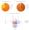
Fig 7. Comparative analysis of global proteome of control and YY1 knockdown cells.
THP-1 cells were transfected with equal mix of two different siRNAs (50 nM each) or a scrambled siRNA for 24 hours, followed by differentiation. Cells were then dislodged in cell dissociation solution (without enzyme) and mass spectrometric analysis was perfomed as described in “Materials and methods”. (A) Pie-chart illustrating the distribution of proteins significantly upregulated by YY1 knockdown, categorized based on average of log2 values. (B) Pie-chart demonstrating the distribution of proteins significantly downregulated by YY1 knockdown, categorized based on average of log2 values. Two sample, two tailed T-test was used to measure statistical significance with p-value <0.05 considered significant. (C) Volcano plot of proteins showing significant fold changes in YY1 knockdown cells as compared to control cells. Proteins significantly upregulated by YY1 knockdown are shown as blue dots, those that were downregulated, as red dots and those that are not modulated as grey dots. Data is from three independent experiments. The p-value cut off of 0.05 or - log10(p-value) of 1.301 is represented by the horizontal dashed line. B denotes YY1 knockdown macrophages; A denotes control macrophages.
Fig 8
Fig 8. Gene ontology analysis of significantly modulated proteins in YY1 knockdown dTHP-1 cells.
Gene ontology (GO) enrichment analysis was performed on top 30 most significantly upregulated and downregulated proteins in YY1 knockdown dTHP-1 cells and classified into three categories: (A) Biological process, (B) Molecular function, and (C) Cellular component. Yellow represents the proteins upregulated by YY1 knockdown and blue represents the proteins downregulated by YY1 knockdown.

Similar articles

Cited by

  • Yin Yang 1: Role in Leishmaniasis.
    Nandan D, Longowal DK, Reiner N. Nandan D, et al. Cells. 2025 Jul 25;14(15):1149. doi: 10.3390/cells14151149. Cells. 2025. PMID: 40801584 Free PMC article. Review.

References

    1. Maxfield L, Crane JS. Leishmaniasis. In: StatPearls [Internet]. Treasure Island (FL): StatPearls Publishing; 2022. [cited 2022 Jul 27]. Available from: http://www.ncbi.nlm.nih.gov/books/NBK531456/
    1. Steverding D. The history of leishmaniasis. Parasit Vectors. 2017;10(1):82. doi: 10.1186/s13071-017-2028-5 - DOI - PMC - PubMed
    1. Serafim TD, Coutinho-Abreu IV, Dey R, Kissinger R, Valenzuela JG, Oliveira F, et al.. Leishmaniasis: the act of transmission. Trends Parasitol. 2021;37(11):976–87. doi: 10.1016/j.pt.2021.07.003 - DOI - PubMed
    1. Leishmaniasis [Internet]. [cited 2023 Oct 30]. Available from: https://www.who.int/news-room/fact-sheets/detail/leishmaniasis
    1. Velez R, Gállego M. Commercially approved vaccines for canine leishmaniosis: a review of available data on their safety and efficacy. Trop Med Int Health. 2020;25(5):540–57. doi: 10.1111/tmi.13382 - DOI - PubMed