. 2025 May 15;20(5):e0323576.
doi: 10.1371/journal.pone.0323576. eCollection 2025.

Effect of UV-A on endophyte colonisation of Arabidopsis thaliana

Affiliations

Effect of UV-A on endophyte colonisation of Arabidopsis thaliana

Aleksandra Giza et al. PLoS One. .

Abstract

UV-A, an important part of sunlight radiation, is typically absent in experiments on plant-endophyte interactions. We examined the impact of UV-A in the 350-400 nm range (UV-A1 waveband) on the plant interactions with fungal endophytes belonging to different taxonomic groups: Paraphoma chrysanthemicola, Phomopsis columnaris, Diaporthe eres, Mucor sp., and yeast Sporobolomyces ruberrimus. Physiologically relevant levels of UV-A did not substantially affect the colonisation of shoots and roots by endophytes. UV-A upregulated the expression of genes involved in the establishment of symbiosis. Specifically, the expression of PDF1.2 was affected by P. chrysanthemicola and S. ruberrimus only under UV-A conditions. Additionally, UV-A exposure upregulated the mRNA levels of ICS1 and PAL1, genes important for plant responses to stress factors. Inoculation with P. chrysanthemicola and S. ruberrimus led to increased expression of the ICS1 gene. We did not observe significant interactions between the effects of UV-A and the presence of endophytes on other examined plant traits, including plant fresh weight, root system architecture, and expression of plant photoreceptor genes. For these physiological parameters, the effects of the presence of endophytes did not depend on UV-A supplementation. Our findings indicate that while UV-A does not substantially influence plant colonisation by the endophytes, it does trigger the upregulation of plant defence genes and affects the shoot growth of Arabidopsis.


Conflict of interest statement

The authors have declared that no competing interests exist.

Figures

Fig. 1
Fig. 1. Fungi mycelia used in the study, cultivated on the PDA medium
(a) Paraphoma chrysanthemicola, (b) Phomopsis columnaris, (c) Diaporthe eres, (d) Mucor sp., and yeasts (e) Sporobolomyces ruberrimus. The top view is in the upper row, the bottom view is in the lower row. Roots of Arabidopsis mock-inoculated (f) or inoculated with endophytes Paraphoma chrysanthemicola (g), Phomopsis columnaris (h), Diaporthe eres (i), Mucor sp. (j), Sporobolomyces ruberrimus (k), grown in the presence or absence of UV-A radiation, stained with Wheat Germ Agglutinin lectin conjugated with Texas Red. Laser-scanning confocal images show the transmitted light channel merged with the channel of red fluorescence (599–630 nm). Plants not inoculated with the endophyte serve as a control for natural autofluorescence. Scale bars = 50 μm. (l) A phylogenetic tree with the highest log likelihood inferred using the Maximum Likelihood method and Tamura-Nei model of nucleotide substitutions. The scale bar shows the length of the branch representing the amount of genetic change of 0.10.
Fig. 2
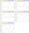
Fig. 2. Endophyte inoculation of Arabidopsis thaliana, grown in the presence or absence of UV-A, assessed by the qPCR method.
The ratio of fungal DNA to plant DNA in roots and shoots of plants inoculated with Paraphoma chrysanthemicola (a), Phomopsis columnaris (b), Diaporthe eres (c), Mucor sp. (d), Sporobolomyces ruberrimus (e). The experiment was performed in four biological replicates. Brackets show the significant differences in means of log-transformed qPCR measurements between inoculated and mock-inoculated samples, from the same organ and light conditions (* 0.01 < p ≤ 0.05, ** 0.001 < p ≤ 0.01, *** p ≤ 0.001, adjusted for multiple comparisons with Hommel’s method). The set of all tested contrasts is in the S2 Appendix, Table S1.
Fig. 3
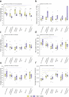
Fig. 3. Fresh weight of shoots
(a), shoot anthocyanin content (b), fresh weight of roots (c), root system length (d), average root volume (e), and average root diameter (f) of 18-day-old plants, either mock-inoculated (control) or inoculated with Paraphoma chrysanthemicola, Phomopsis columnaris, Diaporthe eres, Mucor sp., Sporobolomyces ruberrimus. Plants were grown in the presence or absence of UV-A. The experiment was performed in 9 biological replicates (plates) for every combination of inoculation type and light conditions. Black brackets show the significant differences in means between plants inoculated with the same endophyte, but subject to different light conditions. Yellow (VIS) and violet (VIS + UV) brackets show significant differences in means between inoculated samples and the mock-inoculated control (* 0.01 < p ≤ 0.05, ** 0.001 < p ≤ 0.01, *** p ≤ 0.001, adjusted for multiple comparisons with the Benjamin-Hochberg method). The set of all tested contrasts is in the S2 Appendix, Table S2.
Fig. 4
Fig. 4. Relative transcript levels of CHS
(a), PAL1 (b), ICS1 (c), and PDF1.2 (d) genes in leaves of 18-day-old plants, either mock-inoculated (control) or inoculated with Paraphoma chrysanthemicola, Phomopsis columnaris, Diaporthe eres, Mucor sp., Sporobolomyces ruberrimus. Plants were grown in the presence or absence of UV-A. The experiment was performed in 9 biological replicates. Yellow (VIS) and violet (VIS + UV) brackets show significant differences in means between inoculated samples and the mock-inoculated control (* 0.01 < p ≤ 0.05, ** 0.001 < p ≤ 0.01, *** p ≤ 0.001, adjusted for multiple comparisons with the Benjamin-Hochberg method). The set of all tested contrasts is in the S2 Appendix, Table S3.
Fig. 5
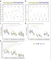
Fig. 5. Chlorophyll content
(a), flavonol content (b), MDA (c) and photosynthetic parameters (d-i) in Arabidopsis thaliana pot cultures inoculated with Sporobolomyces ruberrimus grown in the presence or absence of UV-A. (a - c) The experiment was repeated six times. Each dot represents a measurement obtained from a single plant (in a – b, the mean of values recorded for the 6th, 7th, and 8th leaf). The results of the statistical analysis are in the S2 Appendix, Table S4. (d - f) Induction curves of Arabidopsis pot cultures inoculated with S. ruberrimus with standard error. d – Y(II), e – NPQ, f – ETR. Dark-adapted plants were irradiated with the initial Saturating Pulse (SP) of 10 000 µmol m-2·s-1 for 600 ms, then with a delay of 50 s, Actinic Light (AL) of 75 µmol m-2·s-1 was applied, followed by a set of SP. (g – i) Light curves of Arabidopsis pot cultures inoculated with S. ruberrimus with standard error. g – Y(II), h – NPQ, i – ETR. Actinic light of increasing irradiance (0, 11, 18, 27, 58, 100, 131, 221, 344, 536, 830 µmol m-2·s-1) was applied within 30 s phases. Saturating pulses lasted 600 ms.
Fig. 6
Fig. 6. Endophyte inoculation of Arabidopsis thaliana, wild type, fah1, and tt4 mutants, grown in the presence or absence of UV-A, assessed by the qPCR method.
(a-b) The ratio of fungal DNA to plant DNA in roots and shoots of plants inoculated with Paraphoma chrysanthemicola (a) and Sporobolomyces ruberrimus (b). The experiment was performed in three biological replicates. Brackets show the significant differences in means of log-transformed qPCR measurements between inoculated and mock-inoculated samples light conditions (* 0.01 < p ≤ 0.05, ** 0.001 < p ≤ 0.01, *** p ≤ 0.001, adjusted for multiple comparisons with Hommel’s method). The set of all tested contrasts is in the S2 Appendix, Table S6. (c - d) Fresh weight of shoots (c) and roots (d) 18-day-old plants, either mock-inoculated (control) or inoculated with Paraphoma chrysanthemicola, or Sporobolomyces ruberrimus. Weight was measured 128 h after inoculation (e) Main root length increment of Arabidopsis thaliana, wild type, fah1, and tt4 mutants, 100 hours post inoculation with Paraphoma chrysanthemicola, or Sporobolomyces ruberrimus. Plants were grown in the presence or absence of UV-A. (c – e) The experiment was performed in 8 biological replicates (plates) for every combination of inoculation type and light conditions. Black brackets show the significant differences in means between plants inoculated with the same endophyte, but subject to different light conditions. Yellow (VIS) and violet (VIS + UV) brackets show significant differences in means between inoculated samples and the mock-inoculated control (* 0.01 < p ≤ 0.05, ** 0.001 < p ≤ 0.01, *** p ≤ 0.001, adjusted for multiple comparisons with the Benjamin-Hochberg method). The set of all tested contrasts is in the S2 Appendix, Table S7.
Fig. 7
Fig. 7. Supplementation of PAR with UV-A did not substantially affect the interaction between Arabidopsis thaliana and endophytic fungi.
Both UV-A and endophytes affected the expression of genes related to the salicylic acid pathways. The microorganisms did not affect plant UV-A perception. No endophyte-induced alteration in UV-A receptor expression and anthocyanin accumulation were observed. UV-A did not alter microorganism-dependent changes in root patterning. The minus represents no significant effect of UV-A or endophyte or interaction of both factors. The asterisk represents a significant effect as detected with ANOVA.

Similar articles

References

    1. Roeber VM, Bajaj I, Rohde M, Schmülling T, Cortleven A. Light acts as a stressor and influences abiotic and biotic stress responses in plants. Plant Cell Environ. 2021;44(3):645–64. doi: 10.1111/pce.13948 - DOI - PubMed
    1. Aphalo P, Albert A, Bjorn L, Mcleod A, Robson T, Rosenqvist E. Beyond the visible: A handbook of best practice in plant UV photobiology. Aphalo P, editor. Helsinki: University of Helsinki. 2012.
    1. Robson TM, Aphalo PJ, Banaś AK, Barnes PW, Brelsford CC, Jenkins GI, et al.. A perspective on ecologically relevant plant-UV research and its practical application. Photochem Photobiol Sci. 2019;18(5):970–88. doi: 10.1039/c8pp00526e - DOI - PubMed
    1. Verdaguer D, Jansen MAK, Llorens L, Morales LO, Neugart S. UV-A radiation effects on higher plants: Exploring the known unknown. Plant Sci. 2017;255:72–81. doi: 10.1016/j.plantsci.2016.11.014 - DOI - PubMed
    1. Jansen MAK, Biswas DK. Natural variation in UV-B protection amongst Arabidopsis thaliana accessions. EJFA. 2012;24(6). doi: 10.9755/ejfa.v24i6.14681 - DOI

MeSH terms

LinkOut - more resources