. 2025 May 15;388(6748):769-775.
doi: 10.1126/science.adq5233. Epub 2025 May 15.

Norepinephrine changes behavioral state through astroglial purinergic signaling

Affiliations

Norepinephrine changes behavioral state through astroglial purinergic signaling

Alex B Chen et al. Science. .

Abstract

Both neurons and glia communicate through diffusible neuromodulators; however, how neuron-glial interactions in such neuromodulatory networks influence circuit computation and behavior is unclear. During futility-induced behavioral transitions in the larval zebrafish, the neuromodulator norepinephrine (NE) drives fast excitation and delayed inhibition of behavior and circuit activity. We found that astroglial purinergic signaling implements the inhibitory arm of this motif. In larval zebrafish, NE triggers astroglial release of adenosine triphosphate (ATP), extracellular conversion of ATP into adenosine, and behavioral suppression through activation of hindbrain neuronal adenosine receptors. Our results suggest a computational and behavioral role for an evolutionarily conserved astroglial purinergic signaling axis in NE-mediated behavioral and brain state transitions and position astroglia as important effectors in neuromodulatory signaling.


Conflict of interest statement

Competing interests: The authors declare no competing interests.

Figures

Figure 1.
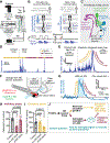
Figure 1.. Futility triggers a biphasic behavioral and neural response through NE neuron activation.
(A) Schematic of virtual reality behavioral experiments with real-time swim detection and visual feedback. (B) Diagram illustrating the difference between closed loop (visual feedback in response to swims) and open loop (no visual feedback) conditions. (C) Schematic: the known cell types involved in futility-induced passivity. (D) Swim trace of an example trial demonstrating closed and open loop swim behavior. (E) Average closed loop and passivity-triggered open loop tail angle demonstrating an initial increase in swim amplitude (excitatory phase) followed by inhibition of swimming (inhibitory phase) in open loop. (F) Neural activity, imaged with a confocal microscope while NE neurons were optogenetically activated using a fiber optic. (G) Optogenetic stimulation-triggered average of neural activity in motor areas demonstrating fast excitation and delayed inhibition, similar to the behavioral futility response. (H,I) Effect of blocking α1-adrenergic receptors (100 μM prazosin) or β-adrenergic receptors (100 μM propranolol) on (H) open loop passivity and (I) open-loop swim vigor. Panel H,I: N = 16 (control), 14 (α1-AR block), 15 (β-AR block) fish. Panel H: P > 0.9999 (control vs. β-AR block), P = 0.0006 (control vs. α1-AR block) Panel I: P = 0.001 (control vs. β-AR block), P > 0.9999 (control vs. α1-AR block). Kruskal-Wallis. (J) Model of parallel noradrenergic channels that contribute to the excitatory and inhibitory phases of the futility response and central problem statement. All error bars and shaded error regions represent s.e.m.
Figure 2.
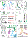
Figure 2.. Futility drives astroglial release of ATP.
(A) Experimental schematic: two-color light-sheet imaging of extracellular ATP and astroglial calcium (Tg(gfap:GRABATP; gfap:jRGECO1a) fish) along with fictive behavioral recording. (B) Fluorescence micrographs of simultaneously collected GRABATP and jRGECO1a signals in a fish in baseline condition or during a futile swim. (C) Two examples of motor nerve electrical activity, GRABATP and jRGECO1a signals during open loop periods. Swim, calcium, and ATP traces are manually offset along the vertical axis to allow for better visualization. (D) Futile-swim triggered astroglial calcium and extracellular ATP signals averaged across fish (N = 7). (E) Futile swim-triggered GRABATP signal in fish treated with an α1-AR blocker (100 μM prazosin) or vehicle, and in fish expressing hPMCA2 in astroglia. N = 4 (control), 5 (α1-AR block), 5 (hPMCA2). P = 0.0065 (control vs. α1-AR block), 0.0038 (control vs. hPMCA2). Kruskal-Wallis test on AUC from 0 to 60s. (F) Experimental schematic: ex vivo confocal imaging during puffing of an α1-AR agonist (10 μM methoxamine) or vehicle in the presence of a neural activity blocker (160 mg/L MS-222, a sodium channel inhibitor). Tg(gfap:GRABATP) fish. (G) GRABATP signal in fish in experiments described in (F), triggered on puff and onset-aligned. N = 5, both conditions. P = 0.013, Mann-Whitney on AUC from 0 to 60 s. (H) Experimental schematic: in vivo widefield imaging during chemogenetic activation of Tg(gfap:rTRPV1-eGFP) fish with 200 nM capsaicin in the presence of a neural activity blocker (170 mg/L MS-222). (I) GRABATP signal in fish treated with capsaicin as described in (F), triggered capsaicin administration and onset-aligned. All error bars and shaded error regions represent s.e.m.
Figure 3.
Figure 3.. ATP promotes passivity via extracellular metabolism into adenosine.
(A) Experimental schematic: Behavioral recording of freely swimming fish treated with 100 μM NPE-ATP or vehicle then subjected to inescapable ultraviolet (360 nm) light, which uncages ATP. (B) Light onset-triggered swim speed of fish treated with NPE-ATP or vehicle. (C) Percent of light ON period spent passive for vehicle control and NPE-ATP treated fish. N = 9 (control), 10 (NPE-ATP). P = 0.0138, Mann-Whitney. (D) Effect of P2 receptor blocker (100 μM suramin) or vehicle on open loop passivity in head-fixed behavior. N = 14 (control), 14 (suramin). P = 0.0395, Mann-Whitney. (E) Diagram illustrating extracellular biochemical ATP-to-adenosine pathway through enzymes Cd39 and Cd73, and pathway inhibition by competitive Cd39 inhibitor ARL 67156 and Cd73 inhibitor AMPCP. (F) Effect of Cd73 block (100 μM AMPCP) on open loop passivity in head-fixed behavior. N = 16 (control), 16 (AMPCP). P = 0.0008, Mann-Whitney. (G) Percent passivity following onset of optogenetic stimulation for fish treated with ARL 67156 or vehicle. N = 9 (control), 7 (ARL 67156). P = 0.0002, Mann-Whitney. (H) Extracellular adenosine (GRABADO1.0) signal of a fish in baseline (left) and during a futile swim (right). (I) Futile-swim-triggered average of GRABADO1.0 signal in fish treated with vehicle, α1-AR blocker (100 μM prazosin) or Cd39 inhibitor (1 mM ARL 67156). N = 4 (control), 7 (ARL 67156), 6 (prazosin); P = 0.002 (control vs. ARL), 0.004 (control vs. prazosin), Kruskal-Wallis on AUC from 0 – 60 s following futile swim. (J) Effect of vehicle or adenosine receptor blocker (100 μM caffeine) on proportion of open loop spent passive (left) and open loop struggle rate (right). N = 15 (control), 16 (caffeine); P = 0.0215 (proportion passivity), 0.2238 (struggle rate), Mann-Whitney. (K,L) Effect of an A1R blocker (DPCPX) and an A2AR blocker (SCH-58261) (K) or an A2BR blocker (MRS 1754) (L) on proportion of open loop spent passive. Panel K: N = 21 (control), 15 (DPCPX), 16 (SCH-58261); N = 19 (control), 17 (MRS 1754). All error bars and shaded error regions represent s.e.m.
Figure 4.
Figure 4.. Adenosine persistently activates the swim-suppressing region L-MO.
(A) Schematic: astroglial communication with L-MO, and mutual inhibition between L-MO and motor regions. (B) Example of L-MO neuronal activity anticorrelation to swim vigor in one fish. (C) Struggle-evoked L-MO activity before and after caffeine (100 μM), mean across five fish; N = 5; P = 0.004, Mann-Whitney on AUC from 0 – 60 s following struggle. (D) Mean of futility-triggered passivity durations before and after caffeine treatment. N = 5; P = 0.0397, paired t-test. (E) Schematic: activating astroglia in Tg(gfap:TRPV1-eGFP;elavl3:JRGECO1a) fish while imaging L-MO activity using light-sheet microscopy. (F) L-MO activity in 4 example fish treated with either vehicle control (top row) 100 μM caffeine (an adenosine receptor blocker, bottom row) or vehicle control, either in baseline untreated condition (left) or with capsaicin (right). (G) Summary of rate of L-MO activation across all fish and conditions. N = 4 (control), 5 (caffeine); P = 0.9633 (baseline control vs. caffeine), 0.0016 (capsaicin control vs. caffeine), two-way ANOVA with Šídák’s multiple comparisons test. (H) Model: futility-triggered NE release drives astroglial ATP release. ATP is metabolized extracellularly into adenosine, and adenosine activates A2 adenosine receptors in L-MO to increase L-MO activity and suppress swimming. (I) Model: futility-related NE-MO firing drives fast excitation (yellow). NE mediates delayed inhibition (red) through astroglial activation, ATP release, and ATP-to-adenosine metabolism; eventually, inhibition overcomes fast excitation to drive the inhibitory phase of passivity. Thus, an astroglial noradrenergic-to-purinergic pathway mediates feedforward inhibition of the passivity response. All error bars and shaded error regions represent s.e.m.

Update of

References

    1. Bennett MV, Nakajima Y, Pappas GD, Physiology and ultrastructure of electrotonic junctions. I. Supramedullary neurons. J. Neurophysiol. 30, 161–179 (1967). - PubMed
    1. Bhattacharya A, Aghayeva U, Berghoff EG, Hobert O, Plasticity of the Electrical Connectome of C. elegans. Cell 176, 1174–1189.e16 (2019). - PMC - PubMed
    1. Smith SJ, Sümbül U, Graybuck LT, Collman F, Seshamani S, Gala R, Gliko O, Elabbady L, Miller JA, Bakken TE, Rossier J, Yao Z, Lein E, Zeng H, Tasic B, Hawrylycz M, Single-cell transcriptomic evidence for dense intracortical neuropeptide networks. Elife 8 (2019). - PMC - PubMed
    1. Lovett-Barron M, Andalman AS, Allen WE, Vesuna S, Kauvar I, Burns VM, Deisseroth K, Ancestral Circuits for the Coordinated Modulation of Brain State. Cell 171, 1411–1423.e17. - PMC - PubMed
    1. Marques JC, Li M, Schaak D, Robson DN, Li JM, Internal state dynamics shape brainwide activity and foraging behaviour. Nature 577, 239–243 (2020). - PubMed

LinkOut - more resources