. 2025 Aug 8;66(2):2401564.
doi: 10.1183/13993003.01564-2024. Print 2025 Aug.

Cyclic GMP-AMP synthase expression is enhanced in systemic sclerosis-associated interstitial lung disease and stimulates inflammatory myofibroblast activation

Affiliations

Cyclic GMP-AMP synthase expression is enhanced in systemic sclerosis-associated interstitial lung disease and stimulates inflammatory myofibroblast activation

Sheeline Yu et al. Eur Respir J. .

Abstract

Objective: The lungs of patients with systemic sclerosis-associated interstitial lung disease (SSc-ILD) contain inflammatory myofibroblasts that arise in association with fibrotic stimuli and perturbed innate immunity. The cytosolic DNA-binding receptor cyclic GMP-AMP synthase (cGAS) is implicated in inflammation and fibrosis, but its involvement in SSc-ILD remains unknown. We examined cGAS expression, activity and therapeutic potential in SSc-ILD using human biospecimens, cultured fibroblasts, precision-cut lung slices and a well-accepted animal model.

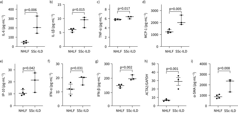
Methods: Expression and localisation of cGAS, cytokines and type 1 interferons were evaluated in SSc‑ILD lung tissues, bronchoalveolar lavage fluid and isolated lung fibroblasts. CGAS activation was assessed in a publicly available SSc-ILD single-cell RNA-sequencing dataset. Production of cytokines, type 1 interferons and α-smooth muscle actin elicited by transforming growth factor-β1 or local substrate stiffness was measured in normal human lung fibroblasts via quantitative reverse transcription PCR, ELISA and immunofluorescence. Small molecule cGAS inhibition was tested in cultured fibroblasts, human precision-cut lung slices and the bleomycin pulmonary fibrosis model.

Results: SSc-ILD lung tissue and bronchoalveolar lavage fluid were enriched for cGAS, cytokines and type 1 interferons. The cGAS pathway showed constitutive activation in SSc-ILD fibroblasts and was inducible in normal human lung fibroblasts by transforming growth factor-β1 or mechanical stimuli. In these settings, and in precision-cut lung slices, cGAS expression was paralleled by the production of cytokines, type 1 interferons and α-smooth muscle actin, which was mitigated by a small molecule cGAS inhibitor. These findings were recapitulated in the bleomycin mouse model.

Conclusion: cGAS signalling contributes to pathogenic inflammatory myofibroblast phenotypes in SSc‑ILD. Inhibiting cGAS or its downstream effectors represents a novel therapeutic approach.


Conflict of interest statement

Conflict of interest: J. Zielonka reports support for the present manuscript from the National Institutes of Health (NIH). A. Ghincea reports support for the present manuscript from the NIH. G. Ishikawa reports support for the present study from the NIH, Pulmonary Fibrosis Foundation and Wit Family Distinguished Scholar in Inflammation Science. M. Hinchcliff reports support for the present manuscript from the NIH. J. Varga reports participation on a data safety monitoring board or advisory board with SRF CONQUER trial, and stock (or stock options) with Neolaia. C. Feghali-Bostwick reports support for the present study from the NIH and grants from K24AR060297. J.L. Gomez reports support for the present study from R01 HL153604 and R03 HL154275. C. Ryu reports support for the present study from the NIH and the Boehringer Ingelheim Discovery Award. E.L. Herzog reports grants from the NIH, Boehringer Ingelheim and Bristol Myers; consulting fees from Boehringer Ingelheim, Merck, Puretech and Iqvia; payment or honoraria for lectures, presentations, manuscript writing or educational events from Boehringer Ingelheim; and participation on a data safety monitoring board or advisory board with Merck and Anne Theodore Foundation. The remaining authors have no potential conflicts of interest to disclose.

Figures

None
Using a comprehensive translational platform to study systemic sclerosis-associated interstitial lung disease (SSc-ILD), we found that the cytosolic DNA receptor cyclic GMP-AMP synthase (cGAS) contributes to inflammatory myofibroblast phenotypes that are attenuated by its inhibition with the small molecule inhibitor G140. α-SMA: α-smooth muscle actin; cGAMP: cyclic GMP-AMP; IFN: interferon; IL: interleukin; IP-10: IFN-γ inducible protein-10; MCP-1: monocyte chemoattractant protein-1; STING: stimulator of interferon genes; TNF-α: tumour necrosis factor-α.
FIGURE 1
FIGURE 1
Systemic sclerosis-associated interstitial lung disease (SSc-ILD) lungs show enhanced expression of cyclic GMP-AMP synthase (cGAS) and its associated inflammatory mediators. a) Immunohistochemistry for cGAS showed enhanced detection in the SSc-ILD lung (right) as compared to the healthy control lung (left). b) A higher percentage of cGAS+ cells per high-powered field (HPF) was observed in the SSc-ILD lung. c–i) Bronchoalveolar lavage fluid (BALF) samples collected from healthy control donors and SSc patients showed that, relative to healthy controls, SSc-ILD patients exhibited increased concentrations of c) interleukin (IL)-6, d) IL-1β, e) tumour necrosis factor-α (TNF-α), f) monocyte chemoattractant protein-1 (MCP-1), g) interferon (IFN)-γ inducible protein-10 (IP-10), h) IFN-α and i) IFN-β.
FIGURE 2
FIGURE 2
Systemic sclerosis-associated interstitial lung disease (SSc-ILD) lung fibroblasts are enriched for cyclic GMP-AMP synthase (cGAS) expression. a) Violin plot of single-cell RNA-sequencing analysis (derived from Gene Expression Omnibus dataset GSE128169) from the lungs of patients with SSc-ILD, demonstrating median expression of cGAS-related genes across multiple cell populations, which was highest among immune and endothelial cells. b) Among stromal cells, fibroblasts (purple) demonstrated enhanced expression of these genes. c) Quantitative reverse transcription PCR and d) ELISA measurements of normal human lung fibroblasts (NHLFs) and SSc-ILD fibroblasts showed increased transcription and protein expression of cGAS in SSc-ILD fibroblasts. e) Immunofluorescence evaluation of NHLFs (left) and SSc-ILD fibroblasts (right) showed the increased presence of α-smooth muscle actin (α-SMA) and cytoplasmic aggregation of cGAS in SSc-ILD fibroblasts (versus the nuclear localisation of cGAS in NHLFs). Scale bars: 100 μm. NK: natural killer; GAPDH: glyceraldehyde-3-phosphate dehydrogenase.
FIGURE 3
FIGURE 3
Systemic sclerosis-associated interstitial lung disease (SSc-ILD) fibroblasts express high concentrations of cyclic GMP-AMP synthase (cGAS)-associated soluble mediators and α-smooth muscle actin (α-SMA). As compared to normal human lung fibroblasts (NHLFs), SSc-ILD fibroblasts expressed high protein levels of a) interleukin (IL)-6, b) IL-1β, c) tumour necrosis factor-α (TNF-α), d) monocyte chemoattractant protein-1 (MCP-1), e) interferon (IFN)-γ inducible protein-10 (IP-10), f) IFN-α and g) IFN-β. Relative to normal cells, SSc-ILD fibroblasts expressed increased h) α-SMA transcripts via quantitative reverse transcription PCR and i) protein via ELISA. ACTA2: actin α2, smooth muscle; GAPDH: glyceraldehyde-3-phosphate dehydrogenase.
FIGURE 4
FIGURE 4
Small molecule cyclic GMP-AMP synthase (cGAS) inhibition mitigates inflammatory fibrotic responses in systemic sclerosis-associated interstitial lung disease (SSc-ILD) fibroblasts. a) Immunofluorescence staining of naïve (left) and G140-treated (right) SSc-ILD fibroblasts showed reduced detection of both cytoplasmic cGAS aggregates and α-smooth muscle actin (α-SMA) in treated cells. Scale bars: 100 μm. These G140-treated cells displayed decreased expression of b) interleukin (IL)-6, c) IL-1β, d) tumour necrosis factor-α (TNF-α), e) monocyte chemoattractant protein-1 (MCP-1), f) interferon (IFN)-γ inducible protein-10 (IP-10), g) IFN-α and h) IFN-β. Treated cells also displayed a reduction in the i) transcription and j) protein expression of α-SMA. k) An MTT assay showed that G140 did not influence the viability of these fibroblasts. Data were normalised to the untreated condition. ACTA2: actin α2, smooth muscle.
FIGURE 5
FIGURE 5
A small molecule cyclic GMP-AMP synthase (cGAS) inhibitor suppresses inflammatory fibroblast activation induced by transforming growth factor-β1 (TGF-β1) and CpG co-stimulation. a) Immunofluorescence of normal human lung fibroblasts (NHLFs) co-stimulated with TGF-β1 and CpG in the absence (left) or presence (right) of G140 showed that treatment diminished the presence of cGAS aggregates in the cytoplasm. Scale bars: 100 μm. Although treatment with G140 did not change levels of b) interleukin (IL)-6, it attenuated expression of c) IL-1β, d) tumour necrosis factor-α (TNF-α), e) monocyte chemoattractant protein-1 (MCP-1), f) interferon (IFN)-γ inducible protein-10 (IP-10), g) IFN-α and h) IFN-β, as well as i) mRNA transcription and j) protein expression of α-SMA. k) An MTT assay showed that these findings were unrelated to changes in cell viability. Data were normalised to the TGF-β1+CpG condition. ACTA2: actin α2, smooth muscle.
FIGURE 6
FIGURE 6
A small molecule cyclic GMP-AMP synthase (cGAS) inhibitor mitigates stiffness-induced fibroblast activation. Normal human lung fibroblasts (NHLFs) were cultured on tunable hydrogels that approximate the stiffness of the normal (1 kPA) and fibrotic (25 kPA) lung. As compared to NHLFs cultured in the untreated 25 kPA condition, those grown on 25 kPA hydrogels treated with G140 exhibited decreased levels of a) interleukin (IL)-1β, b) interferon (IFN)-γ inducible protein-10 (IP-10), c) IFN-α and d) IFN-β; expression of g) monocyte chemoattractant protein-1 (MCP-1) remained unchanged. This inhibitor also led to a reduction in the f) transcription and g) expression of α-smooth muscle actin (α-SMA). h) An MTT assay showed that these findings were unrelated to changes in cell viability. Data were normalised to their respective untreated or treated 1 kPA condition. ACTA2: actin α2, smooth muscle.
FIGURE 7
FIGURE 7
A small molecule cyclic GMP-AMP synthase (cGAS) inhibitor abrogates inflammatory fibrotic responses in human precision-cut lung slices (PCLS). a) Representative images of Masson's trichrome staining of human PCLS sections from those subjected to an inflammatory fibrotic cocktail (IFC) in the absence (left) or presence (right) of G140. b) Collagen quantification via ImageJ software revealed a reduction in collagen content following treatment with G140. Treatment with this inhibitor mitigated expression of c) interleukin (IL)-6, d) IL-1β, e) tumour necrosis factor-α (TNF-α), f) monocyte chemoattractant protein-1 (MCP-1), g) interferon (IFN)-γ inducible protein-10 (IP-10) and h) IFN-α, and transcription of i) actin α2, smooth muscle (ACTA2). Data were normalised to the IFC condition. j) Heatmap of bulk RNA-sequencing between IFC cultured PCLS in the absence or presence of G140 revealed 126 differentially expressed genes. k) Unbiased pathway enrichment analysis. Apo-2L: apo-2 ligand; FDR: false discovery rate; HCV: hepatitis C virus; HMGB1: high mobility group box-1; MHC: major histocompatibility complex; SLE: systemic lupus erythematosus; TLR: Toll-like receptor; TNFSF10: tumour necrosis factor receptor superfamily member-10.
FIGURE 8
FIGURE 8
A small molecule cyclic GMP-AMP synthase (cGAS) inhibitor ameliorates bleomycin (Bleo)-induced pulmonary fibrosis. a) Representative images of Masson's trichrome staining of lung sections from bleomycin-challenged mice in the absence (left) or presence (right) of G140. Collagen quantification via b) ImageJ software and c) Sircol assay revealed a reduction in collagen content following treatment with G140. Bleomycin-exposed mice treated with G140 displayed reduced bronchoalveolar lavage fluid (BALF) concentrations of d) interleukin (IL)-6, e) tumour necrosis factor-α (TNF-α), f) interferon (IFN)-γ inducible protein-10 (IP-10), g) IFN-α and h) IFN-β, while levels of i) IL-1β, j) monocyte chemoattractant protein-1 (MCP-1) and k) white blood cell (WBC) counts remained unchanged.

Update of

Comment in

References

    1. Denton CP, Khanna D. Systemic sclerosis. Lancet 2017; 390: 1685–1699. doi: 10.1016/S0140-6736(17)30933-9 - DOI - PubMed
    1. Herzog EL, Mathur A, Tager AM, et al. Review: interstitial lung disease associated with systemic sclerosis and idiopathic pulmonary fibrosis: how similar and distinct? Arthritis Rheumatol 2014; 66: 1967–1978. doi: 10.1002/art.38702 - DOI - PMC - PubMed
    1. Steen VD, Medsger TA. Changes in causes of death in systemic sclerosis, 1972–2002. Ann Rheum Dis 2007; 66: 940–944. doi: 10.1136/ard.2006.066068 - DOI - PMC - PubMed
    1. Rahaghi FF, Hsu VM, Kaner RJ, et al. Expert consensus on the management of systemic sclerosis-associated interstitial lung disease. Respir Res 2023; 24: 6. doi: 10.1186/s12931-022-02292-3 - DOI - PMC - PubMed
    1. Raghu G, Montesi SB, Silver RM, et al. Treatment of systemic sclerosis-associated interstitial lung disease: evidence-based recommendations. An official American Thoracic Society clinical practice guideline. Am J Respir Crit Care Med 2023; 209: 137–152. doi: 10.1164/rccm.202306-1113ST - DOI - PMC - PubMed

MeSH terms

LinkOut - more resources