. 2025 May 15;16(1):4515.
doi: 10.1038/s41467-025-59657-7.

Tick-borne flavivirus exoribonuclease-resistant RNAs contain a double loop structure

Affiliations

Tick-borne flavivirus exoribonuclease-resistant RNAs contain a double loop structure

Conner J Langeberg et al. Nat Commun. .

Abstract

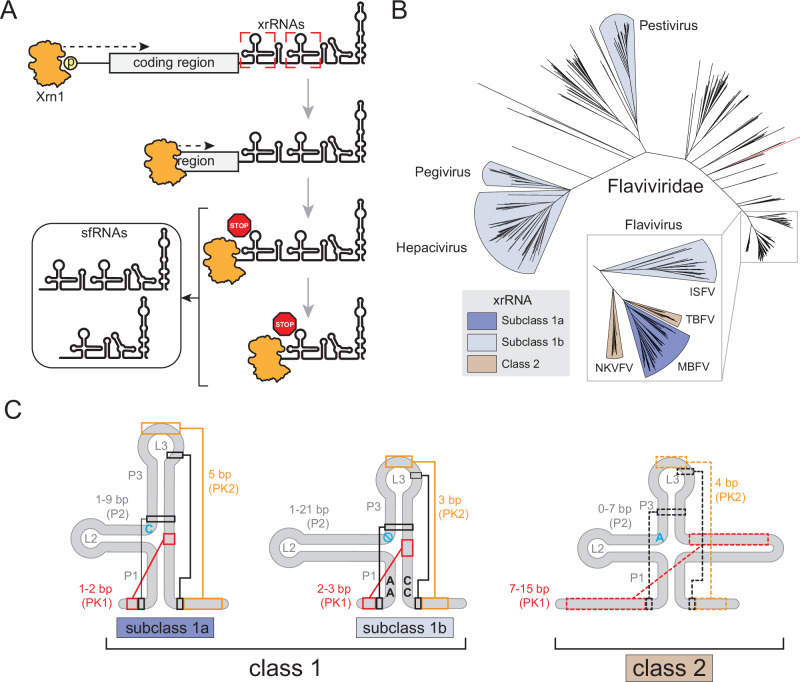
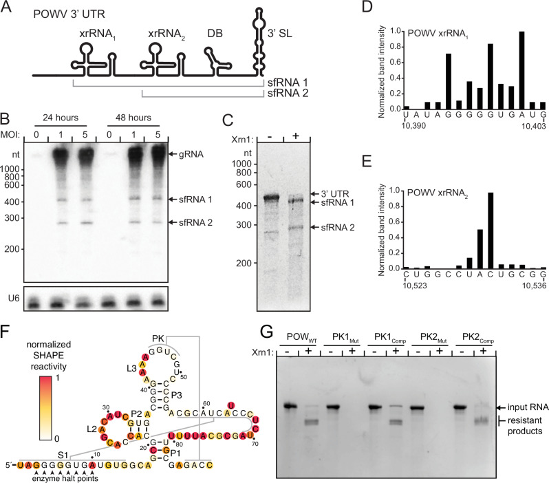
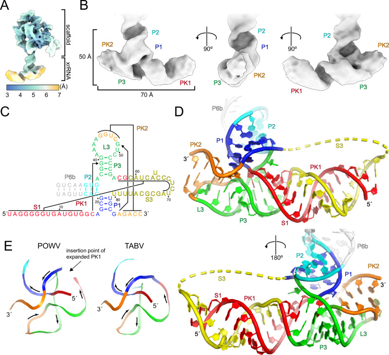
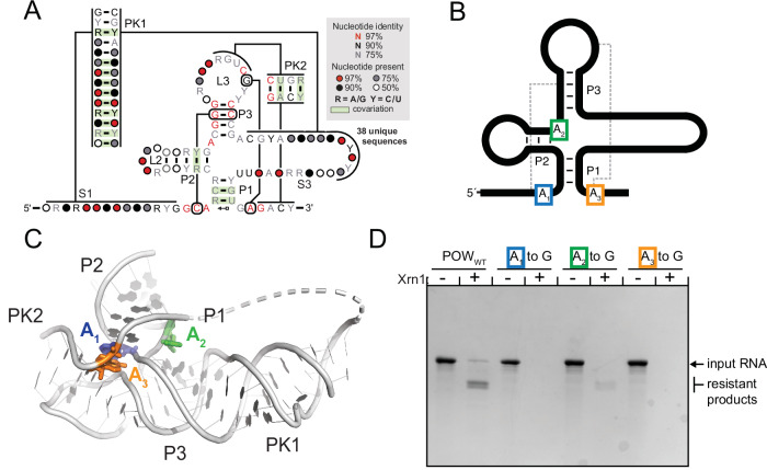
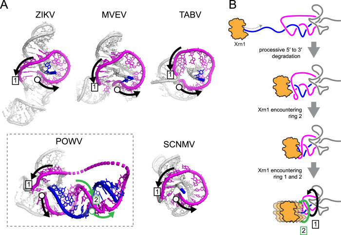
Viruses from the Flaviviridae family contain human relevant pathogens that generate subgenomic noncoding RNAs during infection using structured exoribonuclease resistant RNAs (xrRNAs). These xrRNAs block progression of host cell's 5' to 3' exoribonucleases. The structures of several xrRNAs from mosquito-borne and insect-specific flaviviruses reveal a conserved fold in which a ring-like motif encircles the 5' end of the xrRNA. However, the xrRNAs found in tick-borne and no known vector flaviviruses have distinct characteristics, and their 3-D fold was unsolved. Here, we verify the presence of xrRNAs in the encephalitis-causing tick-borne Powassan Virus. We characterize their secondary structure and obtain a mid-resolution map of one of these xrRNAs using cryo-EM, revealing a unique double-loop ring element. Integrating these results with covariation analysis, biochemical data, and existing high-resolution structural information yields a model in which the core of the fold matches the previously solved xrRNA fold, but the expanded double loop ring is remodeled upon encountering the exoribonuclease. These results are representative of a broad class of xrRNAs and reveal a conserved strategy of structure-based exoribonuclease resistance achieved through a unique topology across a viral family of importance to global health.


Conflict of interest statement

Competing interests: The authors have no competing interests.

Figures

Fig. 1
Fig. 1. Flaviviridae sfRNA biogenesis and xrRNA structural determinants.
A Cartoon representation of sfRNA production and function through Xrn1-mediated degradation of the viral genome. B Overview of the phylogenetic relationship within Flaviviridae based on the sequence of viral protein NS5. The tree files, alignments, and analysis were reanalyzed from data published by Bamford et al. and Mifsud et al. C Structural features of Flaviviridae xrRNAs are shown on the cartoon representations of the secondary structures of each class or subclass. Known long-range tertiary interactions are indicated with colored lines and boxes in class 1. Analogous predicted interactions in class 2 are indicated with dashed colored lines and boxes. The blue letters indicate conserved junction nucleotides, blue no symbol in subclass 1b denotes the absence of a nucleotide in this position. Source data is provided as a Source Data file.
Fig. 2
Fig. 2. Identification characterization of POWV xrRNAs.
A Cartoon representation of POWV 3’ UTR. Abbreviations: DB – dumbbell, 3′ SL – 3′ stem loop. B Northern blot of POWV-infected Vero cells at the indicated MOI and time post infection. Probes were specific for the 3′ UTR of POWV or the U6 RNA loading control. Three independent experiments were performed, results of one is shown. C In vitro degradation by Xrn1 of in vitro transcribed RNA of the POWV Spooner isolate 3′ UTR. The partially degraded but stable RNAs appear as bands in the lane with Xrn1, demonstrating the presence of xrRNAs. The gel is representative of experiments performed in triplicate. D, E Stop site analysis of POWV xrRNA1 and xrRNA2. RNA treated with Xrn1 was analyzed using reverse transcription primer extension. The intensity of the resultant bands is graphed as a function of nucleotide position. F Secondary structure diagram of the POWV xrRNA1 in the context of the full 3′ UTR with the results of 1M7 chemical probing. G In vitro RNA resistance assay demonstrating the functional importance of both PK1 and PK2. A diagram of the mutations is found in Fig. S3. The gel is representative of experiments performed in triplicate. Source data is provided as a Source Data file.
Fig. 3
Fig. 3. Cryo-EM structure of the POWV class 2 xrRNA.
A Local resolution map of the scaffolded POWV xrRNA. Resolution is colored as denoted in the inset key B. Cryo-EM map of the POWV xrRNA, the locations of secondary structure features are indicated. C Secondary structure diagram of the POWV xrRNA, colored to match D and E. D Structural model of the POWV xrRNA colored to match C. The yellow dashed backbone represents 9 nucleotides that were not visible in the map and hence not built. Structural features are labeled. E Comparison of POWV and TABV ring topologies colored corresponding to C.
Fig. 4
Fig. 4. Sequence and structure conservation of class 2 xrRNAs from flavivirus.
A Consensus secondary structure model of class 2 xrRNAs calculated by R-Scape,. B Diagram denoting the location of three perfectly conserved A bases that were mutated to test their functional significance. C. Structure of the POWV xrRNA with the three conserved A bases indicated in B and C. D. In vitro RNA resistance assay demonstrating the functional importance of the three perfectly conserved (A) nucleotides in class 2 xrRNAs. The gel is representative of experiments performed in triplicate. Source data are provided as a Source Data file.
Fig. 5
Fig. 5. A conserved ring-like fold present across all xrRNA structures.
A The ring-like structure (magenta) encircling the 5′ end (blue) of the xrRNA from ZIKV (PDB: 5TPY), Murray Valley Encephalitis virus (MVE) (PDB: 4PQV), TABV (PDB: 7K16), POWV (this study), and Sweet Clover Necrotic Mosaic Virus (SCNMV) (PDB: 6D3P). The open circle denotes the start of the ring structure, the boxed 1 indicates the completion of one loop. In POWV, the RNA loops around the 5′ end a second time, indicated with green arrows and a boxed 2. B Proposed mechanistic model of Xrn1 encountering a class 2 xrRNA based on the three-dimensional structure and biochemical experiments. Depicted is the double loop consisting of the primary ring-like motif found in all solved xrRNAs to date (1) and the second ring resulting from the extended PK1 interaction in class 2 xrRNAs (2).

Similar articles

Cited by

References

    1. Mackenzie, J. S., Gubler, D. J. & Petersen, L. R. Emerging flaviviruses: the spread and resurgence of Japanese encephalitis, West Nile and dengue viruses. Nat. Med.10, S98–S109 (2004). - PubMed
    1. Garcia-Blanco, M. A., Ooi, E. E. & Sessions, O. M. RNA viruses, pandemics and anticipatory preparedness. Viruses14, 2176 (2022). - PMC - PubMed
    1. ME, J. W., Adair, K. & Brierley, L. RNA Viruses: A case study of the biology of emerging infectious diseases. Microbiol. Spectr.1, OH-0001-2012 (2013). - PMC - PubMed
    1. Pierson, T. C. & Diamond, M. S. The continued threat of emerging flaviviruses. Nat. Microbiol5, 796–812 (2020). - PMC - PubMed
    1. Valderrama, A., Diaz, Y. & Lopez-Verges, S. Interaction of Flavivirus with their mosquito vectors and their impact on the human health in the Americas. Biochem. Biophys. Res Commun.492, 541–547 (2017). - PubMed

LinkOut - more resources