. 2025 May 15;16(1):4520.
doi: 10.1038/s41467-025-59798-9.

Structural dynamics and permeability of the TRPV3 pentamer

Affiliations

Structural dynamics and permeability of the TRPV3 pentamer

Shifra Lansky et al. Nat Commun. .

Abstract

TRPV3 belongs to the large superfamily of tetrameric transient receptor potential (TRP) ion channels. Recently, using high-speed atomic force microscopy (HS-AFM), we discovered a rare and transient pentameric state for TRPV3 that is in equilibrium with the tetrameric state, and, using cryo-EM, we solved a low-resolution structure of the TRPV3 pentamer, in which, however, many residues were unresolved. Here, we present a higher resolution and more complete structure of the pentamer, revealing a domain-swapped architecture, a collapsed vanilloid binding site, and a large pore. Molecular dynamics simulations and potential of mean force calculations of the pentamer establish high protein dynamics and permeability to large cations. Subunit interface analysis, together with thermal denaturation experiments, led us to propose a molecular mechanism of the tetramer-to-pentamer transition, backed experimentally by HS-AFM observations. Collectively, our data demonstrate that the TRPV3 pentamer is in a hyper-activated state with unique, highly permissive permeation properties.


Conflict of interest statement

Competing interests: The authors declare no competing interests.

Figures
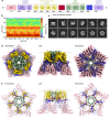
Fig. 1
Fig. 1. TRPV3 pentamer structure.
a TRPV3 subunit architecture, with ARD (pink), linker domain and pre-S1 (purple), VSLD (S1–S4, yellow), S4–S5 linker (red), PD (S5–SF–S6, green), TRP helix (blue), and CTD (purple). b Euler diagram showing the orientation distribution of TRPV3 pentamer particles used for cryo-EM 3D-density reconstruction. Note the abundance (red) of particles around 0π elevation corresponding to side-views. c Representative 2D class averages of the TRPV3 pentamer. Note the first two side-view classes. d Cryo-EM 3D-density map and (e) structural model of the TRPV3 pentamer in top (left), side (center), and bottom (right) views, colored according to the domains in (a). Arrows in (d, e) indicate S4–S5-linker.
Fig. 2
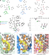
Fig. 2. The TRPV3 channel pore.
a Pore profile of the closed tetramer (green, top), open tetramer (blue, middle), and pentamer (pink, bottom). b Surface representation of the pores as in (a). c PMF, w(z), representing the free energy as a function of the distance from the pore center of mass, for the translocation of sodium (green), Tris+ (red), NMDG+ (purple), and 2-MAE+ (yellow) cations through the closed tetramer (top), open tetramer (middle), and pentamer (bottom). The dashed lines represent the average positions of G638 (SF), I674 (gate), and E682 (exit) during the simulations. Error bars represent the variance of the collected forces within each 0.1 Å wide bin of the PMF calculation.
Fig. 3
Fig. 3. Rotation of the VSLD in the open tetramer and pentamer structures constricts the vanilloid binding pocket.
a Analysis of the helix CoM positions (labeled S1–S6) in the pentamer subunit (pink, right), the open tetramer (blue, center), and the closed tetramer (green, left). b Overlay of the helix positions in the three structures, where the central pore axis is used for lateral alignment and the position of pore-helix S6 for angular alignment (arbitrarily set to 0°). In the open tetramer (blue), all helices move outwards, and the VSLD helices (S1–S4) undergo a collective rotation of 3°, with respect to the closed tetramer. In the pentamer (pink), the helices move further outwards, and the VSLD helices undergo a collective rotation of 24°, with respect to the closed tetramer, corresponding to a rotation of 6° with respect to the next subunit in the pentamer. c Close-up view of the vanilloid pocket, located at the interface between the VSLD (color) of one subunit and the PD (gray) of a neighboring subunit, of the closed tetramer (green, left), open tetramer (blue, center), and pentamer (pink, right). In the closed tetramer, the vanilloid lipid (yellow) is present in the vanilloid binding pocket, ‘glueing’ the two adjacent subunits together via five hydrophobic interactions from each subunit. In the open tetramer and pentamer, the pocket volumes are constricted, leading to clashes of the vanilloid lipid (shown semitransparent) with the surrounding protein structures and ejection of the lipid from the vanilloid pocket.
Fig. 4
Fig. 4. State-dependent inter-subunit interface interactions.
ad Open book representation of the closed tetramer (a, green), open tetramer (b, blue), inactivated tetramer (c, yellow), and pentamer (d, pink), inter-subunit contact areas. Residues forming contacts are shown as spheres and colored according to domain interaction groups PD–VSLD (yellow), PD–linker (red), PD–PD (green), ARD–CD (purple), and, in the case of the open tetramer only, ARD-IDR (orange). eh Inter-subunit interactions of the PD of one subunit (gray) with the PD, VSLD, and S4–S5 linker of an adjacent subunit in the closed tetramer (e, green), open tetramer (f, blue), inactivated tetramer (g, yellow), and pentamer (h, pink). Solid and dashed arrows indicate, respectively, many (solid) and lesser (dashed) interactions between two adjacent helices. ij Plots of the total (i) or TMD (j) interactions, formed by one subunit with the adjacent subunit, for the closed tetramer (green circles), open tetramer (blue triangles), inactivated tetramer (yellow crosses), and pentamer (red dots). The background coloring in (j) indicates the VSLD (yellow), S4/S5 linker (red), and PD (green) regions of the adjacent subunit (chain B). k Bar graph comparing the number of ARD–CD, PD–VSLD, PD–linker, PD–PD, and total number of interactions one subunit forms with an adjacent subunit (left), as well as the total number of inter-subunit interactions per oligomer (right), for the closed tetramer (green), open tetramer (blue), inactivated tetramer (yellow), and pentamer (pink). l Energy of interaction per two adjacent subunits (left) or per the total oligomer (right), as computed by equilibrium MD simulations, for the closed tetramer (green), open tetramer (blue), and pentamer (pink).
Fig. 5
Fig. 5. Thermal stability of TRPV3 in response to activators and control compounds.
NanoDSF thermal denaturation data of TRPV3 in the presence of different concentrations of (a) 2-APB, b camphor, c propofol, d DPTHF, e capsaicin, and (f) DMSO. Each panel shows the raw data NanoDSF curves (top left), their first derivative (bottom left; Tm1 and Tm2 are indicated by downwards and upwards arrowheads, respectively, in (ac) and by shaded bars in (df) where no Tm1/Tm2 changes were observed), bar graph (top right) and curve plot (bottom right) comparing Tm1 and Tm2 following addition of different concentrations of additive. Tm values are presented as mean ± s.e.m. obtained from n = 3 to 5 biologically independent repeats. Statistical significance was assessed with the one-tailed Welch’s t-test. **P < 0.025; ***P < 0.005.
Fig. 6
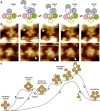
Fig. 6. Proposed mechanism for pentamer formation.
a Schematic of the proposed tetramer-to-pentamer transition mechanism implicating dissociation of a subunit–subunit interface in the tetramer, in order to accommodate the insertion of a fifth subunit. VSLDs are represented by the outer (darker colors) while PDs are represented by the inner and smaller (lighter colors) moon shapes. Each subunit is colored in a different color. b HS-AFM video sequences capturing proposed intermediate states of the tetramer-to-pentamer transition, viewed from the intracellular side, displaying the ARD domains. c Proposed reaction coordinate for pentamer formation from the closed tetramer state through passage of many intermediate states, including the open tetramer, inactivated tetramer, and various transition states, as indicated in (a).

References

    1. Himmel, N. J. & Cox, D. N. Transient receptor potential channels: current perspectives on evolution. Proc. R. Soc. B Biol. Sci.287, 1–9 (2020). - PMC - PubMed
    1. Cosens, D. J. & Manning, A. Abnormal electroretinogram from a Drosophila mutant. Nature224, 285–287 (1969). - PubMed
    1. Ferreira, L. G. B. & Faria, R. X. TRPing on the pore phenomenon: What do we know about transient receptor potential ion channel-related pore dilation up to now? J. Bioenerg. Biomembr.48, 1–12 (2016). - PubMed
    1. Zheng, J. & Ma, L. Structure and function of the thermoTRP channel pore. Curr. Top. Membr.74, 233–257 (2014). - PubMed
    1. Chung, M. K., Güler, A. D. & Caterina, M. J. TRPV1 shows dynamic ionic selectivity during agonist stimulation. Nat. Neurosci.11, 555–564 (2008). - PubMed

LinkOut - more resources