. 2025 May 15;16(1):4531.
doi: 10.1038/s41467-025-59785-0.

Tandem ketone reduction in pepstatin biosynthesis reveals an F420H2-dependent statine pathway

Affiliations

Tandem ketone reduction in pepstatin biosynthesis reveals an F420H2-dependent statine pathway

Jingjun Mo et al. Nat Commun. .

Abstract

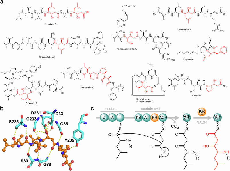
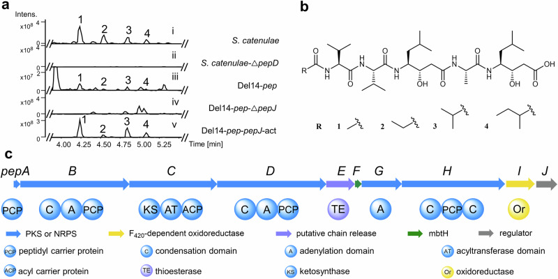
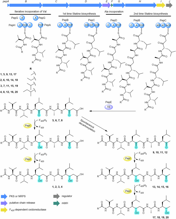
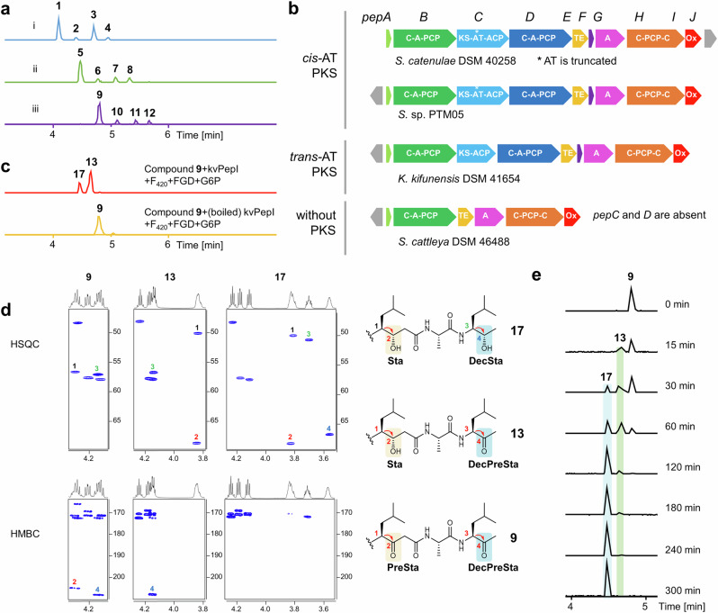
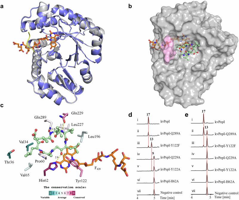
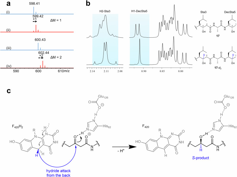
Pepstatins are potent inhibitors of aspartic proteases, featuring two statine residues crucial for target binding. However, the biosynthesis of pepstatins, especially their statine substructure, remains elusive. Here, we discover and characterize an unconventional gene cluster responsible for pepstatin biosynthesis, comprising discrete nonribosomal peptide synthetase and polyketide synthase genes, highlighting its trans-acting and iterative nature. Central to this pathway is PepI, an F420H2-dependent oxidoreductase. The biochemical characterization of PepI reveals its role in the tandem reduction of β-keto pepstatin intermediates. PepI first catalyzes the formation of the central statine, then produces the C-terminal statine moiety. The post-assembly-line formation of statine by PepI contrasts with the previously hypothesized biosynthesis involving polyketide synthase ketoreductase domains. Structural studies, site-directed mutagenesis, and deuterium-labeled enzyme assays probe the mechanism of F420H2-dependent oxidoreductases and identify critical residues. Our findings uncover a unique statine biosynthetic pathway employing the only known iterative F420H2-dependent oxidoreductase to date.


Conflict of interest statement

Competing interests: The authors declare no competing interests.

Figures

Fig. 1
Fig. 1. Representative statine-containing natural products, pepstatin binding mode, and the proposed pathway for statine biosynthesis.
a Pepstatin A and representative known compounds with Sta/Sta-like residues (highlighted in red). b Interactions between pepstatin A and cathepsin D (PDB ID:1LYB). Only hydrogen bonds between pepstatin A (orange) and residues in the protein binding pocket (cyan) are shown. c The prior hypothesis on statine biosynthesis involving a modular PKS KR domain.
Fig. 2
Fig. 2. Identification of pepstatins and the pep biosynthetic gene cluster.
a UPLC-HRMS analysis (base peak chromatogram (BPC)) of pepstatin congeners (1-4) produced by Streptomyces catenulae DSM40258 (i); Knockingout pepD abolished pepstatin 1-4 production (ii); Pepstatin congeners (1-4) produced by heterologous expression of BGC pep in Del14 (Del14-pep) (iii); The production of 1-4 significantly decreased by pepJ deletion in Del14-peppepJ (iv); The production of 1-4 increased in Del14-pep-pepJ-act by promoter exchange of pepJ (v). b The chemical structures of pepstatins isolated from Streptomyces catenulae DSM40258. c The schematic representation of the pepstatin BGC from S. catenulae DSM40258.
Fig. 3
Fig. 3. Proposed pepstatin biosynthesis pathway.
Fatty acids are likely activated to fatty acyl-CoA by acyl-CoA synthase and transferred to the carrier protein PepA, whereas PepG, PepH, PepB, PepC and PepD build up the peptide chain. The pentapeptide chain is subsequently released by PepE. Oxidation status changes are highlighted in turquoise to exhibit the two ketoreduction reactions catalyzed by PepI.
Fig. 4
Fig. 4. PepI catalyzes tandem ketone reductions.
a UPLC-HRMS analysis of pepstatins and unreduced β-keto intermediates in the pepI deletion mutant. (i) EICs ([M + H]+, blue) of 1 (644.42), 2 (658.44), 3 (672.45), 4 (686.47) from the improved heterologous expression of pep (pepJ_act); (ii) EICs ([M + H]+, green) of 5 (640.39), 6 (654.40), 7 (668.42), 8 (682.44) and (iii) EICs ([M + H]+, purple) of 9 (596.40), 10 (610.42), 11 (624.44), 12 (638.45) from the pepI deletion mutant based on Del14-pep- pepJ-act. b Representative pep-like actinobacterial pathways classified into three types mainly distinguished by containing cis-AT PKS, trans-AT PKS, or without PKS. c In vitro characterization of kvPepI. EICs ([M + H]+, red) of 13 (598.42) and 17 (600.43) produced by kvPepI reaction with 9 and EIC ([M + H]+, orange) of 9 from the control reaction using boiled kvPepI and 9 were shown. d HSQC and HMBC slices of 9, 13, and 17, showing the structural changes at the Sta residue, decarboxylated Sta residue (DecSta), the precursor of Sta (PreSta), and the precursor of DecSta (DecPreSta). e Time-course of kvPepI using 9 as the substrate, supplemented with F420, FGD, and G6P. BPCs of the reactions were shown.
Fig. 5
Fig. 5. Structural analysis, molecular modeling, and biochemical assays of kvPepI and mutants.
a Superposition of the kvPepI structure with (gray) and without (slate) F420 bound. Changes in the overall structure of the protein are minimal (Cα RMSD of 0.15 Å over all non-hydrogen atoms), except for the stabilization of a loop (residues 187 – 190; yellow line) that serves as a lid over the bound cofactor. b Hypothetical binding pose of U-shaped 9 in kvPepI – F420 complex structure. The missing loop (pink) was modeled using AlphaFold,. The orientation and conformation of 9 were modeled based on experimental data regarding the position of hydride transfer, as well as loss-of-activity-conferring amino acid mutations (details see experimental section). F420 (orange) and 9 (pale green) are shown as sticks (atom color: carbon black, nitrogen blue, oxygen red). c Schematic 3D representation of kvPepI – F420 interactions with modeled pose of 9. Hydrogen bonds are depicted with dotted yellow lines with distances given in Å, while all other residues form hydrophobic interactions with the substrate. The residues are colored according to the conservation score calculated using the ConSurf server,. Reactions using 9 (d) or 13 (e) as the substrate with kvPepI (i), Q289A (ii), Y122F (iii), Q229A (iv), Y122A (v), H62A (vi), and boiled kvPepI as negative control (vii). BPCs of reactions were shown.
Fig. 6
Fig. 6. Deuterium-labeled assays and mechanistic considerations for PepI.
a MS analysis of 13 and 17 in the kvPepI and 9 reactions without (i: 13, iii: 17, blue) or with (ii: 13-d1, iv: 17-d2, red) F420-5-d1 provided by the coupled hexokinase assay using d-Glucose-1-d1. b The 2 Da increase in the molecular mass indicated 17-d2 was the deuterated derivative of 17. The position of deuteration was deduced by comparing the 1H-NMR spectra.The methyl group H1-DecSta5 in 17-d2 showed a singlet whereas in 17 it was a doublet, together with the disappearance of the H2-DecSta5 signal in 17-d2, indicating the H2-DecSta5 was deuterated. Similarly, the H3-Sta3 was deuterated, as evidenced by the disappearance of H3-Sta3 signal and splitting pattern change of H2-Sta3. c Proposed reaction mechanism derived from the experimental observations indicates the orientation of the pepstatin peptide backbone in relation to His62 and F420 to achieve the observed S-product. R represents the lactyloligoglutamate tail of F420.

Similar articles

References

    1. Kunimoto, S., Aoyagi, T., Morishima, H., Takeuchi, T. & Umezawa, H. Mechanism of inhibition of pepsin by pepstatin. J. Antibiot.25, 251–255 (1972). - PubMed
    1. Aoyagi, T., Morishima, H., Nishizawa, R., Kunimoto, S. & Takeuchi, T. Biological activity of pepstatins, pepstanone A and partial peptides on pepsin, cathepsin D and renin. J. Antibiot.25, 689–694 (1972). - PubMed
    1. Marks, N., Grynbaum, A. & Lajtha, A. Pentapeptide (pepstatin) inhibition of brain acid proteinase. Science181, 949–951 (1973). - PubMed
    1. Matúz, K., Mótyán, J., Li, M., Wlodawer, A. & Tőzsér, J. Inhibition of XMRV and HIV-1 proteases by pepstatin A and acetyl-pepstatin. FEBS J.279, 3276–3286 (2012). - PMC - PubMed
    1. Seelmeier, S., Schmidt, H., Turk, V., Helm, K. & von der Human immunodeficiency virus has an aspartic-type protease that can be inhibited by pepstatin A. Proc. Natl. Acad. Sci. USA85, 6612–6616 (1988). - PMC - PubMed

MeSH terms

LinkOut - more resources