

Electromagnetic waves destabilize the SARS-CoV-2 Spike protein and reduce SARS-CoV-2 Virus-Like particle (SC2-VLP) infectivity
- PMID: 40374718
- PMCID: PMC12081674
- DOI: 10.1038/s41598-025-01896-1
Electromagnetic waves destabilize the SARS-CoV-2 Spike protein and reduce SARS-CoV-2 Virus-Like particle (SC2-VLP) infectivity
Abstract
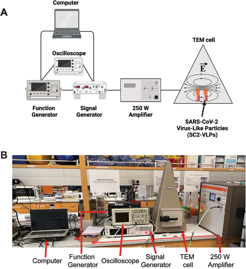
Infection and transmission of Severe Acute Respiratory Syndrome Coronavirus 2 (SARS-CoV-2) continues to pose a global public health concern. Using electromagnetic waves represents an alternative strategy to inactivate pathogenic viruses such as SARS-CoV-2. However, whether electromagnetic waves reduce SARS-CoV-2 infectivity is unclear. Here, we adapted a coplanar waveguide (CPW) to identify frequencies that could potentially neutralize SARS-CoV-2 virus-like particles (SC2-VLPs). Treatment of SC2-VLPs at frequencies between 2.5 and 3.5 GHz and an electric field of 413 V/m reduced infectivity. Exposure of SC2-VLPs to a frequency of 3.1 GHz -and to a lesser extent, 5.9 GHz- reduced their binding to antibodies targeting the SARS-CoV-2 Spike S1 receptor-binding domain (RBD) but did not alter the total levels of Spike, Nucleocapsid, Envelope, or Membrane proteins in virus particles. These results suggest that electromagnetic waves alter the conformation of Spike, thereby reducing viral attachment and entry. Overall, this data provides proof-of-concept in using electromagnetic waves for sanitation and prevention efforts to curb the transmission of SARS-CoV-2 and potentially other pathogenic enveloped viruses.
Keywords: Coplanar waveguide; Electromagnetic waves; SARS-CoV-2; Sanitation; Spike; Transmission.
© 2025. The Author(s).
Conflict of interest statement
Declarations. Competing interests: The authors declare no competing interests.
Figures
Update of
-
Electromagnetic waves destabilize the SARS-CoV-2 Spike protein and reduce SARS-CoV-2 Virus-Like Particle (SC2-VLP) infectivity.bioRxiv [Preprint]. 2024 Sep 12:2024.09.11.612487. doi: 10.1101/2024.09.11.612487. bioRxiv. 2024. Update in: Sci Rep. 2025 May 15;15(1):16836. doi: 10.1038/s41598-025-01896-1. PMID: 39314332 Free PMC article. Updated. Preprint.
Similar articles
-
Electromagnetic waves destabilize the SARS-CoV-2 Spike protein and reduce SARS-CoV-2 Virus-Like Particle (SC2-VLP) infectivity.bioRxiv [Preprint]. 2024 Sep 12:2024.09.11.612487. doi: 10.1101/2024.09.11.612487. bioRxiv. 2024. Update in: Sci Rep. 2025 May 15;15(1):16836. doi: 10.1038/s41598-025-01896-1. PMID: 39314332 Free PMC article. Updated. Preprint.
-
V367F Mutation in SARS-CoV-2 Spike RBD Emerging during the Early Transmission Phase Enhances Viral Infectivity through Increased Human ACE2 Receptor Binding Affinity.J Virol. 2021 Jul 26;95(16):e0061721. doi: 10.1128/JVI.00617-21. Epub 2021 Jul 26. J Virol. 2021. PMID: 34105996 Free PMC article.
-
Construction and immunogenicity of SARS-CoV-2 virus-like particle expressed by recombinant baculovirus BacMam.Microbiol Spectr. 2024 Aug 6;12(8):e0095924. doi: 10.1128/spectrum.00959-24. Epub 2024 Jun 25. Microbiol Spectr. 2024. PMID: 38916311 Free PMC article.
-
Structural basis of severe acute respiratory syndrome coronavirus 2 infection.Curr Opin HIV AIDS. 2021 Jan;16(1):74-81. doi: 10.1097/COH.0000000000000658. Curr Opin HIV AIDS. 2021. PMID: 33186231 Review.
-
Severe acute respiratory syndrome-coronavirus-2 spike (S) protein based vaccine candidates: State of the art and future prospects.Rev Med Virol. 2021 May;31(3):e2183. doi: 10.1002/rmv.2183. Epub 2020 Oct 15. Rev Med Virol. 2021. PMID: 33594794 Free PMC article. Review.
References
-
- Zaki, A. M., Van Boheemen, S., Bestebroer, T. M., Osterhaus, A. D. M. E. & Fouchier, R. A. M. Isolation of a novel coronavirus from a man with pneumonia in Saudi Arabia. N Engl. J. Med.367, 1814–1820 (2012). - PubMed
MeSH terms
Substances
Grants and funding
LinkOut - more resources
Full Text Sources
Medical
Miscellaneous
