

Time-restricted feeding attenuated hypertension-induced cardiac remodeling by modulating autophagy levels in spontaneously hypertensive rats
- PMID: 40374761
- PMCID: PMC12081920
- DOI: 10.1038/s41598-025-01587-x
Time-restricted feeding attenuated hypertension-induced cardiac remodeling by modulating autophagy levels in spontaneously hypertensive rats
Abstract
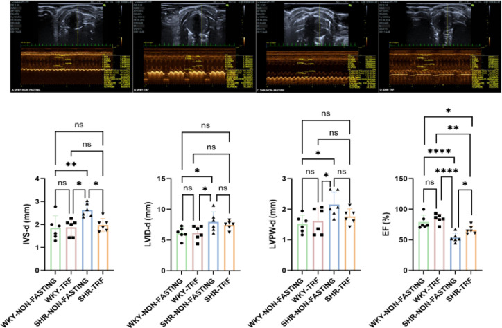
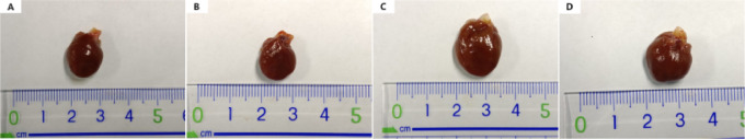
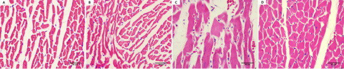
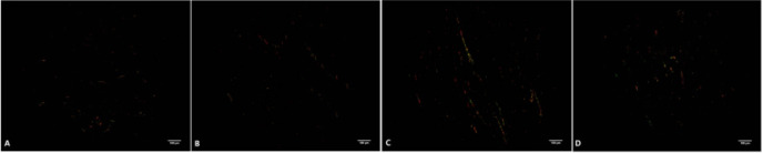
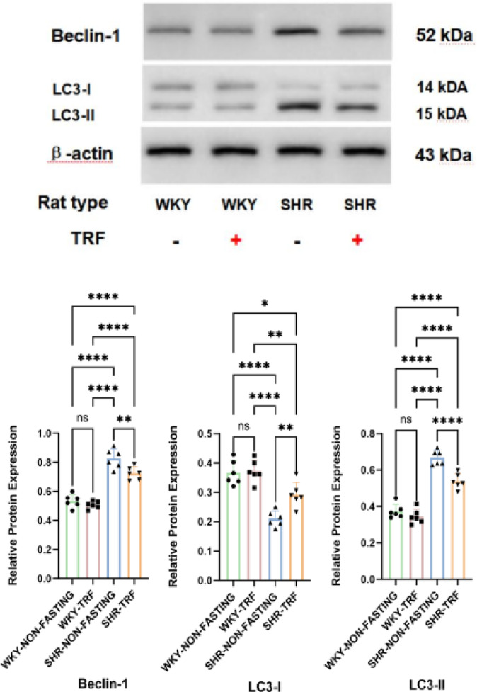
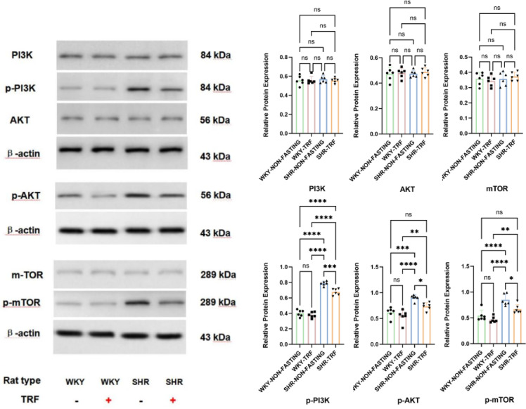
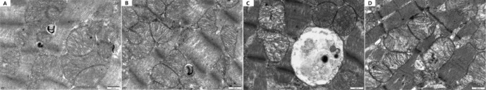
To investigate whether time-restricted feeding (TRF) can alleviate cardiac remodeling in spontaneously hypertensive rats (SHRs) by regulating autophagy levels. A 16-week TRF intervention was conducted on Wistar Kyoto (WKY) rats and SHRs, with dietary intake confined to the interval from 9:00 am to 5:00 pm each day. The study examined the impact of TRF on blood pressure (BP), cardiac morphology and function, and the expression levels of key proteins involved in autophagy and its associated signaling cascades. Transmission Electron Microscopy (TEM) was utilized to further evaluate autophagic changes in left ventricular (LV) tissues. TRF significantly mitigated systolic blood pressure (SBP), diastolic blood pressure (DBP), and mean blood pressure (MBP) in SHRs. Additionally, TRF improved ejection fraction (EF) and diminished interventricular septal thickness at end-diastole (IVS-d). The study further revealed that TRF enhanced the expression of microtubule-associated protein-I light chain 3 (LC3-I), while reducing that of microtubule-associated protein-II light chain 3 (LC3-II). Moreover, TRF suppressed the expression levels of Beclin-1, phosphorylated phosphoinositide 3-kinase (p-PI3K), phosphorylated protein kinase B (p-AKT), and phosphorylated mechanistic target of rapamycin (p-mTOR) in the LV tissues. TEM analysis confirmed that TRF could inhibit autophagy levels in the LV tissues. TRF can attenuate cardiac remodeling in SHRs by regulating autophagy levels.
Keywords: Autophagy; Cardiac remodelling; Hypertension; Spontaneously hypertensive rats; Time-restricted feeding.
© 2025. The Author(s).
Conflict of interest statement
Declarations. Competing interests: The authors declare no competing interests. Consent for publication: Not applicable.
Figures

Similar articles
-
Augmentation of autophagy by atorvastatin via Akt/mTOR pathway in spontaneously hypertensive rats.Hypertens Res. 2015 Dec;38(12):813-20. doi: 10.1038/hr.2015.85. Epub 2015 Jul 30. Hypertens Res. 2015. PMID: 26224487
-
Effect of farnesyltransferase inhibition on cardiac remodeling in spontaneously hypertensive rats.Int J Cardiol. 2013 Oct 9;168(4):3340-7. doi: 10.1016/j.ijcard.2013.04.038. Epub 2013 May 9. Int J Cardiol. 2013. PMID: 23664044
-
Effects of electroacupuncture at Taichong (LR 3) and Baihui (DU 20) on cardiac hypertrophy in rats with spontaneous hypertension.J Tradit Chin Med. 2019 Aug;39(4):502-508. J Tradit Chin Med. 2019. PMID: 32186097
-
Time-restricted feeding reduced blood pressure and improved cardiac structure and function by regulating both circulating and local renin-angiotensin systems in spontaneously hypertensive rat model.PLoS One. 2025 Apr 3;20(4):e0321078. doi: 10.1371/journal.pone.0321078. eCollection 2025. PLoS One. 2025. PMID: 40179126 Free PMC article.
-
Large blood pressure variability and hypertensive cardiac remodeling--role of cardiac inflammation.Circ J. 2009 Dec;73(12):2198-203. doi: 10.1253/circj.cj-09-0741. Epub 2009 Oct 29. Circ J. 2009. PMID: 19875896 Review.
References
-
- Di Palo, K. E. & Barone, N. J. Hypertension and heart failure: prevention, targets, and treatment. Cardiol. Clin.40 (2), 237–244. 10.1016/j.ccl.2021.12.011 (2022). - PubMed
-
- Nwabuo, C. C. & Vasan, R. S. Pathophysiology of hypertensive heart disease: beyond left ventricular hypertrophy. Curr. Hypertens. Rep.22 (2), 11. 10.1007/s11906-020-1017-9 (2020). - PubMed
MeSH terms
Substances
LinkOut - more resources
Full Text Sources
Medical
Miscellaneous
