. 2025 May 15;15(1):16900.
doi: 10.1038/s41598-025-99215-1.

A combined in silico and MD simulation approach to discover novel LpxC inhibitors targeting multiple drug resistant Pseudomonas aeruginosa

Affiliations

A combined in silico and MD simulation approach to discover novel LpxC inhibitors targeting multiple drug resistant Pseudomonas aeruginosa

Awadh Alanazi et al. Sci Rep. .

Abstract

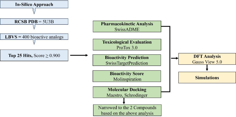
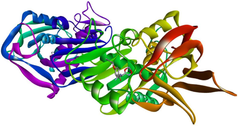
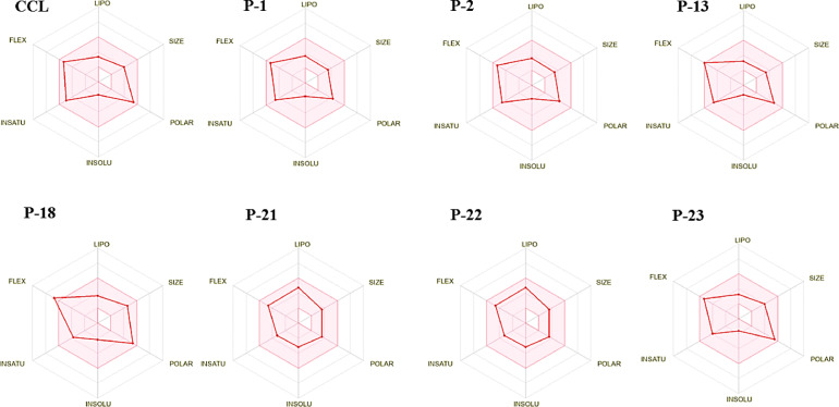
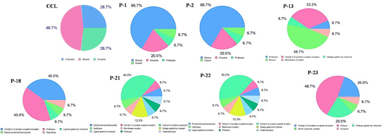
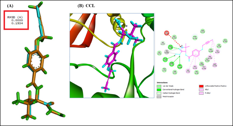
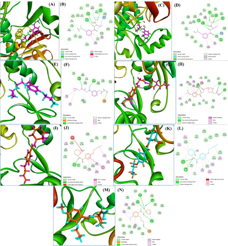
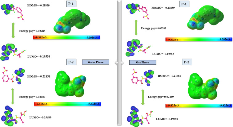
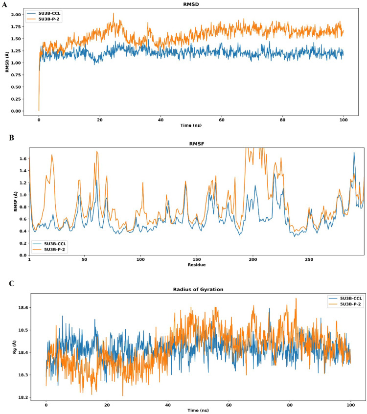
Pseudomonas aeruginosa (P. aeruginosa), a member of the ESKAPE family, is the major cause of infections leading to increased morbidity and mortality due to multidrug resistance (MDR). One of the main proteins involved in the Raetz pathway is LpxC, which plays a significant role in anti-microbial resistance (AMR). Our study aimed to identify a novel compound to combat MDR due to the LpxC protein. It involved in silico methods comprising molecular docking, simulations, ADMET profiling, and DFT calculations. First, an ADMET and bioactivity evaluation of the 25 top-hit compounds retrieved from ligand-based virtual screening was performed, followed by molecular docking. The results revealed compound P-2 as the lead compound, which was further subjected to DFT analysis and molecular dynamics (MD) simulations. With these analyses, our in silico study identified P-2, 3-[(dimethylamino)methyl]-N-[(2 S)-1-(hydroxyamino)-1-oxobutan-2-yl]benzamide as a potential lead compound that may behave as a very potent inhibitor of LpxC for the development of targeted therapies against MDR P. aeruginosa.

Keywords: ADMET; Bioactivity; DFT; In silico; LpxC; MD simulation; MDR; Molecular docking; Toxicity.


Conflict of interest statement

Declarations. Competing interests: The authors declare no competing interests.

Figures

Fig. 1
Fig. 1
Schematic workflow of the in silico approach of the current study.
Fig. 2
Fig. 2
The 3D structure of the protein LpxC (PDB ID: 5U3B), visualized on discovery studio visualizer.
Fig. 3
Fig. 3
Bioavailability radar of the top-hit inhibitory analogs in colored zones revealing the reliable physiochemical space for oral bioavailability.
Fig. 4
Fig. 4
Off-target prediction of the top-hit compounds, assessed through swiss target prediction.
Fig. 5
Fig. 5
(A) RMSD value of the superimposed original CCL (PubChem CID: 59323957; cyan) and re-docked pose (orange). (B) Visualization of the 3D allosteric pocket of the target protein (PDB ID: 5U3B) and 2D interactions of the CCL within the binding cavity of the target protein.
Fig. 6
Fig. 6
Binding mode of the complex 5U3B with the best inhibitory compounds. (A,C,E,G,I,K,M) show the visual 3D representations of the binding cavity of 5U3B with the top-scoring ligands P-1, P-2, P-13, P-18, P-21, P-22, and P-23, respectively. (B,D,F,H,J,L,N) depict the 2D plot of the binding interactions between the top inhibitors and LpxC (PDB ID: 5U3B).
Fig. 7
Fig. 7
Optimized structural geometry showing the FMO and MEPS of compounds P-1 and P-2.
Fig. 8
Fig. 8
Graphical representation of the simulation. (A) RMSD fluctuation of the CCL (blue) and P-2 (orange), showing the duration on the X-coordinate and RMSD on the Y-coordinate. (B) RMSF of the CCL (blue) and P-2 (orange), showing residues on the X-coordinate and RMSF on the Y-coordinate. (C) RGyr of the CCL (blue) and P-2 (orange), showing time duration on the X-coordinate and RGyr on the Y-coordinate.
Fig. 9
Fig. 9
Principal component analysis of the lead compound (P-2) in comparison to the CCL. (A) Combined scatter plots for both complexes (blue = CCL and orange = P-2). (B,D) Individual scatter plots of CCL and P-2, respectively in complex with the target protein 5U3B. (C,E) Plots colored to depict Gibb’s energy.
Fig. 10
Fig. 10
DCCM and FEL of the complexes. (A,B) DCCM of the CCL and P-2, respectively, in complex with the target protein 5U3B. (C,D) FEL of the CCL and P-2 in the form of Gibbs energy.
Fig. 11
Fig. 11
Graphical representation of MM/GBSA analysis, denoting time on the X-coordinate and energy in kcal/mol on the Y-coordinate.

Similar articles

References

    1. Horcajada, J. P. et al. Epidemiology and treatment of multidrug-resistant and extensively drug-resistant pseudomonas aeruginosa infections. Clin. Microbiol. Rev.32, (2019). - PMC - PubMed
    1. Amera, G. M. et al. Computer aided ligand based screening for identification of promising molecules against enzymes involved in peptidoglycan biosynthetic pathway from Acinetobacter baumannii. Microb. Pathog.147, 104205 (2020). - PubMed
    1. Tacconelli, E. et al. Discovery, research, and development of new antibiotics: the WHO priority list of antibiotic-resistant bacteria and tuberculosis. Lancet Infect. Dis.18, 318–327 (2018). - PubMed
    1. Beswick, E., Amich, J. & Gago, S. Factoring in the complexity of the cystic fibrosis lung to understand Aspergillus fumigatus and Pseudomonas aeruginosa interactions. Pathog (Basel Switzerland)9, (2020). - PMC - PubMed
    1. Shevade, A. & Naik, S. Mitigation of antimicrobial resistance (AMR) in G20. Indian Public. Policy Rev.4, 1–29 (2023).

MeSH terms

LinkOut - more resources