. 2025 Jun;17(6):1417-1451.
doi: 10.1038/s44321-025-00244-0. Epub 2025 May 15.

Genotype-specific differences in infertile men due to loss-of-function variants in M1AP or ZZS genes

Affiliations

Genotype-specific differences in infertile men due to loss-of-function variants in M1AP or ZZS genes

Nadja Rotte et al. EMBO Mol Med. 2025 Jun.

Abstract

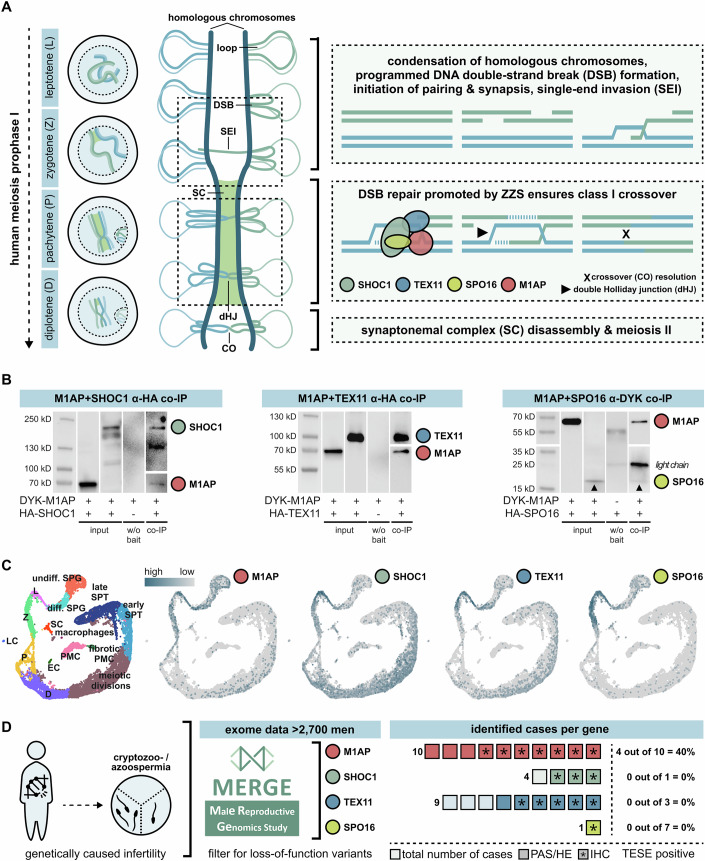
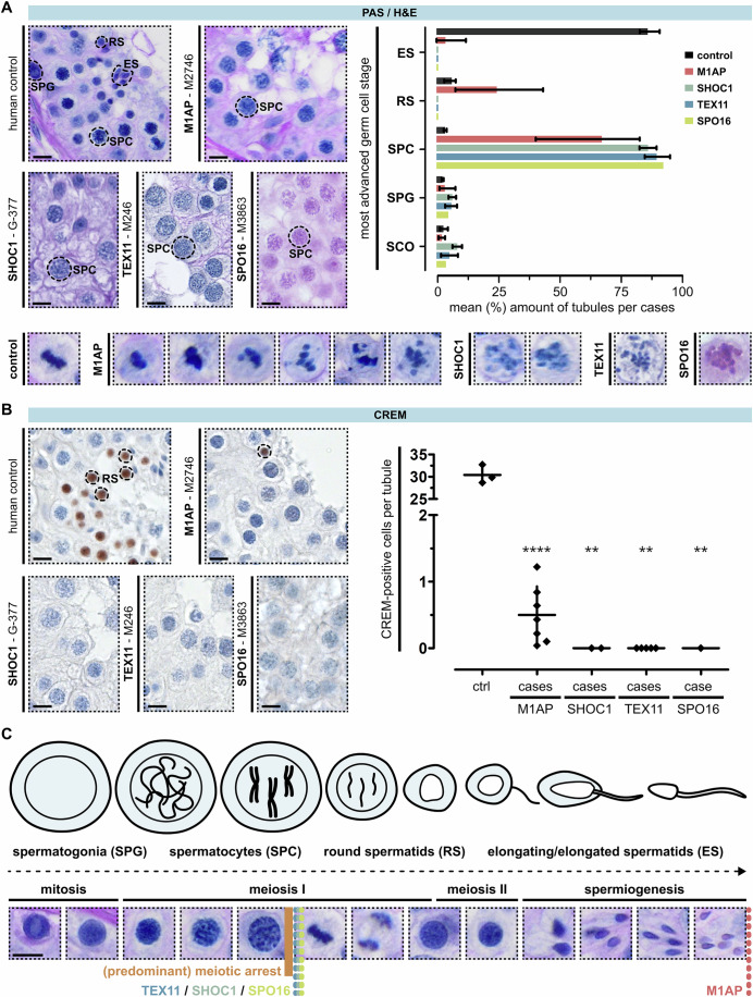
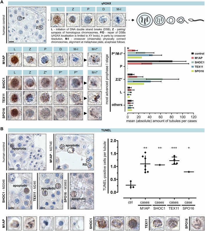
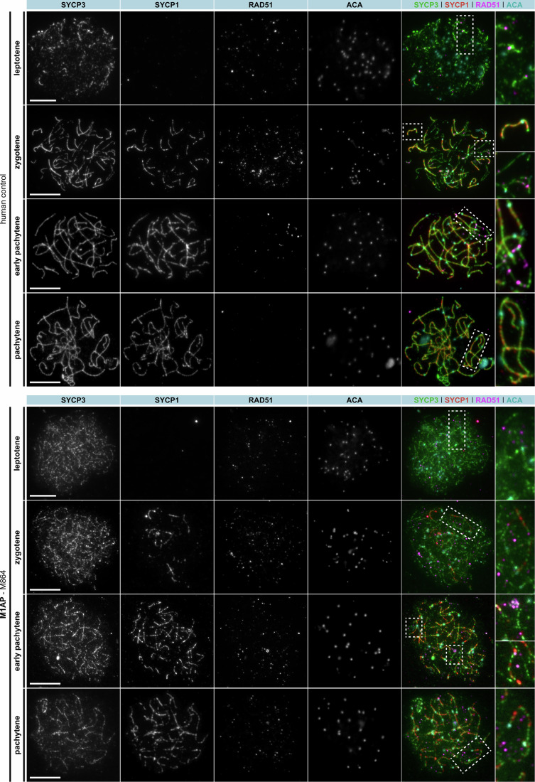
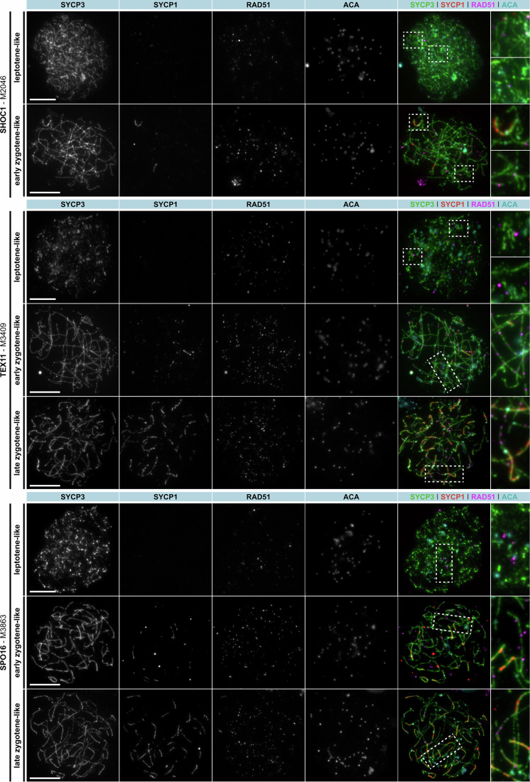
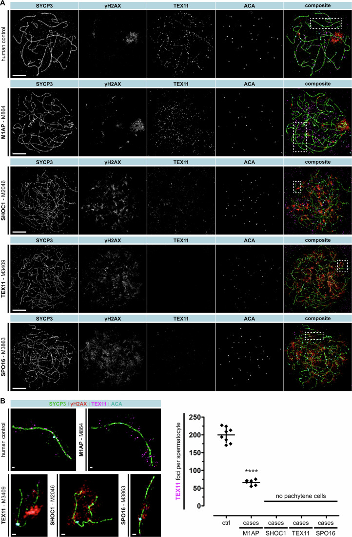
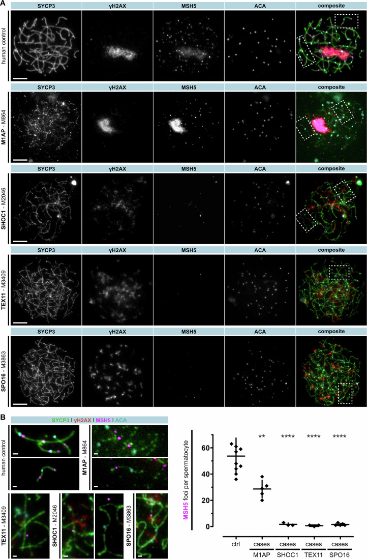
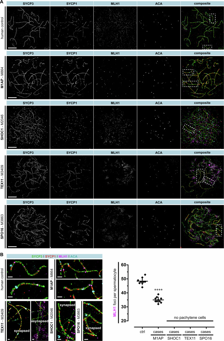
Male infertility has been linked to M1AP. In mice, M1AP interacts with the ZZS proteins SHOC1/TEX11/SPO16, promoting DNA class I crossover formation during meiosis. To determine whether M1AP and ZZS proteins are involved in human male infertility by recombination failure, we screened for biallelic/hemizygous loss-of-function (LoF) variants in the human genes to select men with presumed protein deficiency (N = 24). After in-depth characterisation of testicular phenotypes, we identified gene-specific meiotic impairments: men with ZZS deficiency shared an early meiotic arrest. Men with LoF variants in M1AP exhibited a predominant metaphase I arrest with rare haploid round or even elongated spermatids. These differences were explained by different recombination failures: deficient ZZS function led to incorrect synapsis of homologous chromosomes, unrepaired DNA double-strand breaks, and incomplete recombination. Abolished M1AP led to a reduced number of recombination intermediates and class I crossover. Medically assisted reproduction resulted in the birth of a healthy child, offering the possibility of fatherhood to men with LoF variants in M1AP. Our study establishes M1AP as an important, but non-essential, functional enhancer in meiotic recombination.

Keywords: Crossover; Infertility; M1AP; Meiosis; Recombination.


Conflict of interest statement

Disclosure and competing interest statement. The authors declare no competing interests.

Figures

Figure 1
Figure 1. Human meiotic recombination in men depends on ZZS function—and M1AP interacts with all three complex components.
(A) Schematic representation of human prophase I and meiotic recombination. To simplify, the global arrangement of just one pair of homologous chromosomes is depicted and homologues are differentiated with two colours (blue, green). The molecular mechanisms of class I crossover resolution are represented in a simplified manner within the dotted boxes. In leptotene (L), homologous chromosomes duplicate, condensate, and align. DNA double-strand breaks (DSBs) are initiated. During zygotene (Z), homologous chromosomes pair up and the initiation of synapsis is supported by the dynamic assembly of a ladder-like structure—the synaptonemal complex (SC). This complex provides a structural basis for meiotic recombination and in pachytene (P), the chromosomes are fully synapsed. DSB repair results in at least one crossover (CO) per chromosome pair (= obligatory crossover principle) and highly depends on SHOC1, TEX11, and SPO16 (= ZZS complex) activity. An integration of M1AP in this process was shown in mice (Li et al, 2023). Cells have completed DSB repair in diplotene (D), the SC disassembles, and homologues are physically connected by chiasmata. (B) Co-immunoprecipitation (IP) proved the interaction of human M1AP (detected by N-terminal DYK-tag) with each of the ZZS complex proteins (detected by C-terminal HA tag) co-transfected in HEK293T cells. Input and prey proteins served as positive and negative controls. Respective co-IP Western blot panels can be read from left to right as following: 1st lane = marker, 2nd = co-transfection of both plasmids, detection of M1AP in input sample, 3rd = co-transfection of both plasmids, detection of ZZS in input sample, 4th = transfection of pure prey protein and detection after immunoprecipitation (negative control), 5th = co-transfection of both plasmids, detection of both proteins from co-IP sample. In the last panel, the upturned arrowhead indicates the faint bands of SPO16 for better visualisation. Due to the low detection signal of SPO16, antibody chains of the co-IP specific anti-DYKDDDDK antibody, detected with anti-mouse HRP secondary antibody, are visible too. Experiments were conducted in three independent biological replicates. (C) Uniform manifold approximation and projection (UMAP) plot of obstructive azoospermic controls (N = 3) adapted from Di Persio et al, . Through mRNA expression profiling, individual stages of human spermatogenesis were clustered and visualised. Expression data of human M1AP, SHOC1, TEX11, and SPO16 mRNA was compiled by querying the dataset. (D) For each gene, variants leading to dysfunctional proteins were searched for in the MERGE study dataset to associate a deficient genotype with a distinct phenotype. Overall, ten men with LoF variants in M1AP, four men with LoF variants in SHOC1 (N = 3 from MERGE and N = 1 from GEMINI), nine men with LoF variants in TEX11, and one man with a LoF variant in SPO16 were selected. If possible, material aiming for testicular sperm extraction (TESE) was used for subsequent analyses including periodic acid-Schiff (PAS) or haematoxylin and eosin (H&E) staining (both highlighted in bolder colour), immunohistochemical staining (IHC = ∗), and spermatocyte spreads (one sample per gene of interest). A positive TESE result was only seen in men with LoF variants in M1AP (N = 4). Source data are available online for this figure.
Figure 2
Figure 2. Testicular phenotyping of men with loss-of-function variants in M1AP, SHOC1, TEX11, and SPO16.
(A) PAS staining of testicular tissue with full spermatogenesis and infertile men with LoF variants in genes encoding the ZZS proteins and M1AP. One representative case per gene is depicted (M1AP: M2746 (also shown in Appendix Fig. S6), SHOC1: G-377, TEX11: M246, SPO16: M3863). For all men, the most advanced germ cell type per tubule was assessed and is shown in the representative image and the bar graph. Data are presented as mean (%) and range, quantifying one biopsy per case (CTRL = 3 cases, M1AP = 7, SHOC1 = 2, TEX11 = 5, SPO16 = 1). In addition, intact (control, M1AP) metaphase and aberrant (M1AP, SHOC1, TEX11, SPO16) metaphase-like cells were observed (detail view). (B) Haploid germ cells were analysed by CREM localisation. Only men with LoF variants in M1AP had CREM-positive round spermatids. Compared to the controls, the total amount of these cells was significantly reduced. P values were determined between the control and M1AP group by an unpaired two-tailed t test (****P = 1.9765*10−10) or between the control and SHOC1, TEX11 or SPO16 group per one sample two-tailed t test (**P = 0.0016). Data is presented as the mean and standard deviation, quantifying one biopsy per case (CTRL = 3, M1AP = 7, SHOC1 = 2, TEX11 = 5, SPO16 = 1). (C) Schematic illustration and corresponding PAS staining of human spermatogenesis. Coloured dotted lines represent the identified gene-specific germ cell arrest. SPG spermatogonia, SPC spermatocytes, RS round spermatids, ES elongated spermatids. Figure EV3E also shows part of the panel. The scale bar represents 10 µm. Source data are available online for this figure.
Figure 3
Figure 3. Investigation of meiotic recombination and arrest in men with loss-of-function variants in M1AP, SHOC1, TEX11, and SPO16.
(A) Localisation of γH2AX in a control demonstrates the marker’s specific staining pattern during each substage of meiosis prophase I. Analysis of this marker in the infertile men revealed genotype-specific aberrations and meiotic arrest: M1AP = partial metaphase (M-I) arrest, SPO16/TEX11/SHOC1 = zygotene- (Z*) to early pachytene-like (P*) arrest, with occasional metaphase-like cells (M-I*). Data are presented as mean and range, quantifying one biopsy per case (CTRL = 3 cases, M1AP = 7, SHOC1 = 2, TEX11 = 5, SPO16 = 1. (B) TUNEL assay showed increased apoptosis in patients independent of the genetic background (dot plot). Most TUNEL-positive cells already showed hallmarks of apoptosis (detail view), and only men with LoF variants in M1AP showed TUNEL-negative metaphase cells with correctly aligned chromosomes, round, and elongated spermatids. P values were determined between the control and M1AP or TEX11 group by an unpaired two-tailed t test (**P = 0.0026, ***P = 0.0003, respectively) or between the control and SHOC1 or SPO16 group per one sample two-tailed t test (**P = 0.0096, *P = 0.0219, respectively). Data are presented as the mean and standard deviation, quantifying one biopsy per case (CTRL = 3 cases, M1AP = 7, SHOC1 = 1, TEX11 = 4, SPO16 = 1). The scale bar represents 10 µm. Source data are available online for this figure.
Figure 4
Figure 4. Human spermatocyte spreads showed persistent RAD51 foci throughout prophase I in the man with loss-of-function variant in M1AP.
Human spermatocyte spreads were stained for synaptonemal complex formation (= SYCP3, green + SYCP1, red), centromeric regions (= ACA, cyan), and single-strand invasion (RAD51 = magenta). The LoF variant in M1AP led to persistence of RAD51 foci during pachytene progression. The scale bar represents 10 µm. Source data are available online for this figure.
Figure 5
Figure 5. Human spermatocyte spreads showed persistent RAD51 foci in men with loss-of-function variants in the ZZS genes throughout incomplete prophase I progression.
Human spermatocyte spreads were stained for synaptonemal complex formation (= SYCP3, green + SYCP1, red), centromeric regions (= ACA, cyan), and single-strand invasion (RAD51 = magenta). LoF variants in SHOC1, TEX11, or SPO16 led to persistence of RAD51 until late zygotene-like stage. The scale bar represents 10 µm. Source data are available online for this figure.
Figure 6
Figure 6. Reduced TEX11 foci in a man with loss-of-function variant in M1AP (M864).
(A) Human spermatocyte spreads were stained for synaptonemal complex formation (= SYCP3, green), DNA double-strand breaks (γH2AX = red), centromeric regions (= ACA, cyan), and ZZS recruitment (TEX11 = magenta). Pachytene-like cells with progressed DNA double-strand breaks and γH2AX-positive XY bodies of a control and M864, deficient for M1AP, were compared. LoF in SHOC1 (M2046), TEX11 (M3409), and SPO16 (M3863) was associated with an early meiosis I defect including incomplete DSB repair and disturbed assembly of early recombination intermediates leading to a lack of TEX11 foci on the chromosomal axes. (B) Along the chromosomal axis of wild-type spermatocytes, 199.88 ± 20.16 TEX11 foci were counted per cell, while M1AP deficiency significantly reduced the recruitment of TEX11 to the axes (66.00 ± 7.16 foci per cell). In contrast, men deficient for SHOC1 (M2046), TEX11 (M3409), or SPO16 (M8363) completely lacked pachytene cells, and no quantification was performed in these cases. P value was determined between the control and M1AP group by an unpaired two-tailed t test (****P = 6.2408*10−9). Data are presented as the mean and standard deviation, quantifying individual pachytene spermatocytes of one case each (CTRL = eight spermatocytes, M1AP = six spermatocytes). The scale bars represent 10 µm or 1 µm for magnification. Source data are available online for this figure.
Figure 7
Figure 7. Human spermatocyte spreads showed reduced or absent MSH5 foci in men with loss-of-function variants in M1AP or the ZZS genes.
(A) Human spermatocyte spreads were stained for synaptonemal complex formation (= SYCP3, green), DNA double-strand breaks (γH2AX = red), centromeric regions (= ACA, cyan), and ZMM recruitment (MSH5 = magenta). A control and men deficient for M1AP (M864), SHOC1 (M2046), TEX11 (M3409), or SPO16 (M8363) were compared. (B) Distinct MSH5 foci were seen on chromosome axes of pachytene cells in the control and M864. The LoF variant in M1AP led to a significant reduction of MSH5 foci, while LoF variants in SHOC1, TEX11, or SPO16 led to a nearly complete absence of MSH5 foci. Bleed-through was observed between the red and magenta channels. Therefore, only MSH5 foci that were distinct from the XY body (γH2AX) were counted because the γH2AX signal was superimposed on the MSH5 foci. P value was determined between the control and each LoF group by an unpaired two-tailed t test (**P = 0.0023, ****P = 5.7658*10−5 (SHOC1), 4.8489*10−5 (TEX11), 1.5895*10−6 (SPO16)). Data are presented as the mean and standard deviation, quantifying individual pachytene (CTRL, M1AP) or most progressed zygotene-like spermatocytes (ZZS) of one case each (CTRL = nine spermatocytes, M1AP = five spermatocytes, SHOC1/TEX11 = three spermatocytes, SPO16 = five spermatocytes). The scale bars represent 10 µm and 1 µm for magnification. Source data are available online for this figure.
Figure 8
Figure 8. A man with loss-of-function variant in M1AP showed reduced MLH1 foci.
(A) Human spermatocyte spreads were stained for synaptonemal complex formation (= SYCP3, green + SYCP1, red), centromeric regions (= ACA, cyan), and designated crossover sites (= MLH1, magenta). A control and M864, deficient for M1AP (also seen in Fig. 9A), M2046 (SHOC1), M3409 (TEX11), and M3863 (SPO16) were compared. (B) MLH1 localisation was seen on synapsed chromosome axes of pachytene spermatocytes in the control and M864. The total number of MLH1 foci per spermatocyte was reduced in M864. In contrast, men deficient for SHOC1 (M2046), TEX11 (M3409), or SPO16 (M8363) completely lacked pachytene-like cells. Instead, spermatocytes showed asynapsed (SHOC1) or incomplete synapsed (TEX11 and SPO16) chromosomes where SYCP1 localisation was lacking or reduced. No MLH1 was seen because cells arrested in a zygotene-like stage, and no quantification was performed in these cases. P value was determined between the control and M1AP group by an unpaired two-tailed t test (****P = 4.6836*10−10). Data are presented as the mean and standard deviation, quantifying individual pachytene spermatocytes of one case each (CTRL nine spermatocytes, M1AP ten spermatocytes). The scale bars represent 10 µm and 1 µm for magnification. Source data are available online for this figure.
Figure 9
Figure 9. A man with loss-of-function variant in M1AP showed at least one class I crossover per chromosome.
(A) A pachytene spermatocyte of M864 deficient for M1AP was stained for SYCP3 (green), SYCP1 (red), ACA (centromere, cyan), and MLH1 (magenta). Panel is also seen in Fig. 5A. (B) Super resolution structured illumination microscopy (SR-SIM) of the spread spermatocyte shown in (A). Homologous chromosomes were digitally separated to mark the MLH1 foci (dotted circles), which represent the sites of class I crossover. In this spermatocyte, each pair of homologues has at least one MLH1 focus and, thus, the obligatory crossover principle is met. The scale bars represent 10 µm and 1 µm for magnification.
Figure 10
Figure 10. Proof-of-principle: M1AP-associated infertility can be overcome by medically assisted reproduction (MAR).
(A) One man (M2746) with a loss-of-function variant in M1AP was diagnosed with cryptozoospermia and predominant meiotic arrest. Three cycles of intracytoplasmic sperm injection (ICSI) with ejaculate-derived spermatozoa resulted in the birth of a healthy boy. Segregation analysis showed the autosomal-recessive inheritance pattern of the frameshift variant c.676dup. (B) Illustration of how predominant meiotic arrest caused by M1AP-associated infertility still enables fatherhood.
Figure EV1
Figure EV1. Full-length M1AP is mandatory for the protein-protein interaction with TEX11.
(A) The protein tolerance landscape of M1AP illustrates the respective regions selected for mutagenesis for cloning truncated versions (Q108*: c.322 C > T p.Gln108Ter, C335*: c.1005 T > A p.Cys335Ter, L433*: c.1297 C > T, c.1298 T > A p. Leu433Ter). Positions were selected in tolerant regions (blue, green) of M1AP to prevent destruction of the protein’s function. The construct c.676dup p.W226L*4 has been described in (Wyrwoll et al, 2020). (B) Western blot analysis of the input lysates of the co-transfection of full-length TEX11 (WT, detected by C-terminal HA tag) with full-length (WT) and truncated M1AP constructs (detected by N-terminal DYK-tag) confirm the expression in HEK293T cells. (C) Co-immunoprecipitation (IP) proved the interaction of human WT M1AP with WT TEX11. In contrast, no truncated M1AP was detected upon co-transfection with TEX11, pointing towards an absence of protein-protein interaction and thereby specifying the M1AP-TEX11 WT interaction. Experiments were replicated in three biological replicates. Source data are available online for this figure.
Figure EV2
Figure EV2. M1AP splice site variant identified in M3609.
(A) Amplified minigene cDNA encompassing—c.1073_1074+10del or respective wild-type (control/WT) sequence. (B) Schematic illustration of variant effect on genomic, transcriptomic, and protein level combined with sequencing results for each minigene product reveals aberrant splicing. In the WT minigene construct, M1AP exon 7 (Ex7) is encompassed by two known exons of rat Insulin 2, exon 3 and exon 4 (rat Ins2 Ex3/Ex4). In M3609, the variant led to two splicing products: one (MUT1) showed the recognition of a cryptic splice site leading to a frameshift and premature stop codon in M1AP exon 8. The second (MUT2) resulted in skipping of exon 7 and a premature stop codon in exon 8. (C) Both splicing products lead to the loss of amino acids, presumably effecting M1AP’s function and interaction.
Figure EV3
Figure EV3. SHOC1 splice site variant identified in M3260 with predominant meiotic arrest and rare round spermatids.
(A) SHOC1 (NM_173521.5) has 26 exons and its corresponding protein comprises 1444 amino acids. Amplified minigene cDNA encompassing—c.1939+2 T > C (M3260) or respective wild-type (control/WT) sequence. (B) Schematic illustration of variant effect on genomic, transcriptomic, and protein level combined with sequencing results for each minigene product reveals aberrant splicing. In the WT minigene construct, SHOC1 exon 13 (Ex13) is encompassed by two known exons of rat Insulin 2, exon 3 and exon 4 (rat Ins2 Ex3/Ex4). In M3260, the variant resulted in in-frame skipping of exon 13 and a predictive loss of 59 amino acids representing 4% of the total protein. (C) This affects the distant helicase hits region but not the highly conserved ‘SHOC1 homology region’ (amino acids 937–1105, NP_775792; Macaisne et al, 2008). This region contains an XPF endonuclease-like central and a helix-hairpin-helix (HhH2) domain and is important for the XPF-ERCC1-like complex formation between SHOC1 and SPO16 (De Muyt et al, ; Zhang et al, 2019). Yeast studies highlighted that the N-terminal part of Zip2 is linked to the chromosome axis and the other ZMM components through Zip4 interaction, while the XPF domain interacts exclusively with Spo16 (De Muyt et al, 2018). Given that M3260 expresses all exons of SHOC1 except for exon 13, the interaction with SPO16 and in parts with the ZMM proteins, such as TEX11, remains intact. However, a changed protein conformation due to the loss of exon 13 could influence some of these interactions and explain the observed testicular phenotype (D) of predominant meiotic arrest with rare round spermatids that were positive for CREM-staining. H3S10 staining showed only aberrant metaphase I-like spermatocytes (M-I*). TUNEL staining showed an increased number of apoptotic spermatocytes similar to patients with complete LoF variants in M1AP, SHOC1, TEX11, or SPO16. In γH2AX staining, single tubules contained pachytene-like cells (P) with a clearly distinguishable XY body were observed, which is in line with the presence of round spermatids. In addition, also aberrant pachytene-like cells were present. (E) The specific type of arrest of M3260 is described as a metaphase I arrest (MM-I) with rare round spermatids (RS) (panel taken from Fig. 2C). The scale bar represents 100 µm and 10 µm, respectively.
Figure EV4
Figure EV4. SPO16 loss-of-function variant identified in M3863.
(A) Sanger sequencing of M3863 revealed the frameshift variant c.266del leading to premature stop codon (p.Leu89Trpfs*15). (B) Such a truncated protein would lack the highly conserved helix-hairpin-helix (HhH2) domain, which is important for the XPF-ERCC1-like complex formation between SHOC1 and SPO16 (De Muyt et al, ; Zhang et al, 2019). (C) Conversation analysis of the SPO16 variant. (D) The premature stop codon would truncate 42.5% of the complete protein.
Figure EV5
Figure EV5. Euploidy analysis of M2746, his child, and the child’s mother.
Genome sequencing data was queried and read counts were normalised by dividing the median read count of each chromosome by the median read count of all autosomes. Normalised (log2) read counts of autosomes (0.97 to 1.06) and of gonosomes (0.49 to 0.51) gave no evidence for chromosome aneuploidies.

Similar articles

References

    1. Arango NA, Huang TT, Fujino A, Pieretti-Vanmarcke R, Donahoe PK (2006) Expression analysis and evolutionary conservation of the mouse germ cell-specific D6Mm5e gene. Dev Dyn 235:2613–2619 - PubMed
    1. Arango NA, Li L, Dabir D, Nicolau F, Pieretti-Vanmarcke R, Koehler C, McCarrey JR, Lu N, Donahoe PK (2013) Meiosis I arrest abnormalities lead to severe oligozoospermia in meiosis 1 arresting protein (M1ap)-deficient mice. Biol Reprod 88:1–11 - PMC - PubMed
    1. Baker SM, Plug AW, Prolla TA, Bronner CE, Harris AC, Yao X, Christie DM, Monell C, Arnheim N, Bradley A et al (1996) Involvement of mouse Mlh1 in DNA mismatch repair and meiotic crossing over. Nat Genet 13:336–342 - PubMed
    1. Bolcun-Filas E, Handel MA (2018) Meiosis: the chromosomal foundation of reproduction. Biol Reprod 99:112–126 - PubMed
    1. Börner GV, Hochwagen A, MacQueen AJ (2023) Meiosis in budding yeast. Genetics 225:1–33 - PMC - PubMed

LinkOut - more resources