. 2025 May 15;8(1):758.
doi: 10.1038/s42003-025-08175-9.

SEC24D depletion induces osteogenic differentiation deficiency by inactivating the ATF6/TGF-β/Runx2 regulatory loop

Affiliations

SEC24D depletion induces osteogenic differentiation deficiency by inactivating the ATF6/TGF-β/Runx2 regulatory loop

Jing Zhang et al. Commun Biol. .

Abstract

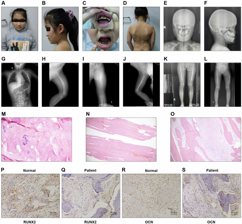
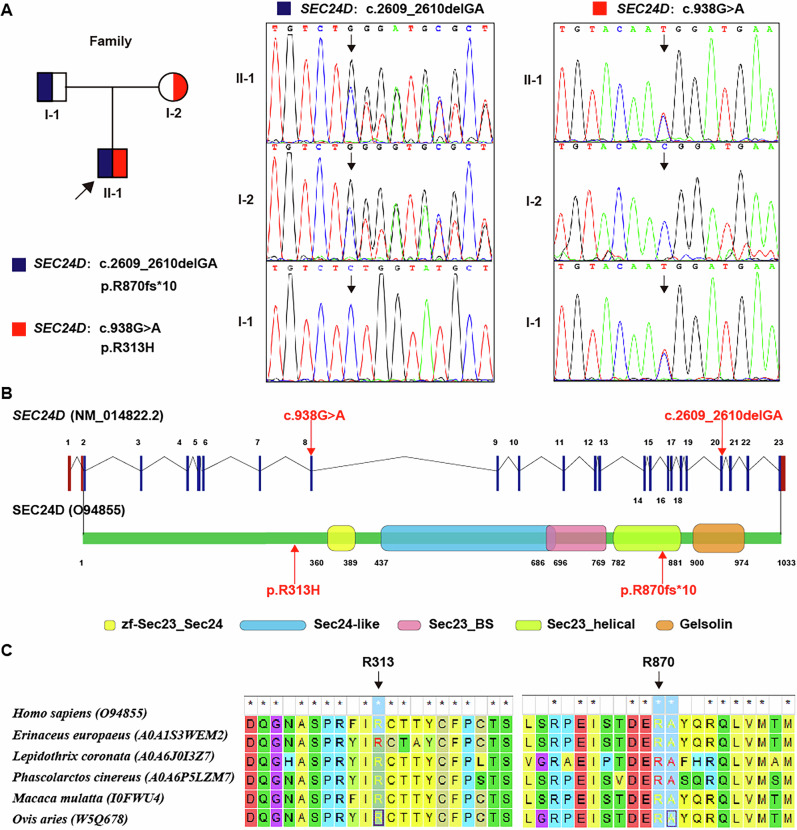
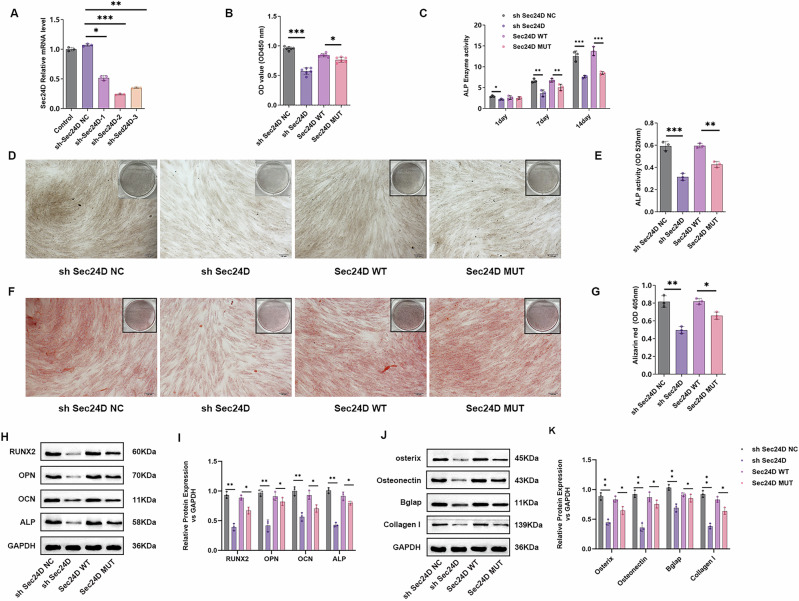
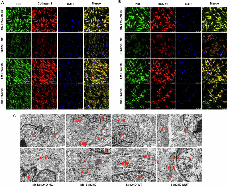
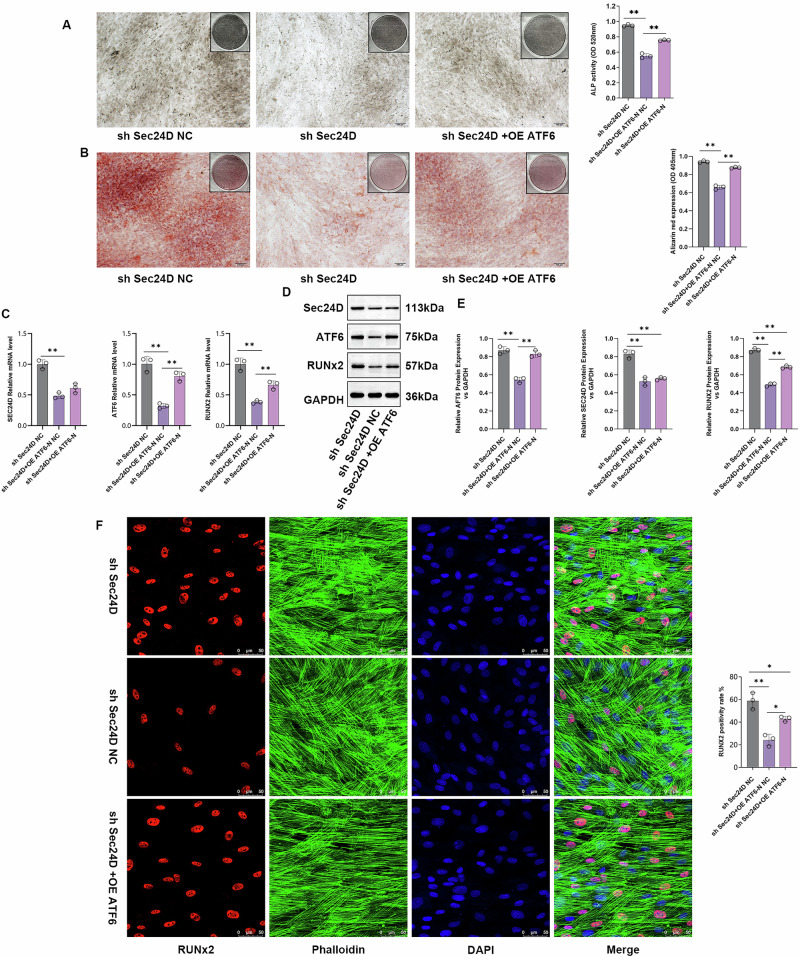
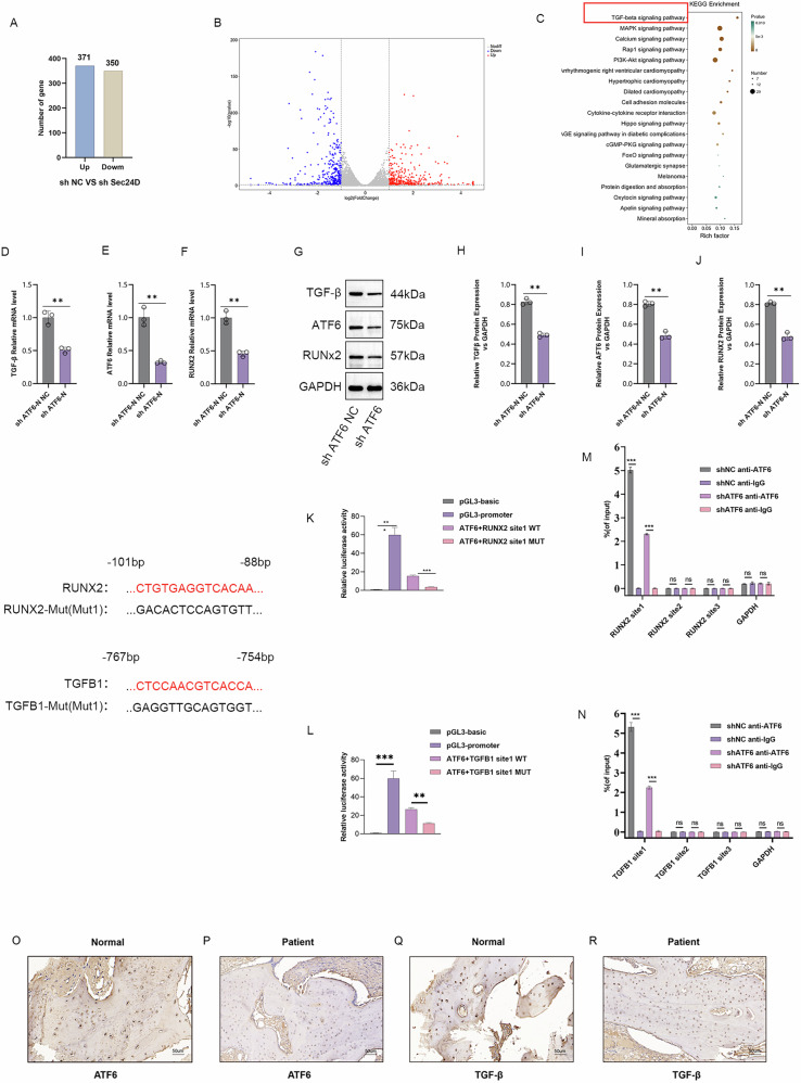
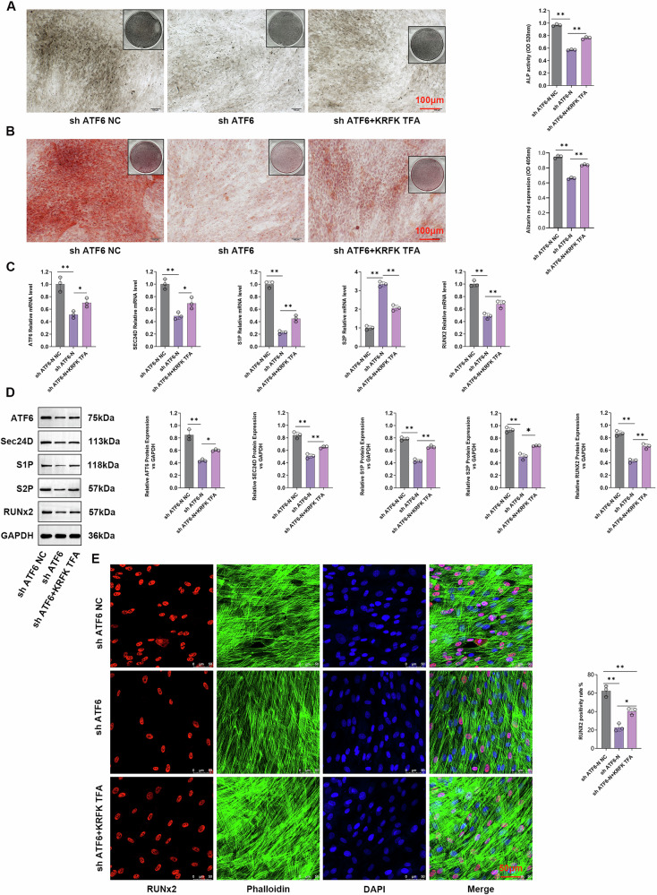
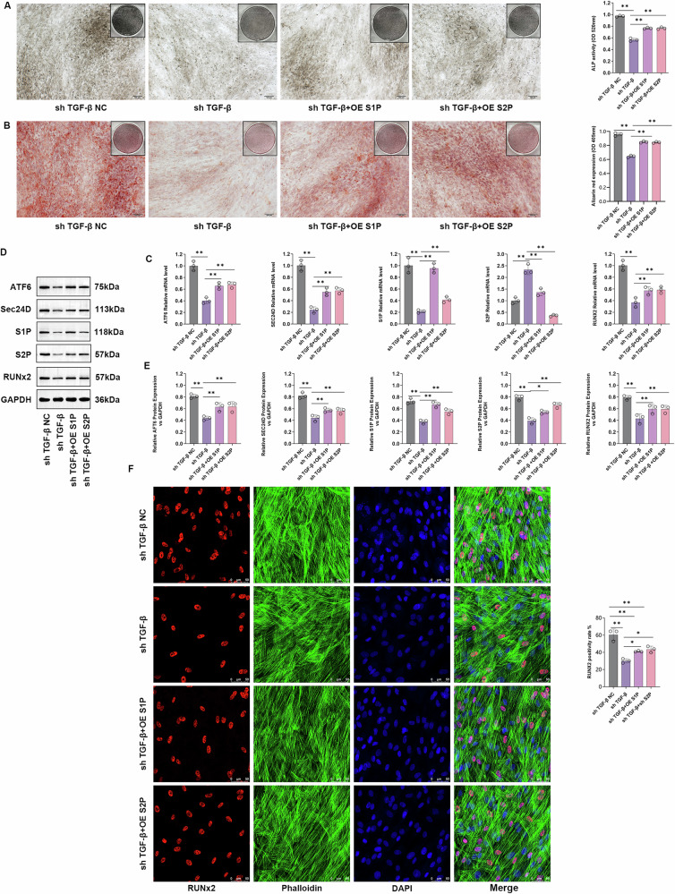
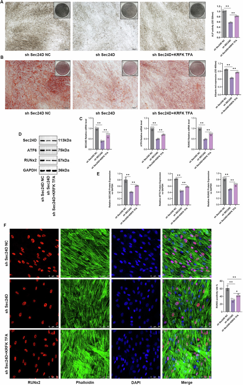
Protein coat complexes strongly influence intracellular cargo trafficking. Coatopathies represent a wide range of genetic conditions caused by mutations in protein coat components. The SEC24D gene, which encodes a Sec24 isoform that constitutes a cargo-specific capturer in the COPII coat, is responsible for a rare type of autosomal recessive osteogenesis imperfecta. We report an OI patient. Clinical and imaging findings suggested that the patient had OI. Genetic detection by whole-exome sequencing (WES) identified a compound heterozygous SEC24D variants, including c.2609_2610delGA (p. R870fs*10) and c.938G>A (p. R313H). In silico analysis suggested that the missense R313H mutation most likely affects protein stability and secondary structure. In vitro studies showed that knockdown or mutation of SEC24D affected the osteogenic differentiation of mesenchymal stem cells (MSCs) and inducted ER stress. Transcriptomic sequencing suggested that the TGF-β pathway mediated the destructive effect of SEC24D depletion on osteogenic differentiation. Further experiments confirmed that ATF6 participated in regulating the TGF-β pathway and osteogenic biomarkers by SEC24D. This study identified a SEC24D variation causing OI, which expanded the mutation spectrum of this gene. Further studies on the mechanism of action showed that SEC24D defects may induce osteogenic differentiation deficiency by inactivating the ATF6/TGF-β/Runx2 regulatory loop.


Conflict of interest statement

Competing interests: The authors declare no competing interests. Ethics approval and consent to participate: This study was approved by the Ethics Committee of Shijiazhuang Obstetrics and Gynecology Hospital (approval No. 20240057), and written informed consent was obtained from all participants.

Figures

Fig. 1
Fig. 1. The examination of patients included in this study.
AD Photos of the patient’s face, teeth, and back. EL X-ray examination of the patient. MO HE examination of femoral tissues. P-S. Immunohistochemistry detection of RUNX2 and OCN protein expression levels in femoral tissues of patients and normal people.
Fig. 2
Fig. 2. Genetic analysis of the patient’s causative variation.
A A compound heterozygous variation in the SEC24D gene was identified. B The location at the nucleic acid and peptide chain levels of the SEC24D gene. C The cross-species conservation of the two residues identified by MEGA7 analysis.
Fig. 3
Fig. 3. Knockdown or mutation of SEC24D affects the osteogenic differentiation of mesenchymal stem cells.
A Expression validation of different transfected groups. B The viability of different transfected groups. C ELISA detection of ALP in different transfected groups. D ALP staining in different transfected groups. E Statistical analysis of ALP staining in different transfected groups. F Alizarin Red staining in different transfected groups. G Statistical analysis of Alizarin Red staining in different transfected groups. H Western blot detection of ALP, RUNX2, OPN, and OCN protein expression levels in different transfected groups. I Statistical analysis of ALP, RUNX2, OPN, and OCN protein expression. J Western blot analysis of Bglap, Colla1, Osteonectin, and Osteris protein expression in different transfected groups. K Statistical analysis of Bglap, Colla1, Osteonectin, and Osteris protein expression. n = 3 independent experiments. The error bars are equivalent throughout the Figure.
Fig. 4
Fig. 4. The SEC24D mutation downregulates RUNX2 and promotes ER stress.
A Immunofluorescence staining of different transfection groups for protein disulfide isomerase (PDI)/Colla1. B Immunofluorescence staining of different transfection groups for protein disulfide isomerase (PDI)/RUNX2. C Transmission electron microscopy observation of different transfection groups. In the sh SEC24D-NC group, a track-like dense and coherent arrangement with ribosomes attached to the ER surface was observed. In the sh-SEC24D group, swelling, fragmentation, and disrupted coherence with detached ribosomes were observed. A lower ER status was detected in the SEC24D-MUT group than in the SEC24D-WT group.
Fig. 5
Fig. 5. SEC24D Regulates Osteogenesis by Modulating the Expression of RUNX2 and ATF6-N.
A ALP staining and quantification in different transfection groups. B Alizarin Red staining and quantification in different transfection groups. C Relative expression levels of SEC24D, ATF6-N, and RUNX2 mRNA in different transfection groups. D Protein expression levels of SEC24D, ATF6-N, and RUNX2 in different transfection groups. E Western blot (WB) statistical analysis. F Immunofluorescence staining and quantification of Ghostine/RUNX2 in different transfection groups. n = 3 independent experiments. The error bars are equivalent throughout the Figure.
Fig. 6
Fig. 6. Transcriptomic Sequencing to Observe the Potential Effect of SEC24D Knockdown on Osteogenic Differentiation, revealing that ATF6-N regulates the Transcription of RUNX2 and TGF-β.
A Venn diagrams. B Heatmaps. C KEGG analysis. D, E, F Expression levels of ATF6-N, TGF-β, and RUNX2 mRNA in different transfection groups. GJ WB statistical analysis and relative protein expression levels of ATF6-N, TGF-β, and RUNX2 in different transfection groups. K, L Luciferase reporter assay, and M-N ChIP Experiment demonstrate that ATF-N can act on the transcription of Runx2 and TGFβ, respectively. OR Immunohistochemistry detection of ATF6 and TGF-β protein expression levels in bone tissues of patients and normal people. n = 3 independent experiments. The error bars are equivalent throughout the Figure.
Fig. 7
Fig. 7. Involvement of the TGF-β signaling pathway in the SEC24D-regulated osteogenic differentiation of RUNX2 cells.
A ALP staining and statistics of different transfection groups. B Alizarin red staining and statistical analysis of the different transfection groups. C Relative expression levels of SEC24D, ATF6-N, and RUNX 2 mRNA in different transfection groups. D Differently transfected histone expression levels and WB statistical analysis. E Immunofluorescence staining and statistics of the ghost pen cyclic peptide/RUNX2 ratio in different transfection groups. n = 3 independent experiments. The error bars are equivalent throughout the Figure.
Fig. 8
Fig. 8. TGF-β regulates the expression of S1P and S2P.
A ALP staining and quantification in different transfectio n groups. B Safranin O staining and quantification in different transfection groups. C Relative expression levels of S1P, S2P, ATF6, SEC24D, and RUNX2 mRNA in different transfection groups. D Relative protein expression levels of S1P, S2P, ATF6, SEC24D, and RUNX2 in different transfection groups. E WB statistical analysis. F Immunofluorescence staining of different transfection groups using GhostPepper peptide/RUNX2. n = 3 independent experiments. The error bars are equivalent throughout the Figure.
Fig. 9
Fig. 9. The involvement of the TGF-β signaling pathway in the regulation of RUNX2 osteogenic differentiation mediated by SEC24D.
A ALP staining and statistical analysis of different transfection groups. B Alizarin Red staining and statistical analysis of different transfection groups. C Relative expression levels of SEC24D, ATF6, and RUNX2 mRNA in different transfection groups. D Protein expression levels of SEC24D, ATF6, and RUNX2 in different transfection groups. E Western blot (WB) analysis. F Immunofluorescence staining and statistical analysis of different groups transfected with Ghost Pepper/RUNX2. n = 3 independent experiments. The error bars are equivalent throughout the Figure.

Similar articles

References

    1. Verma, K., Verma, M., Chaphalkar, A. & Chakraborty, K. Recent advances in understanding the role of proteostasis. Fac. Rev.10, 72 (2021). - PMC - PubMed
    1. Bonifacino, J. S. & Lippincott-Schwartz, J. Coat proteins: shaping membrane transport. Nat. Rev. Mol. Cell Biol.4, 409–414 (2003). - PubMed
    1. Kreis, T. E. & Pepperkok, R. Coat proteins in intracellular membrane transport. Curr. Opin. Cell Biol.6, 533–537 (1994). - PubMed
    1. Dell’Angelica, E. C. & Bonifacino, J. S. Coatopathies: Genetic disorders of protein coats. Annu Rev. Cell Dev. Biol.35, 131–168 (2019). - PMC - PubMed
    1. Miller, E. A. & Schekman, R. COPII - a flexible vesicle formation system. Curr. Opin. Cell Biol.25, 420–427 (2013). - PMC - PubMed

MeSH terms

Substances

LinkOut - more resources